Book of abstracts - www.isfri2021.uj.edu.pl
←
→
Page content transcription
If your browser does not render page correctly, please read the page content below
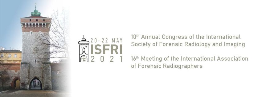
Book of abstracts
 20-22 May, 2021
 Kraków, Poland

 www.isfri2021.uj.edu.pl

 ver 2.12
Scientific Committee

Natalie L. Adolphi, University of New Mexico, USA
Summer Decker, University of South Florida, USA
Edel Doyle, RMIT University, Melbourne, Australia (IAFR Chair)
Guillaume Gorincour, Aix-Marseille Université, France
Hideki Hyodoh, Hokkaido University, Japan
Jeroen Kroll, Maastricht UMC+, the Netherlands
Peter Mygind Leth, University of Southern Denmark, Denmark
Chris O'Donnell, the Victorian Institute of Forensic Medicine, Melbourne, Australia
Lars Oesterhelweg, Charité Universitätsmedizin Berlin, Germany
Rick R. van Rijn, Academisch Medisch Centrum Universiteit van Amsterdam, the Netherlands
Guy N. Rutty, University of Leicester, UK
Michael Thali, University of Zurich, Switzerland
Mark Viner, University of London, UK
Krzysztof Woźniak, Jagiellonian University Medical College, Poland (ISFRI Chair)

 Organizing Committee

Filip Bolechała
Piotr Kluza
Artur Moskała
Ewa Rzepecka-Woźniak
Marek Sanak
Krzysztof Woźniak - Chairman

 2
Agenda

20 May 2021
08:55-09:00 Congress Opening & Gill Brogdon HONORARY LECTURE:

Chairpersons: Natalie Adolphi, Krzysztof Woźniak

 "Humanitarian Forensic Action and Forensic Radiology and Imaging",

 Duarte Nuno Vieira, Institute of Legal Medicine and Institute of Bioethics,
 Coimbra University Centre for Humanitarian and Human Rights Forensic
 Research and Training, Universidade de Coimbra.

9.55-10.05 Break

10:05-11:05 PLENARY SESSION I - COVID - 19 THROUGH CONTINENTS

Chairperson: Michael Thali

 I/1. Post-mortem CT lung findings in SARS-CoV-2 RT PCR positive cases with
 autopsy correlation, Chris O’Donnell, Forensic Pathology, Victorian Institute of
 Forensic Medicine,

 I/2. PMCT lung findings in SARS-COV-2 cases: correlation with lung
 histopathological and autopsy results, Laura Filograna, Diagnostic Imaging,
 Policlinico Universitario Tor Vergata Roma,

 I/3. Postmortem CT lung findings of Covid-19: A review of 14 decedents and
 potential triage implications, Emily Helmrich, Office of the Medical
 Investigator, University of New Mexico.

11.05-11.15 Break

 3
11:15-12:15 PLENARY SESSION II - TRAUMATOLOGY

Chairperson: Guillaume Gorincour

 II/1. Effects of blood loss on liver attenuation on postmortem CT – What role
 does glycogen play?, Jakob Heimer, Forensic Medicine and Imaging, Institute
 of Forensic Medicine, University of Zurich,

 II/2. Location specific sensitivity and specificity of PMCT in blunt force skull
 fractures, Mikkel Henningsen, Section of Forensic Pathology, Department of
 Forensic Medicine, University of Copenhagen,

 II/3. Case report of a skeleton find in forest – characterizing skull fracture
 aspects, Wolf Schweitzer, Forensic Medicine and Imaging, Zurich Institute of
 Forensic Medicine,

 II/4. Can post-explosion perforations with beveling be revealed with PMCT?,
 Jarosław Berent, Department of Forensic Medicine, Medical University of Łódź,

 II/5. Strikes, Cuts and Cross-sections – Dismemberment in PMCT, Lars
 Oesterhelweg, Institute of Forensic Medicine, Charité – Universitätsmedizin
 Berlin,

 II/6. Imaging of intimate partner violence, Thomas Ruder, University Hospital
 Bern Inselspital.

12:20-12:50

 Synergistic Role of Newer Techniques for Forensic and Postmortem CT
 Examinations, Alain Blum, Guilloz Imaging Department, Nancy University
 Hospital, Nancy, France.

 4
13:00-14:00 PLENARY SESSION III - TECHNICAL DEVELOPMENT 1

Chairpersons: Guy Rutty, Lars Ebert

 III/1. PMCT from head to toe - how to approach a stack of 4000 images,
 Thomas Ruder, University Hospital Bern Inselspital,

 III/2. Automatic generation of images for the visualization of gas, bone and
 foreign bodies from PMCT, Lars Ebert, 3D Center Zurich, Institute of Forensic
 Medicine, University of Zurich,

 III/3. Volumetric segmentation in post-mortem computed tomography
 imaging using deep learning techniques, Akos Dobay, Forensic Imaging, Zurich
 Institute of Forensic Medicine,

 III/4. The Forensic Holodeck Technical Setup – Recommendations after 7
 years, Till Sieberth, Institute of Forensic Medicine, University of Zurich,

 III/5. MR Microscopy of Optic Nerve Sheath Hemorrhage in Non-Accidental
 Injuries, Barry Daly, Diagnostic Radiology, University of Maryland.

 5
21 May 2021
09:00-10:00 PLENARY SESSION IV

Chairpersons: Rick R. van Rijn, Krzysztof Woźniak

 Honorary Lecture:

 "Postmortem Angiography - the Past, Present and Future", Silke Grabherr,
 University Centre of Legal Medicine (CURML), Lausanne and Geneva.

10.00-10.15 Break

10:15-11:15 PLENARY SESSION V - TECHNICAL DEVELOPMENT 2

Chairperson: Peter Mygind Leth

 V/1. The effect of radiographic expertise - Improving image acquisition on
 PMCT, Sara Tangmose Larsen, Section of Forensic Pathology, Department of
 Forensic Medicine, University of Copenhagen,

 V/2. 3D whole body spectral imaging in line with PMCT examinations, Soeren
 Kottner, Zurich Institute of Forensic Medicine, University of Zurich,

 V/3. Local Temperature Estimation in Postmortem Muscle Tissue by in situ
 Magnetic Resonance Spectroscopy, Niklaus Zoelch, Institute of Forensic
 Medicine, University of Zurich,

 V/4. High-Resolution 7 Tesla MRI in forensic medicine, Eva Deininger-
 Czermak, Institute of Forensic Medicine, University of Zurich,

 V/5. Importance of postmortem MRI of the brain for forensic practice, Yohsuke
 Makino, The University of Tokyo,

 V/6. Remote consultations in Forensic Anthropology and Entomology:
 examples of conveniences and pitfalls, Edda E. Guareschi, Medical, Molecular
 and Forensic Sciences, Murdoch University.

11.15-11.30 Break

 6
11:30-12:30 PLENARY SESSION VI - DVI AND FORENSIC ANTHROPOLOGY 1

Chairperson: Chris O’Donnell

 VI/1. The biggest Shipwreck of the Mediterranean Sea as the basis for a
 multidisciplinary approach between pathology, anthropology and radiology in
 the evaluation of commingled skeletal remains, Alberto Amadasi, Institute of
 Legal Medicine and Forensic Sciences of the Charité - Universitätsmedizin
 Berlin,

 VI/2. Personal Identification Using Part-to-Part Comparison of L1-L5 Vertebra
 from AMCT and PMCT Scans, Summer Decker, Radiology, University of South
 Florida,

 VI/3. Standardized Image Acquisition and Reporting in Pediatric
 Paleoradiology: Interdisciplinary Approach, Katherine Van Schaik, Department
 of Radiology, Harvard Medical School,

 VI/4. Investigating the average thickness and density of the human
 neurocranium, Samantha Rowbotham, Human Identification Service, Victorian
 Institute of Forensic Medicine,

 VI/5. Multidisciplinary study of a South American natural mummy: a practical
 example, Fabrice Dedouit, Medico-legal department, Chu Toulouse.

12.30-12.45 Break

12.45-14.00 2021 ISFRI Members General Assembly

 7
22 May 2021
09:30-10:30 PLENARY SESSION VII - IAFR SESSION - THE DEFINITION OF “THE
FORENSIC RADIOGRAPHER” AROUND THE WORLD, PART 1

Chairperson: Jeroen Kroll

 VII/1. The roles of the forensic radiographer in Finland (20 min.), Fox
 Marttinen, Finland,

 VII/2. The roles of the forensic radiographer in Denmark (20 min.), Christina
 Caroee Ejlskov Pedersen, Department of Forensic Medicine, Aarhus, Denmark,

 VII/3. The roles of the forensic radiographer in Australia (20 min.), Anthony
 Buxton, Australia.

10.30-10.40 Break

10:40-11:40 PLENARY SESSION VIII - IAFR SESSION - THE DEFINITION OF “THE
FORENSIC RADIOGRAPHER” AROUND THE WORLD, PART 2

Chairperson: Edel Doyle

 VIII/1. The roles of the forensic radiographer in the UK (20 min.), Claire
 Robinson & Amy Lee Brookes, UK,

 VIII/2. The roles of the forensic radiographer in Switzerland (20 min.),
 Alejandro Dominguez, Switzerland,

 VIII/3. The roles of the forensic radiographer in Japan (20 min.), Tomoya
 Kobayashi, Japan.

10:40-12:00 PLENARY SESSIONS VII / VIII – IAFR SESSIONS: QUESTIONS AND
ANSWERS

Chairperson: Mark Viner

12.00-12.10 Break

 8
12:10-13:10 PLENARY SESSION IX

Chairperson: Lars Oesterhelweg

 IX/1. A Comparison of Post Mortem CT and External Examination of the Neck
 in Suspected Hanging Cases, Danielle Chew, Staffordshire University,

 IX/2. Exploring Metabolic Changes in Post-mortem Muscle Tissue by In-Situ
 Magnetic Resonance Spectroscopy, Dominic Gascho, Institute of Forensic
 Medicine, University of Zurich,

 IX/3. Imaging Findings of Contraband Smuggling in 2021, Barry Daly,
 University of Maryland,

 IX/4. Personal identification using post-mortem CT image corresponding to
 periodontal disease examination, Hideko Fujimoto, Kyoto Forensic
 Odontology Center,

 IX/5. Anatomical variability of the maxillary sinus and their application in
 human identification, Dorota Lorkiewicz-Muszyńska Department of Forensic
 Medicine, Uniwersytet Zielonogórski,

 IX/6. Forensic face reconstruction based on postmortem computed
 tomography, Marta Barszcz, Department of Forensic Medicine and Doctoral
 School of Medical Sciences and Health Sciences, Jagiellonian University
 Medical College.

13.10-13.20 Break

13.20-13.30 Closing Ceremony

Chairpersons: Summer Decker, Krzysztof Woźniak, Hideki Hyodoh, Fabrice Dedouit

 9
Archives of Forensic Medicine and Criminology

 10
PLENARY SESSION I - COVID - 19 THROUGH CONTINENTS

 I/1 Post-mortem CT lung findings in SARS-CoV-2 RT PCR positive cases
 with autopsy correlation

Authors: O’Donnell Chris1, Iles Linda1, Woodford Noel1,
1Victorian Institute of Forensic Medicine and Department of Forensic Medicine, Monash University

Introduction/purpose
CoVID-19 is a novel viral infection with now well-established clinical course and radiological
findings. There is limited data on post-mortem imaging.

Materials and Methods
In an 11-week period, 39 deceased persons were referred for medicolegal investigation with
pre-existing or subsequent nasopharyngeal swabs showing positivity on SARS-CoV-2 RT PCR
testing. All 39 had routine whole-body CT scans on admission and 12 underwent medicolegal
autopsy. These 39 cases were contrasted with 4 other cases which were negative on
nasopharyngeal swabs despite PMCT findings suggestive of CoVID-19 pneumonia
(designated false positive).

Results
There were 8 patterns of PMCT lung abnormality including typical findings of CoVID-19 alone,
minimal changes of CoVID-19 alone, typical agonal findings alone, mixed typical agonal and
CoVID-19 findings, dense airless lungs, gross decomposition, and features of another
pathology +/- typical agonal changes. Nine of the 12 autopsies showed lung histology of
varying severity, consistent with those reported in CoVID-19 pneumonia. Typical clinical
CoVID-19 lung findings on PMCT were detected in 5 (42%). In 3 of 4 false positive cases, lung
findings showed non-COVID-19 histology but in 1, findings were identical.

Discussion
PMCT CoVID-19 findings in the lungs are not specific and may not be detected in all cases
due to obscuration by expected agonal CT findings in the chest or other pathologies that pre-
dated SARS-CoV-2 infection. PMCT findings may otherwise be subtle and autopsy histological
findings similarly minimal.

Conclusion
Given this spectrum of change on PMCT, the challenge for pathologists and radiologists is to
determine whether death has been caused by, or merely associated with SARS-CoV-2
infection.

Keywords: CoVID-19, SARS-CoV-2, PMCT, lung

 11
PLENARY SESSION I - COVID - 19 THROUGH CONTINENTS

 I/2 PMCT lung findings in SARS-COV-2 cases: correlation with lung
 histopathological and autopsy results
Authors: Filograna Laura1, Manenti Guglielmo1, Oliva Antonio2, Arena Vincenzo3, Di Donna
Carlo1, Nardoni Francesco4, Colosimo Cesare5, Pascali Vincenzo Lorenzo2, Floris Roberto1
1 Diagnostic Imaging, Policlinico Universitario Tor Vergata Roma, Italy
2 Forensic medicine, Catholic University of Rome, Italy
3 Pathology, Catholic University of Rome, Italy
4 Forensic Medicne, Catholic University of Rome, Italy
5 Diagnostic imaging, Catholic University of Rome, Italy

Introduction/purpose
Post-mortem computed tomography (PMCT) has been already applied in deaths related to
SARS-COV-2 infection. The purpose of this study is to investigate a correlation between PMCT
lung findings in autopsy cadavers positive for SARS-COV-2 infection and the severity of SARS-
COV-2 lung disease on histopathological analysis.

Materials and Methods
We reviewed chest PMCT findings with particular attention to lung parenchyma in 8 autopsy
cases positive for SARS-COV-2. Correlation between chest PMCT findings and
histopathological findings was assessed. Correlations with ante-mortem clinical status and
comorbidities were also investigated. The primary cause of death was finally considered.

Results
In the 6/8 cases with severe pulmonary coronavirus disease 2019 (COVID-19) on
histopathological analysis, the pulmonary PMCT findings were massive consolidation (4/8),
and bilateral, diffuse mixed densities with crazy paving pattern (2/8). In the cases 4/8 cases
with massive consolidations, important comorbidities were associated. In these 6/8 cases the
autopsy ascertained cause of death was cardio-respiratory failure due to SARS-COV-2
infection. In the remaining 2/8 cases, pulmonary PMCT findings were ground glass and
consolidations with a prevalent gradient distribution. In these 2/8 cases the histopathological
analysis revealed lung alterations due to oedema and few signs of SARSCOV-2 infection. In
these 2/8 cases the cause of death was not attributed to SARS-COV-2 infection.

Discussion and Conclusion
Chest PMCT findings correlate with the severity of COVID-19 lung disease. Although the
paucity of cases, PMCT findings of the lungs in cadavers positive for SARS-COV-2 infection
might correlate with the cause of death, especially after consideration of clinical antemortem
data.

Keywords Post-mortem CT, COVID-19, SARS-COV-2, lung findings, pulmonary hystopathology.

 12
PLENARY SESSION I - COVID - 19 THROUGH CONTINENTS

 I/3 Postmortem CT lung findings of COVID-10: a review of 14 decedents
 and potential triage implications
Authors: Helmrich Emily1, Decker Lauren1, Adolphi Natalie2, Makino Yohsuke3
1 Office of the Medical Investigator, University of New Mexico, United States
2 Center for Forensic Imaging, Office of the Medical Investigator, University of New Mexico, United States
3 Graduate School of Medicine, University of Tokyo, Japan

Introduction
Computed tomography has significant utility as a diagnostic tool for coronavirus disease 2019
(COVID-19) in the clinical setting. COVID-19 deaths are sometimes examined by forensic
pathologists, often in the setting of an unknown diagnosis. We assessed the utility of
postmortem computed tomography (PMCT) for use as a triage tool for these autopsy
examinations.

Materials and methods
We reviewed PMCT findings in 14 and histopathology in 11 decedents who were positive for
COVID-19.

Results
Eleven decedents (79%) demonstrated mixed densities (near-equal amounts of both ground-
glass opacities [GGOs] and consolidations). Seven decedents (50%) had ill-defined rounded
consolidations. Other common features included traction bronchiectasis (eleven decedents,
79%), interseptal thickenings (ten decedents, 71%) and crazy paving pattern (nine
decedents, 64%). Therefore, the predominant imaging findings were bilateral mixed densities
in either a diffuse or peripheral distribution, with traction bronchiectasis, interseptal
thickenings and/or crazy paving. In particular, we found that traction bronchiectasis, ill-
defined rounded consolidations, and reverse halo sign were useful when distinguishing from
other postmortem changes.

Conclusion
We conclude that triage with a PMCT may aid the forensic pathologist in diagnosing possible
COVID-19 infection prior to autopsy examination.

Keywords
COVID-19, Postmortem CT, Autopsy

 13
PLENARY SESSION II - TRAUMATOLOGY

 II/1 Effects of blood loss on liver attenuation on postmortem CT – What
 role does glycogen play?

Authors: Heimer Jakob1, Chatzaraki Vasiliki1, Schweitzer Wolf1, Thali Michael J.1, Ruder Thomas D.2
1Forensic Medicine and Imaging, Institute of Forensic Medicine, University of Zurich, Switzerland
2 2Department of Diagnostic, Interventional and Pediatric Radiology, Inselspital, Bern University Hospital,
University of Bern, Switzerland

Purpose
To investigate the effects of blood loss on the CT attenuation of the liver.

Methods
A total of 125 cases with blood loss were sex- and age-matched to 125 control cases without
blood loss. Individual liver attenuation was measured on transverse CT images. Bivariate
comparison of means of liver attenuation between both groups as well as multiple regression
with liver attenuation as the response variable and sex, age, BMI, postmortem interval, liver
fat content, and performed reanimation as predictors were performed.

Results
CT attenuation of the liver was significantly higher (61 HU > 49 HU, p
PLENARY SESSION II - TRAUMATOLOGY

 II/2 Location specific sensitivity and specificity of PMCT in blunt force
 skull fractures
Authors: Henningsen Mikkel1, Jacobsen Christina1, Villa Chiara1
1 Section of Forensic Pathology, Department of Forensic Medicine, University of Copenhagen, Denmark

Congruence between PMCT and autopsy is an important area of forensic medical research.
Several papers have examined the ability of PMCT to detect skull fracture and found high
sensitivity and specificity. Almost all reported the presence or absence of a fracture systems
in the base and vault. The aim of this study was to assess the congruence between PMCT and
autopsy in detecting the individual fracture lines in each bone, producing location specific
sensitivity and specificity of PMCT.

We retrospectively included 99 cases of cranial blunt force trauma in adults autopsied
between 2013 and 2019. Cases with severely comminute fractures, missing bone parts,
insufficient reporting, or lack of PMCT were excluded. PMCT was interpreted blindly from
autopsy findings. All fracture lines from both autopsy and PMCT were registered and
compared. Sensitivity and specificity of PMCT were calculated.

We present here only preliminary results from a larger study also including the cranial base
and sutures of the 99 cases. We identified more than 1,800 fracture lines in total. For the
individual bones of the cranial vault, we found sensitivities ranging from 0.68 to 0.83 and
specificities ranging from 0.77 to 0.94

This study adds to the existing knowledge by providing sensitivities and specificities of PMCT
for individual cranial bones by a novel approach of considering individual fracture lines. Our
findings differ from those previously published and will be discussed thoroughly. Our study
indicates that the need for autopsy persists if advanced fracture analysis is warranted.

Keywords
PMCT, skull fracture, diagnostic test accuracy, sensitivity & specificity, autopsy

 15
PLENARY SESSION II - TRAUMATOLOGY

 II/3 Case report of a skeleton find in forest – characterizing skull
 fracture aspects
Authors: Schweitzer Wolf1, Thali Michael1, Bolliger Stephan1, Roidou Magda1
1 Forensic Medicine and Imaging, Zurich Institute of Forensic Medicine, Switzerland

Case: Partly skeletonized human remains which constituted an almost complete skeleton
were found in a forest. The identity was confirmed as being that of an approximately 45-year-
old man. What appeared to us as an outstanding feature was the fracture pattern of the skull,
which seemed to present a strong contrast with the remaining skeleton, which was almost
completely unharmed. The skull fracture pattern, particularly as seen in post-mortem CT
(PMCT) was then compared to skull data from other cases in our database. There, we looked
at cases of blunt trauma and various firearm injuries. Based on this data comparison, we
deemed an explosive trauma to be the best explanation for the findings in the case at hand.
Further investigation then found that the deceased, who had a history of depression, had
indeed been a trained pyrotechnician. Based on this and further results of the examination,
suicide was considered a plausible manner of death.

Details of the skull fractures, and comparison with other cases, will be presented and
discussed.

 16
PLENARY SESSION II - TRAUMATOLOGY

 II/4 Can post-explosion perforations with beveling be revealed with
 postmortem computed tomography (PMCT)?
Authors: Berent Jarosław1, Smędra Anna1
1 Department of Forensic Medicine, Medical University of Lodz, Poland

PMCT is a commonly acknowledged diagnostic method in forensic pathology, but its
usefulness may vary on a case-by-case basis. Fragments moving at high velocity during
explosions may cause different types of bone injuries. Among these, perforating lesions with
beveling are the most interesting ones, from the evidential point of view. Such lesions are
diagnostic for an impact of objects moving at high velocity (high energy objects), such as
projectiles fired from firearms or fragments released during an explosion. Other reasons for
the occurrence of such lesions are so extremely rare that the finding of such lesions during a
forensic autopsy practically confirms that they have been caused by the trauma described.
The purpose of this paper was to examine the usefulness of PMCT in the detection of
postexplosion perforating bone lesions with beveling.
In order to do this, two experimental explosions were carried out (thermobaric load and
penthrite) featuring flat bone models (Synbone®). Both explosions resulted in diagnostic
lesions in the form of perforations with beveling of different sizes. The bone models had
subsequently been CT scanned and a 3D reconstruction was prepared. The conducted
experiment showed that PMCT 3D reconstructions are of limited value when analyzing the
morphology of small lesions. Such bone injuries can be revealed with PMCT only in case of
large lesions. It is not possible to recognize smaller lesions due to the limitations of digital 3D
reconstruction algorithms. Therefore, PMCT cannot replace the classic visual evaluation of
such small lesions during forensic autopsy.

Keywords: explosion, blast, lesion, beveling

 17
PLENARY SESSION II - TRAUMATOLOGY

 II/5 Strikes, cuts and cross-sections - dismemberment in PMCT
Authors: Oesterhelweg Lars1, Dümpelmann Denise1, Hartwig Sven1, Tsokos Michael1
1 Institute of Forensic Medicine Charite - Universitaetsmedizin Berlin, Germany

Dismembered bodies are rare but highly noticed entities in Legal Medicine. When mutilated
or dismembered bodies or body parts have been found, the criminal investigation department
and medico legal services are facing several reconstructive questions. A complete
documentation of all injuries is mandatory for the police to reconstruct the course of events
and is the grounding of high court decisions. PMCT gives us a valuable tool for a complete
head-to-toe documentation and for reconstructions, measurements, and 3D-demonstrations
of injuries and findings. In the forensic literature dismemberment of bodies is classified in
offensive and defensive actions. In offensive dismemberment the disintegration is part of the
act of killing while in defensive dismemberment it is a process to hide or to transport the
deceased or to hinder the identification. In 4 cases the different classifications of
dismemberment will be demonstrated.

 18
PLENARY SESSION II - TRAUMATOLOGY

 II/6 Imaging of intimate partner violence
Authors: Ruder Thomas1
1 Department of Radiology, University Hospital Bern Inselspital, Switzerland

The COVID-19 pandemic has triggered an increase in intimate partner violence (IPV). Reasons
for this rise in IPV include elevated levels of stress from social isolation and economic
hardship as well as the confinement of families at home for extended periods during lock-
downs.
The aim of this presentation is to review injury patterns of IPV on imaging and to outline the
role of radiologists in the detection of IPV.
Based on a review of the radiologic literature, this presentation provides an overview of
frequent patterns and imaging findings in IVP victims.
IPV-related injuries are found both in target areas and in defensive locations. Target areas
include the face, neck and back. Typical defence injuries occur in the forearms and hands. The
presence of acute, healing, and healed fractures is suggestive for IVP.
Imaging can play a crucial role in the detection of IPV and it therefore is important that
radiologists are aware of imaging patterns and red flags for IPV.

 19
PLENARY SESSION III - TECHNICAL DEVELOPMENT 1

 III/1 Post-mortem CT from head to toe – how to approach a stack of
 4000 images
Authors: Ruder Thomas1
1 Department of Radiology, University Hospital Bern Inselspital, Switzerland

Post-mortem computed tomography (PMCT) is often part of the preliminary investigations in
a Coroner’s Inquest. The imaging findings can guide triage and management decisions
regarding subsequent post-mortem investigations. At the time of the PMCT, information
regarding the circumstances of death is often very limited. This means that radiologists and
pathologists who read a PMCT do not know what abnormalities they are looking for and where
they should be looking for abnormalities. Therefore, a thorough review and careful
interpretation of all CT images is crucial. Reading through thousands of images from head to
toe is challenging.
The aim of this presentation is to present a systematic approach for reading and reporting
PMCT using a structured report template.
Structured reporting has several advantages over traditional free-test report. For the
beginner, a report template serves as checklist and ensures the completeness of a report.
Structured reporting also represents a countermeasure against bias from case irrelevant
context information. In addition, it increases both inter-rater consistency and clarity of a
radiology report.
This presentation provides practical tips on how to approach a whole-body post-mortem CT.

 20
PLENARY SESSION III - TECHNICAL DEVELOPMENT 1

 III/2 Automatic generation of images for the visualization of gas, bones
 and foreign bodies from PMCT
Authors: Ebert Lars1, Seckiner Dilan1, Sieberth Till1, Thali Michael2, Franckenberg Sabine3
1 3D Center Zurich, Institute of Forensic Medicine, University of Zurich, Switzerland
2 Institute of Forensic Medicine, University of Zurich, Switzerland
3 Institute for Diagnostic and Interventional Radiology, University Hospital Zurich, Switzerland

Introduction
Post mortem computed tomography (PMCT) can aid in localizing foreign bodies, bone
fractures, and gas accumulations. The visualization of these findings play an important role in
the communication of radiological findings. We developed an algorithm, that allows for the
automated visualization of gas distributions on PMCT image data of the thorax and abdomen.

Materials and Methods
The algorithm calculates different projection images (namely an average projection, a
minimum intensity projection (minIP) and a maximum intensity projection (MIP)) that are
differently colored and layered to build up the final overview image. The algorithm was tested
on a total of 955 PMCT scans of the thorax and abdomen.

Results and Discussion
In all cases, the algorithm managed to generate useful images, visualizing foreign bodies as
well as gas distributions. While this type of visualization cannot replace a real radiological
analysis of the image data, it can provide a quick overview for briefings and image reports.

 21
PLENARY SESSION III - TECHNICAL DEVELOPMENT 1

 III/3 Volumetric segmentation in post-mortem computed tomography
 imaging using deep learning techniques
Authors: Dobay Akos1, Gaspar Dominic1, Kurmann Astrid1, Handschin Philippe1, Garyfalia
Ampanozi1, Franckenberg Sabine1, Affolter Raffael1, Sieberth Till1, Ebert Lars1
1 Forensic Imaging, Zurich Institute of Forensic Medicine, Switzerland

Aims and objectives
Automated segmentation of volumetric data remains a challenging problem, especially in
medical imaging. In this study we present a deep learning algorithm able to segment
automatically multiple organs on post-mortem computed tomography (PMCT) scans.

Materials and methods
A total of 40 post-mortem cases were retrospectively selected from our database at the Zurich
Institute of Forensic Medicine. We excluded cases with signs of advanced decomposition. For
each case the PMCT scan of the dead body was imported in Amira, a software platform for 3D
data visualization and data processing. Two medical students supervised by a certified board
forensic pathologist manually segmented the volume corresponding to the lungs, the liver,
the heart, the kidneys, and the bladder. The volumetric data were subsequently used to train,
validate, and test a deep learning algorithm for automated organ segmentation. The algorithm
was derived from an existing neural network architecture called U-Net and adapted to work
on volumetric PMCT scans.

Results
Based on a visual inspection of the resulting outcome, the algorithm was able to segment the
volume corresponding to the lungs, the liver, the heart, the kidneys, and the bladder. However,
volumetric overlaps between adjacent organs were still visible.

Discussion and Conclusion
In this study, we were able to show that a deep learning algorithm can be used to
automatically segment organs on volumetric PMCT scans. However, the current version of the
algorithm still suffers from a lack of precision based on our visual assessment of the resulting
images.

Keywords
Deep learning, PMCT

 22
PLENARY SESSION III - TECHNICAL DEVELOPMENT 1

 III/4 The forensic holodeck technical setup – recommendations after 7
 years of experience with virtual reality head-mounted displays in
 forensics

Authors: Sieberth Till1, Seckinger Dilan1, Dobay Akos1, Dobler Erika2, Affolter Raffael1,
Ebert Lars1
1 Institute of Forensic Medicine, University of Zurich, Switzerland
2 Zurich Forensic Science Institute, Switzerland

Introduction/Purpose
In 2013 we introduced the use of Virtual Reality in forensic under the name Forensic
Holodeck. Since then we applied it in several case, both during the forensic investigation as
well as in court trails. This presentation will have a look at the equipment we use in addition
to the Virtual Reality set-up.

Materials and Methods
For an all-round documentation for the usage of Virtual Reality in forensic investigations and
court trails we use TV broadcast and streaming grade equipment. This equipment includes
cameras, Mix Effect (M/E) switchers and audio equipment, but also networking equipment for
multiple VR set-ups.

Discussion and Conclusion
After 7 years of using virtual reality in a professional forensic capacity it showed that
advanced equipment is necessary to fulfil customer and court requirements and forensic
standards. This mostly includes audio-visual and broadcasting technology for complete
documentation of the application of virtual reality, but also some other IT equipment, which
should be available for as low as 20’000 US$.

Keywords: Virtual Reality, Forensic Holodeck, 3D Reconstruction, Experience Report

 23
PLENARY SESSION III - TECHNICAL DEVELOPMENT 1

 III/5 MR microscopy of optic nerve sheath hemorrhage in non-
 accidental injures
Authors: Daly Barry1
1 Diagnostic Radiology, University of Maryland, United States

Purpose
There is a known association between non-accidental injuries in children and the presence of
optic nerve sheath hemorrhages. This study explores the practicality and accuracy of using
high-resolution MR microscopy with an orbital surface coil as both an adjunct to traditional
enucleated globe pathology procedures and a potential in-vivo diagnostic tool for optic nerve
hemorrhage.

Materials and Methods
In this pilot study we identified nine deceased children (ages 1-40 months) following
suspected non-accidental head injuries. Using a 7-Tesla MRI scanner and hemorrhage-
sensitive T2* (star) sequences, we obtained high resolution images of ex-vivo globes and
optic nerves. These images were interpreted blindly by two radiologists and compared with
pathology findings. We also tested a prototype orbital surface coil for in-vivo acquisition of
high resolution imaging of the optic nerve in volunteers.
Results: The results show high concordance between MR microscopy and traditional surgical
pathology with agreement in 6 true positive and two true negative cases. MR was negative in
one case of subtle hemorrhage noted at pathology. A prototype orbital surface coil at 3 Tesla
was shown to provide satisfactory high resolution images of the optic nerve in volunteers.

Conclusion
This study supports the potential of high-resolution 7T MR microscopy to detect optic nerve
sheath hemorrhages and to augment or possibly replace traditional autopsy procedures.
A dedicated orbital surface coil in combination with 3 Tesla clinical MR magnets may allow
detection of optic nerve hemorrhage in-vivo, allowing a more definitive imaging diagnosis of
suspected pediatric non-accidental head injuries.

Key Words: Pediatric non-accidental injury, Optic Nerve Sheath Hemorrhage, MR Microscopy

 24
PLENARY SESSION V - TECHNICAL DEVELOPMENT 2

 V/1 The effect of radiographic expertise - Improving image acquisition
 on PMCT
Authors: Slot-Svendsen Knud1, Larsen Sara Tangmose1, Jacobsen Christina1
1 Department of Forensic Medicine, University of Copenhagen, Section of Forensic Pathology, Denmark

Introduction
The acquisition of PMCT scans at our institution was until recently based on bodyweight
dependent standard protocols. The actual scans were performed by forensic technicians, with
limited radiographic knowledge. The adaption of parameters was performed utilizing the
suggestions of the CT-scanner software based on dosage modulations. Image quality was
generally estimated to be sufficient, serving the purpose of facilitating the planning of the
forensic autopsy and possibly providing a preliminary cause of death. Since spring 2020, the
PMCT scans have been performed by a radiographer. The aim of this study is to evaluate how
the radiographic adaptations have improved image quality of PMCT scans.

Materials and Methods
Using a Siemens SOMATOM Definition AS 64-slice CT-scanner, three deceased were scanned
using the previous bodyweight dependent standard protocol and the radiographically
adjusted standard protocol.
The three whole body scans were performed focusing on the cerebrum and abdomen. Image
quality of the PMCTs from these anatomical locations were compared regarding several
parameters.

Results
The preliminary subjective visual difference on the PMCT scans in different anatomic
locations as well as objective measurements show an improvement in image quality using
radiographically adapted standard CT-scan protocols.

Discussion and Conclusion
Based on preliminary visual examinations as well as ongoing objective measurements, the
use of radiographically adapted CT-scan protocols shows that image quality has improved and
thereby may optimize the diagnostic opportunities of PMCT scans prior to forensic autopsies.
Further evaluation of objective data are needed to confirm the improved image quality using
radiographically adjusted standard protocols.

Keywords: Post mortem CT, Image quality, CT protocols

 25
PLENARY SESSION V - TECHNICAL DEVELOPMENT 2

 V/2 3D whole body spectral imaging in line with PMCT examinations
Authors: Kottner Soeren1, Schulz Martin M.2, Berger Florian1, Thali Michael1, Gascho Dominic1
1 Zurich Institute of Forensic Medicine, University of Zurich, Switzerland
2 Institute of Forensic Medicine, Ludwig-Maximilians-University Munich, Germany

Introduction/purpose
Two-dimensional (2D) and three-dimensional (3D) external body documentation is commonly
used to capture the visible part of the electromagnetic spectrum. Although multispectral
photography is used for a wide range of applications in line with forensic investigations, it
seems that, in this context, 3D multispectral imaging is not yet well known nor widely used.

Materials and Methods
A multi-camera setup in conjunction with a medical X-ray computed tomography (CT) scanner
was used to conduct multispectral photogrammetry alongside whole body postmortem CT
examinations. Multispectral imaging was done in the range between 365 and 960 nm. The
multi-camera setup was based on four digital cameras, ultraviolet and near-infrared light
sources as well as additional lens filters.

Results
Multiview 3D reconstruction based on multispectral image data from four example cases was
carried out successfully. The overall quality of the 3D reconstruction varied in regards to the
spectral range of the image data. Generally, ultraviolet-visual datasets improved the visibility
of foreign substances on the skin and highlighted superficial bruising. Near-infrared datasets
showed enhanced visibility of vein patterns and emphasized subcutaneous injuries and
bruises.

Discussion and Conclusion
Multispectral imaging facilitates the detection of latent trace evidence and allows enhancing
the visibility of findings. 3D whole body spectral imaging in line with PMCT examinations
allows combining multispectral information from the external body documentation with
radiological findings from the internal body documentation.

 26
PLENARY SESSION V - TECHNICAL DEVELOPMENT 2

 V/3 Local temperature tstimation in postmortem muscle tissue by in
 situ magnetic resonance spectroscopy
Authors: Zoelch Niklaus1,2, Heimer Jakob1, Thali Michael1, Gascho Dominic1
1Department of Forensic Medicine and Imaging, Institute of Forensic Medicine, University of Zurich, Zurich,
Switzerland
2Hospital of Psychiatry, Department of Psychiatry, Psychotherapy and Psychosomatics, University of Zurich, 8032

Zurich, Switzerland

Keywords: magnetic resonance spectroscopy; muscle; temperature; forensic radiology
Purpose: The objective of this study is to investigate the feasibility of local temperature
estimation by in situ proton magnetic resonance spectroscopy (1H-MRS) in postmortem
muscle tissue. The 1H chemical shift of water has an almost linear dependence on
temperature and therefore allows for temperature measurements if referenced to a
temperature-stable compound.

Methods: 24 decedents with different postmortem interval (range: 4 to 356 h) underwent 1H-
MRS (3T). Single voxel measurements were performed in the vastus intermedius muscle
and/or in the adductor magnus muscle of the left and right thigh. The local temperature was
estimated based on the chemical-shift difference between water and creatine and using
published calibration values.
 =( − −2.0505)/−0.00999
Finally, the agreement between the temperature estimated by MRS (TMRS) and rectal body
temperature (Trectal) was compared.

Results
The median difference between TMRS and Trectal was -0.63 (TMRS lower than Trectal). The
differences ranged from 2 to -6.3 °C, two-thirds of all TMRS measurements were between ±2
°C. Larger differences were observed in cases of open fractures. There was no correlation
between the PMI and the deviation between TMRS and Trectal. Only small deviations between
TMRS measured in the left and right thigh were observed (median: -0.4 °C, max: 1.1, min: -1.2
°C).

Conclusion: In-situ 1H-MRS provides reasonable local temperature estimates in muscle tissue
using creatine as reference over a wide range of estimated PMIs. This indicates that both
creatine and water chemical shift a rather insensitive to pH and ionic strength fluctuations
within the ranges encountered here.

 27
PLENARY SESSION V - TECHNICAL DEVELOPMENT 2

 V/4 High-resolution 7 Tesla MRI in forensic medicine
Authors: Gascho Dominic1, Zoelch Nikolaus1, Sommer Stefan2, Tappero Carlo1, Thali Michael1,
Deininger-Czermak Eva1
1 Institute of Forensic Medicine, University of Zurich, Switzerland
2 Swiss Center for Musculoskeletal Imaging, Balgrist Campus , Switzerland

Introduction/Purpose
Recently, 7 Tesla (7-T) MRI scanner were approved for clinical use, which will facilitate access
to these ultra-high-field MRI scanners for noninvasive examinations and scientific studies on
decedents. 7-T MRI has the potential to provide a higher signal-to-noise ratio (SNR), a
characteristic that can be directly exploited to improve image quality and invest in attempts
to increase resolution. Therefore, evaluating the diagnostic potential of 7-T MRI for forensic
purposes, such as assessments of fatal head injuries, was deemed essential.

Materials and methods
Two decedents with severe head injuries underwent a CT, 3-T, and 7-T MRI examination. The
CT, 3-T and 7-T images were graded regarding the detectability and the delineation of
microlesions, coup/contrecoup and shearing injuries using 3-point Likert scales.

Results
While on CT microlesions and small coup/contrecoup injuries were missed, they were
detectable on 3-T and 7-T MRI. However, the 3-T images showed less delineated lesions in
direct comparison with the 7-T images, thus the detectability and diagnostic confidence was
improved on 7-T MRI.

Discussion and Conclusion
7-T images provided higher detectability of traumatic brain injuries and microlesions.
However, it was able to determine the cause of death on all modalities. Certainly, the
relevance of the higher resolution and the potential to identify even microlesion on 7-T
promises a further development in forensic imaging. Despite the advantages, it has to be
considered that 7-T scanner are only limited accessible, therefore it lacks practicability.

Keywords: Virtual autopsy, Brain injuries, Contrecoup injury, Magnetic resonance imaging

 28
PLENARY SESSION V - TECHNICAL DEVELOPMENT 2

 V/5 Importance of postmortem MRI of the brain for forensic practice
Authors: Makino Yohsuke1, Kojima Masatoshi2, Yoshida Maiko2, Hoshioka Yumi2, Tsuneya
Shigeki1, Inokuchi Go2, Hattori Shinya3, Mukai Hiroki3, Yokota Hajime3, Iwase Hirotaro2
1 Forensic Medicine, The University of Tokyo, Japan
2 Legal Medicine, Chiba University, Japan
3 Radiology, Chiba University Hospital, Japan

Purpose
To evaluate the added value of brain postmortem MRI (PMMR) for practical use in forensic
medicine compared to non-contrast postmortem CT (PMCT).

Materials and Methods
Forty-one consecutive forensic cases with PMCT, PMMR, and autopsy including detailed brain
histopathology were reviewed. Cases were selected in which the causes of death associated
with brain lesions were determined by board-certified forensic pathologists. The findings of
PMCT and PMMR were compared and classified into the following three categories. Category
1, PMMR shows more detailed brain lesions associated with the cause of death than PMCT;
Category 2, PMMR is equivalent to PMCT; Category 3, PMMR is inferior to PMCT.

Results
In total, 14 of 41 causes of death were associated with brain lesions. Of these, 6 (43%) were
classified into Category 1, none into Category 2, and 8 (57%) into Category 3. The Category-
1 findings included traumatic axonal injuries, cerebral contusions, hemorrhagic necrosis, old
hemorrhage, necrosis related to carbon monoxide poisoning, and Wernickeʼs
encephalopathy.

Discussion and Conclusion
There are already many articles dealing with PMMR, but most of them are aimed at replacing
autopsy, and there are quite a few reports on the importance of supporting forensic practices.
In this preliminary case series study, brain PMMR showed added values on forensic practices,
revealing traumatic brain injuries, poisoning-related lesions or malnutrition. Routine PMMR
can change forensic practices, as it is thought to be useful for routine screening of these
normally hard-to-find but sometimes forensic important brain lesions.

Keywords: Postmortem MRI, Forensic pathology, Forensic medicine, Neuroradiology, Brain

 29
PLENARY SESSION V - TECHNICAL DEVELOPMENT 2

 V/6 Remote consultations in forensic anthropology and entomology:
 examples of conveniences and pitfalls in two cases

Authors: Guareschi Edda E.1, Van de Goot Franklyn R.W.2, Ziogos Stevie S.1, Magni Paola A.1,3
1 Discipline of Medical, Molecular & Forensic Sciences, Murdoch University, 90 South Street, Murdoch, Western
Australia 6150, Australia
2 Department of Pathology, VU University Medical Centre, Amsterdam, The Netherlands
3 Murdoch University Singapore, King’s Centre, 390 Havelock Road, Singapore 169662

Introduction/purpose
The recent pandemic has emphasized the global resorting to remote consultation which, in
the last 20 years, has mostly been applied to clinical imaging. An extension to forensic
imaging is soon to be expected. Two cases are presented here, with the aim of both
highlighting the conveniences and the pitfalls of remote consultation in forensic
anthropology and entomology, and of encouraging the discussion about the need of
guidelines.

Materials and Methods
In the forensic anthropology case, two sets of apparently ancient human bones have been
investigated, in order to assess the biological profile and the possible cause of death. In the
entomology case, clothes associated with decomposing remains and colonized by insects
were analysed, in the attempt to estimate the minimum post-mortem interval and the nature
of the textile damage. The examinations were performed through remote consultation, and
were based on discussion and photographs.

Results
The ancient human bones belonged to multiple individuals of both sexes; the distribution and
the pattern of trauma pointed to widespread violence, with a likely fatal outcome. The
evaluation of the damage on clothes was based on the knowledge of the nature of the
clothing and the presence of insects typically found in such products.

Discussion and Conclusion
The remote consultations were essential to solving both forensic cases. However, some formal
and substantial aspects proved challenging, including but not limited to, the legitimacy of the
assignment, the liability of the consultant and the limitations of the provided documentation
in asynchronous examinations.

Keywords: remote, consultation, forensic, anthropology, entomology

 30
PLENARY SESSION VI - DVI AND FORENSIC ANTHROPOLOGY 1

 VI/1 The biggest shipwreck of the Mediterranean Sea as the basis for a
 multidisciplinary approach between pathology, anthropology and
 radiology in the evaluation of commingled skeletal remains
Authors: Amadasi Alberto1, Mazzarelli Debora2, Cappella Annalisa2, Magli Francesca2,
De Angelis Danilo2, Porta Davide2, Cattaneo Cristina2
1 Institute
 of Legal Medicine and Forensic Sciences of the Charité - Universitätsmedizin Berlin, Corporate Member
of Freie Universität Berlin, Humboldt-Universität zu Berlin and Berlin Institute of Health, Germany
2 Dipartimento di Scienze Biomediche per la Salute, , LABANOF, Laboratorio di Antropologia e Odontologia Forense,

Sezione di Medicina Legale, Universite degli Studi di Milano, Italy

The shipwreck of April 18, 2015 is currently the largest mass disaster in the Mediterranean
Sea since the Second World War. It thus represents an exceptional event due to the toll of
human lives as well as for for the recovery and identification procedures. A multidisciplinary
approach was crucial for the collection of all anthroplogical antemortem data. Moreover,
human remains underwent CT scans using an on-site mobile device. The experience
demonstrated the crucial importance of radiological CT investigations (especially if supported
by 3D reconstructions) in the context of mass disasters. In this case, however, the context was
particular in the field of mass disasters, with such a large amount of remains within a small
environment (the internal areas of a boat). This has led, given the considerable variability of
the state of decomposition of the corpses, and the high amount of skeletonized remains, to a
considerable commingling of bone remains. This situation has therefore greatly lowered the
possibility of correctly identify bone remains In particular, the study has shown that a
fundamental contribution for identification purposes can be provided by TAC assessments,
for example in the distinction between different individuals on the basis of the anatomical
position inside the body bag, in the detection of personal effects, recognition of features (eg
pathologies, bony calluses) with bone or soft tissue localization, detection and positioning of
prostheses, recognition of anatomical structures for the reconstruction of the biological
profile, possibility of carrying out centimeter measurements with greater reliability and
repeatability compared to on-site measurements. Moreover, a 3D reconstruction of entire
individuals or skeletal parts before they were subjected to forensic analysis and sampling has
been of utmost importance.

Keywords: dead migrants, identification, biological profile, anthropology – radiology

 31
PLENARY SESSION VI - DVI AND FORENSIC ANTHROPOLOGY 1

 VI/2 Personal identification using part‐to‐part comparison of L1‐L5
 vertebra from AMCT and PMCT scans
Authors: Decker Summer1, Rutty Guy2, Martin Daniel1, Amoroso Jasmin2, Biggs Michael2,
Ford Jonathan1
1 Radiology, University of South Florida, United States
2 East Midlands Forensic Pathology Unit, University of Leicester, United Kingdom

Introduction
The confirmation of identification for an unknown individual is a critical part of forensic
practice. The comparison of ante mortem imaging for the purposes of personal identification
is a common tool in both odontology and radiographic comparisons. This study utilizes AMCT
scans and compares them against PMCT scans.

Materials and Methods
As part of an ongoing project, University of Leicester acquired 14 matching ante mortem scans
for individuals that passed through their facility for PMCT scanning. All scans were
anonymized and sent to the University of South Florida for identification testing. Each scan
was imported into the Mimics Innovation Suite v. 24. Each Lumbar vertebra was then isolated
and modeled via segmentation and thresholding. Each series of 14 AM vertebra were
registered with a target unknown PMCT derived vertebra. A part‐to‐part comparison was
conducted for each vertebra and a match ratio was measured. A threshold of +/‐ 1 mm was
set for the part comparison.

Results
Every unknown PMCT vertebra was correctly matched to the corresponding AMCT vertebra,
signifying complete accuracy for this sample. A ROC curve was calculated to determined
100% sensitivity and specificity with a cutoff match ratio of 0.705. True identifications had
an average match ratio of 0.943 +/‐ 0.539. Negative identifications had an average match
ratio of 0.375 +/‐ 0.1.

Discussion and Conclusion
With the increase usage of post‐mortem CT (PMCT) there is an equal increase in the
availability and opportunity to utilize 3D tools, such as part‐to‐part comparison, for successful
personal identification.

Keywords: PMCT, DVI, Personal Identification

 32
PLENARY SESSION VI - DVI AND FORENSIC ANTHROPOLOGY 1

 VI/3 Standardized image acquisition and reporting in pediatric
 paleoradiology: interdisciplinary approach

Authors: Van Schaik Katherine1, Tsai Andy2, Liston Maria3
1
 Department of Radiology, Harvard Medical School, United States
2
 Department of Radiology, Boston Children's Hospital/Harvard Medical School, United States
3
 Department of Anthropology, University of Waterloo, United States

Introduction/Purpose
This paper evaluates the use of paleoradiology techniques in the analysis of pediatric remains
from bioarchaeological contexts and offers standardized, multi-modality protocols for the
acquisition, evaluation, and publication of findings.

Materials/Methods
An interdisciplinary team of pediatric radiologists, anthropologists, archaeologists, and
historians completed a literature search of three major bioarchaeological journals, PubMed,
and the Yearbook of Mummy Studies, and supplemented this review with protocols and
studies published in radiological journals.

Results
Although radiological techniques are used with increasing frequency in pediatric
bioarchaeology studies, no standardized pediatric imaging acquisition and reporting
protocols exist. In most cases, radiography is not used systematically, and when it is used,
imaging acquisition parameters are not reported, compromising data interpretation. These
issues of inadequate reporting and interpretation are especially acute for plain film
radiography, the most frequently-used modality in archaeological contexts: use of plain film
radiography without clear protocols and interpretive frameworks is problematic, as recent
research (Spies et al. 2021) has demonstrated that interpretation of such films without
adequate training leads to inaccurate assessments of findings.

Discussion/Conclusion
Based on imaging parameters published in the reviewed articles, as well as forensic and
clinical imaging protocols, we propose acquisition and reporting protocols and best-practices
guidelines that are specific for the paleoradiographic assessment of pediatric human remains.
This is, to the authors’ knowledge, the first literature review to focus exclusively on pediatric
paleoradiology and the first proposal of imaging protocols tailored for the assessment of
pediatric remains from bioarchaeological contexts.

Keywords: paleoradiology, pediatric, plain film radiography, imaging protocols,
bioarchaeology

 33
PLENARY SESSION VI - DVI AND FORENSIC ANTHROPOLOGY 1

 VI/4 Investigating the average thickness and density of the human
 neurocranium
Authors: Rowbotham Samantha1, Mole Calvin2, Tieppo Diana3, Blaszkowska Magda4,
Cordner Stephen5, Blau Soren1
1 Human Identification Service, Victorian Institute of Forensic Medicine, Australia
2 Division of Forensic Medicine and Toxicology, University of Cape Town, South Africa
3 Department of Forensic Medicine, Monash University, Australia
4 Centre for Forensic Anthropology, University of Western Australia, Australia
5 International Programs, Victorian Institute of Forensic Medicine, Australia

Introduction
Attempting to interpret the degree of force required to produce neurocranial fractures relies
on understanding details about the event (extrinsic information) and the individual (intrinsic
data). Whilst some of these variables may be available, the intrinsic variables of ‘average’ skull
thickness and density are poorly understood and so cannot be accounted for; limiting
interpretations of the degree of force. The aim of this study was to identify the average
thickness and density of the bones of the human neurocranium.

Methodology
Thickness (mm) and density (Hounsfield units - Hu) were measured at 20 points across the
frontal, left and right parietal, left and right temporal and occipital bones. Measurements were
taken from post-mortem computed tomography head scans of 604 individuals. Descriptive
statistics were applied to establish thickness and density means. Inferential statistics
assessed how age, sex and ancestry influenced measurements.

Results
Mean thickness of the neurocranial bones ranged from 2.11mm (squamosal portion of the
temporal bone) to 19.19mm (petrous portion). Mean density ranged from 77 Hu (mastoid
process) to 1323 Hu (squamosal portion of the temporal bone). Statistically significant
differences were noted in thickness and density between the variables of age, sex and
ancestry.

Discussion/Conclusion
Findings will allow forensic anthropologists, pathologists and radiologists to identify when a
neurocranial bone is abnormally 'thin' or 'thick', or of abnormally ‘high’ or ‘low’ density.
Accounting for such intrinsic variables will augment and improve interpretations of degree of
force in cases of blunt force neurocranial fractures.

Keywords: Neurocranial fractures, skull thickness, skull density, blunt force trauma, degree of
force

 34
PLENARY SESSION VI - DVI AND FORENSIC ANTHROPOLOGY 1

 VI/5 Multidisciplinary study of a South American natural mummy: a
 practical example of potential infectious disease of the central nervous
 system
Authors: Dedouit Fabrice1,2,3, Mokrane Fatima Zohra1,3, Guilbeau-Frugier Céline3,
Savall Frédéric2,3, Buscail Louis4,5, Duranthon Francis3, Theves Catherine3, Crubezy Eric3,
Berry Antoine6,7, Rousseau Hervé1, Telmon Norbert2,3
1 Radiology department, CHU Toulouse-Rangueil, 1 avenue Professeur Jean Poulhès, 31059 Toulouse, France
2 Medico-legal department, CHU Toulouse-Rangueil, 1 avenue Professeur Jean Poulhès, 31059 Toulouse, France
3 Center for Anthropobiology and Genomics of Toulouse (CAGT, UMR 5288), 37 allées Jules Guesde, 31073

Toulouse Cedex, France
4 Université Fédérale Toulouse Midi-Pyrénées, Université Toulouse III Paul Sabatier, INSERM, CRCT, 31330

Toulouse, France
5 Department of Gastroenterology and Pancreatology, Toulouse University Hospital, 31059 Toulouse, France
6 Infectious & Tropical Diseases Service, CHU de Toulouse, place du Dr Baylac, 31300 Toulouse, France
7 UMR1043, Inserm, 31300 Toulouse, France

Introduction
A South American child mummy, discovered in a Colombian cave, and exposed at the Natural
History Museum of Toulouse (France) was investigated by a multidisciplinary team in the
Toulouse University Hospital, to undergo PMCT and transcranial endoscopic investigation. The
aim of this study was to document a potential central nervous system disease (CNS) and
extract biological samples for analysis.

Material and methods
External examination of the mummy, and PMCT were performed to detect any elements to
determine cause of death, any paleo-pathological abnormalities, estimate age at death, and
confirm the process of mummification. Due to imaging cerebral abnormalities, transcranial
endoscopic exploration, cerebral samples of tissues were performed (with histological and
bio-molecular analyses).

Results
External examination: very young mummified female, curled up on herself, with an intentional
cranial deformation. No trauma stigmata was visible.
PMCT: radiological aspect was consistent with a natural mummification, and a potential
infectious process of the CNS because of numerous calcified millimetric lesions observed in
the remaining brain parenchyma and meninges. Such calcifications are nowadays secondary
to TORCH syndrome, cysticercosis, and tuberculosis. Bone age was consistent with 1.9 years
old; dental analysis, between 1.7 and 2.5 years old.
Complementary investigations on endoscopical cerebral samples: HES coloration revealed
meningeal areas of spots of circular birefringent microcalcifications; PCR focused on the
Taenia solium was inconclusive.

Discussion/Conclusion
Several hypotheses were possible concerning the micro calcification origin: a
neurocysticercosis or other CNS calcifying conditions (parasitosis as hookworm and
whipworm or eventually other bacterial, viral, or fungal diseases.

 35
You can also read