Branching worm with dividing internal organs growing in sea sponge
←
→
Page content transcription
If your browser does not render page correctly, please read the page content below
Branching worm with dividing internal
organs growing in sea sponge
3 May 2021

                                                            collections of the Biodiversity Museum at the
                                                             University of Göttingen. For their analysis, they
                                                             combined techniques such as histology, electronic
                                                             optical microscopy, immunohistochemistry,
                                                             confocal laser microscopy, and X-ray computed
                                                             microtomography. This made it possible to obtain
                                                             three-dimensional images both of the worms'
                                                             different internal organs and of the interior of the
                                                             sponges that they inhabit. The scientists show that
                                                             when the body of these animals divides, so do all
                                                             their internal organs, something that has never
                                                             been observed before.

                                                            Furthermore, the three-dimensional models
Fragment of the anterior end of an individual living worm   developed during this research have made it
(Ramisyllis multicaudata) dissected out of its host
                                                            possible to find a new anatomical structure
sponge. Bifurcation of the gut can be seen where the
worm branches. The yellow structure is a differentiation
                                                            exclusive to these animals, which is formed by
of the digestive tube typical of the Family Syllidae.       muscular bridges that cross between the different
Credit: Ponz-Segrelles & Glasby                             organs whenever their body has to form a new
                                                            branch. These muscular bridges are essential
                                                            because they confirm that the bifurcation process
                                                            does not occur in the early stages of life, but once
The marine worm Ramisyllis multicaudata, which              the worms are adults and then throughout their
lives within the internal canals of a sponge, is one        lives. In addition, researchers propose that this
of only two such species possessing a branching             unique "fingerprint" of muscle bridges makes it
body, with one head and multiple posterior ends.            theoretically possible to distinguish the original
An international research team led by the                   branch from the new one in each bifurcation of the
Universities of Göttingen and Madrid is the first to        complex body network.
describe the internal anatomy of this intriguing
animal. The researchers discovered that the                 In addition, this new study investigates the anatomy
complex body of this worm spreads extensively in            of the reproductive units (stolons) that develop in
the canals of their host sponges. In addition, they         the posterior ends of the body when these animals
describe the anatomical details and nervous                 are about to reproduce, and that are characteristic
system of its unusual reproductive units, the               of the family to which they belong (Syllidae). The
stolons, which form their own brain when detached           results show that these stolons form a new brain
for fertilization, allowing them to navigate their          and have their own eyes. This allows them to
environment. The results were published in the              navigate their environment when they are detached
Journal of Morphology.                                      from the body for fertilization. This brain is
                                                            connected to the rest of the nervous system by a
The research team found the host sponges and                ring of nerves that surrounds the intestine.
their guest worms in a remote area in Darwin,
Australia, where these animals live. They collected
samples, some of which are now located in the

                                                                                                           1/3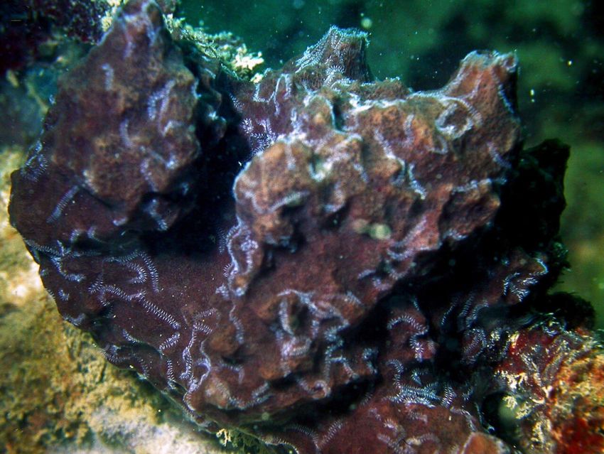
still a long way to go to fully understand how these
fascinating animals live in the wild. For example,
this study has concluded that the intestine of these
animals could be functional, yet no trace of food
has ever been seen inside them and so it is still a
mystery how they can feed their huge branched
bodies. Other questions raised in this study are
how blood circulation and nerve impulses are
affected by the branches of the body." This
research lays the foundations for understanding
how these creatures live and how their incredible
branched body came to evolve.
More information: Guillermo Ponz?Segrelles et
al, Integrative anatomical study of the branched
annelid Ramisyllis multicaudata (Annelida,
The host sponge (Petrosia) where several posterior ends Syllidae), Journal of Morphology (2021). DOI:
of one specimen of the worm Ramisyllis multicaudata can 10.1002/jmor.21356
be seen as white lines crawling on the sponge's surface.
Credit: Glasby
Provided by University of Göttingen
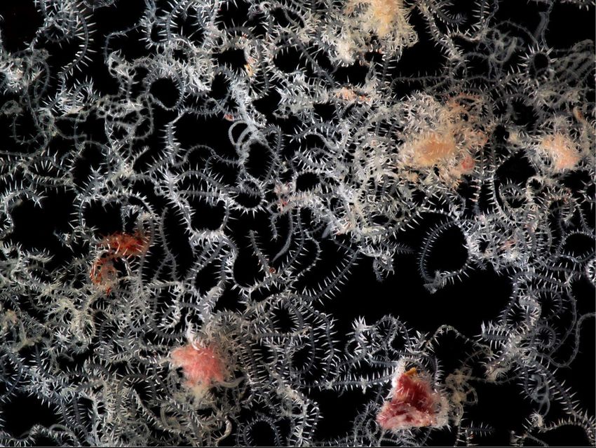
Small fraction of a single living specimen dissected out of
its host sponge as seen through the stereomicroscope.
Some dislodged fragments of sponge tissue can also be
seen. Credit: Ponz-Segrelles, Aguado & Glasby
"Our research solves some of the puzzles that
these curious animals have posed ever since the
first branched annelid was discovered at the end of
the 19th century," explains senior author Dr. Maite
Aguado, University of Göttingen. "However, there is
2/3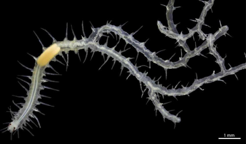
APA citation: Branching worm with dividing internal organs growing in sea sponge (2021, May 3)
                                   retrieved 11 November 2021 from https://phys.org/news/2021-05-worm-internal-sea-sponge.html

                                   This document is subject to copyright. Apart from any fair dealing for the purpose of private study or research, no
                                   part may be reproduced without the written permission. The content is provided for information purposes only.

                                                                                                                                                3/3

Powered by TCPDF (www.tcpdf.org)
You can also read