BTK (Phospho-Tyr223) Colorimetric Cell-Based ELISA Kit
←
→
Page content transcription
If your browser does not render page correctly, please read the page content below
BTK (Phospho-Tyr223)

Colorimetric Cell-Based ELISA Kit

Catalog #: OKAG01497

Please read the provided manual entirely prior to use
as suggested experimental protocols may have
changed.

Research Purposes Only. Not Intended for Diagnostic
or Clinical Procedures.
CONTENTS                                                                                           PAGE
Introduction...................................................................................................3
      Colorimetric Cell-Based ELISAs
      BTK (Phospho-Tyr223) Cell-Based ELISA

Assay Format................................................................................................4

Assay Restrictions........................................................................................5

Antibody Specificity.......................................................................................6
      Anti-BTK (Phospho-Tyr223) Antibody
      Anti-BTK Antibody
      Anti-GAPDH Antibody

Materials Included.......................................................................................10

Storage and Stability...................................................................................10

Buffer Preparation and Recommendations.................................................11

Additional Materials Required.....................................................................13

Health and Safety Precautions...................................................................14

Experiment Design.....................................................................................15

Assay Protocol…........................................................................................16

Short Protocol…………………………………………………………………...19

Data Normalization……………………………………………………………..20

Technical Support.......................................................................................21

Troubleshooting Guide...............................................................................22

ELISA Plate Template…….……………………………………………………23

Notes……………………………………………………………………………..24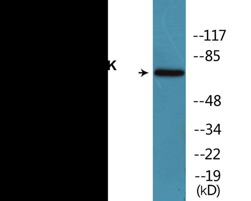
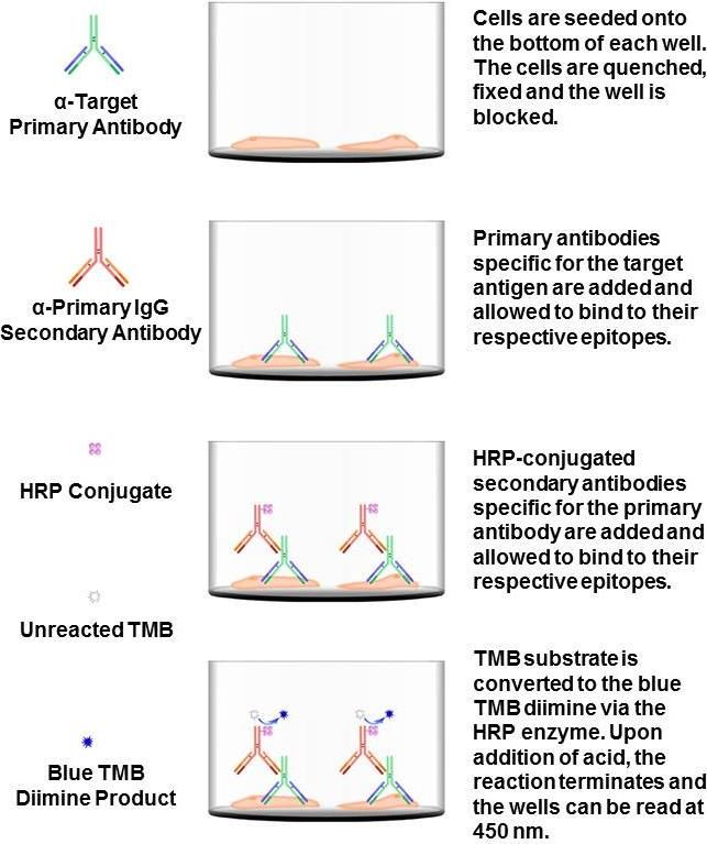
INTRODUCTION
Colorimetric Cell-Based ELISAs

The Colorimetric Cell-Based ELISA Kit allows for the detection of various
target proteins and the effects that certain stimulation conditions have on
target protein expression in different cell lines. Qualitative determination of
target protein concentration is achieved by an indirect ELISA format. In
essence, the target protein is captured by target-specific primary (1°)
antibodies while the HRP-conjugated secondary (2°) antibodies bind the Fc
region of the 1° antibody. Through this binding, the HRP enzyme
conjugated to the 2° antibody can catalyze a colorimetric reaction upon
substrate addition. Due to the qualitative nature of the Cell-Based ELISA,
multiple normalization methods are described: 1) a monoclonal antibody
specific for human GAPDH is included to serve as an internal positive
control in normalizing the target absorbance values. 2) Following the
colorimetric measurement of HRP activity via substrate addition, the crystal
violet whole-cell staining method is used to determine cell density. After
staining, the results can be analyzed by normalizing the absorbance values
to cell amounts, by which the plating difference can be adjusted. 3) If a
phosphorylated target is being detected, an antibody against the non-
phosphorylated counterpart will be provided for normalization purposes.
The absorbance values obtained for the non-phosphorylated target can be
used to normalize the absorbance values for the phosphorylated target.

BTK (Phospho-Tyr223) Colorimetric Cell-Based ELISA

The BTK (Phospho-Tyr223) Cell-Based ELISA Kit is a convenient, lysate-
free, high throughput and sensitive assay kit that can monitor BTK protein
phosphorylation and expression profile in cells. The kit can be used for
measuring the relative amounts of phosphorylated BTK in cultured cells as
well as screening for the effects that various treatments, inhibitors (ie.
siRNA or chemicals), or activators have on BTK phosphorylation.

                                                   BTK (Phospho-Tyr223) | 3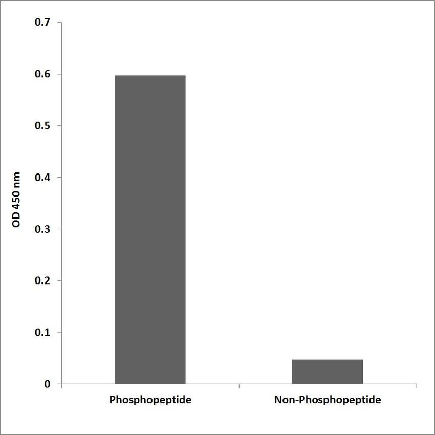
ASSAY RESTRICTIONS

                     BTK (Phospho-Tyr223) | 4
ASSAY RESTRICTIONS
This ELISA kit is intended for research purposes only, NOT
diagnostic or clinical procedures of any kind.
Materials included in this kit should NOT be used past the expiration
date on the kit label.
Reagents or substrates included in this kit should NOT be mixed or
substituted with reagents or substrates from any other kits.
Variations in pipetting technique, washing technique, operator
laboratory technique, kit age, incubation time or temperature may
cause differences in binding affinity of the materials provided.
The assay is designed to eliminate interference and background by
other cellular macromolecules or factors present within any biological
samples. However, the possibility of background noise cannot be fully
excluded until all factors have been tested using the assay kit.
BTK (Phospho-Tyr223) | 5ANTIBODY SPECIFICITY
Anti-BTK (Phospho-Tyr223) Antibody
The Anti-BTK (Phospho-Tyr223) Antibody is a rabbit polyclonal antibody. It
was tested on Western Blots for specificity. The data in Figure 2 shows that
a single protein band was detected. This protein band can be blocked by
the synthesized immunogen peptide.
Figure 2. Western blot analysis of extracts from HeLa cells treated
with Serum 10% 15’, using the Anti-BTK (Phospho-Tyr223) Antibody.
BTK (Phospho-Tyr223) | 6The data in Figure 3 shows that the Anti-BTK (Phospho-Tyr223) Antibody
is highly specific for the phospho-peptide in comparison to the non-
phospho peptide counterpart, through an ELISA.
Figure 3. Enzyme-Linked Immunosorbent Assay (ELISA) for
immunogen phosphor-peptide (left) and non-phospho peptide (right),
using Anti-BTK (Phospho-Tyr223) Antibody.
BTK (Phospho-Tyr223) | 7Anti-BTK Antibody
The Anti-BTK Antibody is a rabbit polyclonal antibody. It was tested on
Western Blots for specificity. The data in Figure 4 shows that a single
protein band was detected. This protein band can be blocked by the
synthesized immunogen peptide.
Figure 4. Western blot analysis of extracts from HeLa cells, using the
Anti-BTK Antibody.
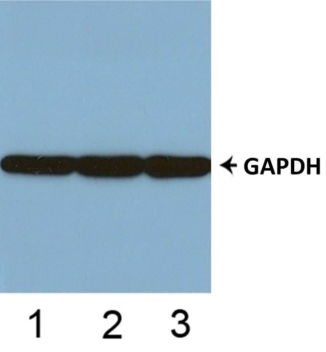
BTK (Phospho-Tyr223) | 8Anti-GAPDH Antibody
The Anti-GAPDH Antibody is a mouse monoclonal antibody. It was tested
on Western Blots with the tissue lysates from human, mouse, and rat for
specificity. The data in Figure 5 shows that a single protein band was
detected from all three lysates.
Figure 5. Western blot analysis of tissue lysates from human (1),
mouse (2) and rat (3).
BTK (Phospho-Tyr223) | 9MATERIALS INCLUDED
Reagent Quantity Container
96-Well Cell Culture
2 Plates -
Clear-Bottom Microplate
10x TBS 24 ml (10x) Clear
Quenching Buffer 24 ml (1x) Clear
Blocking Buffer 50 ml (1x) Clear
10x Wash Buffer 50 ml (10x) Clear
100x Anti-BTK (Phospho-Tyr223)
60 µl (100x) Red
Antibody (Rabbit Polyclonal)
100x Anti-BTK Antibody
60 µl (100x) Purple
(Rabbit Polyclonal)
100x Anti-GAPDH Antibody
60 µl (100x) Green
(Mouse Monoclonal)
HRP-Conjugated
12 ml (1x) Glass
Anti-Rabbit IgG Antibody
HRP-Conjugated
12 ml (1x) Glass
Anti-Mouse IgG Antibody
Primary Antibody Diluent 12 ml (1x) Clear
Ready-to-Use Substrate 12 ml (1x) Brown
Stop Solution 12 ml (1x) Clear
Crystal Violet Solution 12 ml (1x) Glass
SDS Solution 24 ml (1x) Clear
Adhesive Plate Seals 4 Seals -
STORAGE AND STABILITY
Upon receipt, the kit should be stored at 4°C. The un-opened kit will be
stable for up to 6 months from the date of shipment if stored at 4°C. Diluted
Anti-BTK (Phospho-Tyr223) Antibody, Anti-BTK Antibody and diluted Anti-
GAPDH Antibody can each be stored at 4°C for up to two weeks. HRP-
Conjugated Anti-Rabbit IgG Antibody and HRP-Conjugate Anti-Mouse IgG
Antibody will be stable at 4°C for up to six months. The SDS Solution
should be stored at room temperature or warmed up to room temperature if
stored at 4°C.
BTK (Phospho-Tyr223) | 10BUFFER PREPARATION AND RECOMMENDATIONS
Note: Please remember to allow all solutions to warm up to room
temperature prior to use.
1x TBS – 1x TBS is used to wash cells seeded on the plate. 1x TBS can be
prepared by adding 1 volume of 10x TBS provided in the kit to 9 volumes of
ddH2O.
Fixing Solution – This solution is NOT provided. Fixing Solution is used to
fix cells after cell culture. It is prepared by adding formaldehyde to 1x TBS
with light mixing. The 4% formaldehyde is used for adherent cells and 8%
formaldehyde is used for suspension cells and loosely attached cells. 37%
formaldehyde can be purchased from Sigma Cat# F-8775.
Quenching Buffer – This solution is provided as ready-to-use. Quenching
Buffer is used to inactivate the endogenous peroxidase activity of the
seeded cells.
Blocking Buffer – This solution is provided as ready-to-use. Blocking
Buffer is used to block additional binding sites in each well.
Wash Buffer – This buffer is provided as a 10x solution. 1x Wash Buffer
can be prepared by adding 1 volume of 10x Wash Buffer provided in the kit
to 9 volumes of ddH2O.
100x Anti-BTK (Phospho-Tyr223) Antibody – This antibody is a rabbit
polyclonal antibody. This antibody was tested to be specific for the BTK
protein phosphorylated at Tyr223. The supplied antibody is a 100x solution.
Make 1:100 dilutions in Primary Antibody Diluent prior to use. The diluted
primary antibody can be stored at 4°C for up to two weeks.
100x Anti-BTK Antibody – This antibody is a rabbit polyclonal antibody.
This antibody was tested to be specific for the BTK protein. The supplied
antibody is a 100x solution. Make 1:100 dilutions in Primary Antibody
Diluent prior to use. The diluted primary antibody can be stored at 4°C for
up to two weeks.
BTK (Phospho-Tyr223) | 11100x Anti-GAPDH Antibody – This antibody is a mouse monoclonal
antibody. This antibody was tested to be specific for GAPDH. The supplied
antibody is a 100x solution. Make 1:100 dilutions in Primary Antibody
Diluent prior to use. The diluted primary antibody can be stored at 4°C for
up to two weeks.
HRP-Conjugated Anti-Rabbit IgG Antibody – This solution is provided
as ready-to-use. HRP-Conjugated Anti-Rabbit IgG Antibody is used as the
secondary antibody to detect the target-bound, primary rabbit antibodies.
HRP-Conjugated Anti-Mouse IgG Antibody – This solution is provided as
ready-to-use. HRP-Conjugated Anti-Mouse IgG Antibody is used as the
secondary antibody to detect the target-bound, primary mouse antibodies.
Primary Antibody Diluent – This solution is provided as ready-to-use. Use
this solution to dilute the provided antibodies.
Ready-to-Use Substrate – This solution is provided as ready-to-use.
Ready-to-Use Substrate must be warmed to room temperature before use.
Keep away from light as this solution is light-sensitive.
Stop Solution – This solution is provided as ready-to-use. Stop Solution
must be handled with caution as it contains 2N Sulfuric Acid (H2SO4) and is
corrosive. Wear eye protection and gloves when handling.
Crystal Violet Solution – This solution is provided as ready-to-use. Crystal
Violet is an intense stain used to stain cell nuclei. Avoid contact with skin
and clothing.
SDS Solution – This solution is provided as ready-to-use. SDS is used to
solubilize the crystal violet in preparation for cell staining. Store this solution
at room temperature or warm up to room temperature if stored at 4°C.
BTK (Phospho-Tyr223) | 12ADDITIONAL MATERIALS REQUIRED
The following materials and equipment are NOT provided in this kit but are
necessary to successfully conduct the experiment:
Microplate reader able to measure absorbance at 450 nm and/or 595
nm for Crystal Violet Cell Staining (Optional)
Micropipettes with capability of measuring volumes ranging from 1 μl
to 1 ml
37% formaldehyde (Sigma Cat# F-8775) or formaldehyde from other
sources
Deionized or sterile water
Squirt bottle, manifold dispenser, multichannel pipette reservoir or
automated microplate washer
Graph paper or computer software capable of generating or
displaying logarithmic functions
Absorbent papers or vacuum aspirator
Test tubes or microfuge tubes capable of storing ≥1 ml
Orbital shaker
Poly-L-Lysine (Sigma Cat# P4832 for suspension cells)
BTK (Phospho-Tyr223) | 13HEALTH AND SAFETY PRECAUTIONS
Reagents provided in this kit may be harmful if ingested, inhaled or
absorbed through the skin.
Fixing Solution contains formaldehyde. Formaldehyde is known to be
a highly toxic reagent. Personal protection is strongly recommended
while working with this chemical.
Stop Solution contains 2N Sulfuric Acid (H2SO4) and is an extremely
corrosive agent. Please wear proper eye, hand and face protection
when handling this material. When the experiment is finished, be sure
to rinse the plate with copious amounts of running water to dilute the
Stop Solution prior to disposing the plate or strips.
Crystal Violet is an intense stain reagent. Avoid contact stain and
clothing.
BTK (Phospho-Tyr223) | 14EXPERIMENT DESIGN
1) Cell Line: The cell line must express the target protein. This protocol
can be used directly for adherent cells. For suspension cells and
loosely attached cells, two steps are required: 1) Coat the plates with
100 µl of 10 µg/ml Poly-L-Lysine (Sigma Cat# P4832, not included) to
each well of the 96-well plate for 30 minutes at 37°C before
proceeding to Step 1 of Assay Protocol (on page 16). Use 8%
formaldehyde to fix the cells on Step 5 of Assay Protocol.
2) Cell Number and Sensitivity: The number of cells plated onto the
96-well plates depends on the expression level of BTK protein in the
cells, cell size, treatment conditions and incubation time. The cells
used for testing should be around 75-90% confluent. Typically for
HeLa cells, seed 30,000 cells per well overnight for treatment the
following day. The BTK (Phospho-Tyr223) Colorimetric Cell-Based
ELISA Kit can detect Phospho-BTK expression in as little as 5,000
HeLa cells.
3) Cell Treatment: The cells can be treated with inhibitors, activators,
stimulators (ie. chemicals, proteins/peptides) or a combination of the
substances listed above. The cells can be treated with UV and serum
starvation to meet the needs of the end-user.
4) Positive and Negative Controls: Mouse Anti-GAPDH Antibody
(included) should be used to detect the internal positive controls for
normalization of OD values of the target protein. The negative
controls are HRP-Conjugated Anti-Rabbit IgG Antibody and HRP-
Conjugated Anti-Mouse IgG Antibody alone in different wells (without
the primary antibodies). Both positive and negative controls should be
performed in the same plate with the Phospho-BTK target
experiments.
5) Accuracy and Precision: Each condition should be performed in
duplicate or in triplicate.
BTK (Phospho-Tyr223) | 15ASSAY PROTOCOL
Note: Please read the whole manual before performing the experiment.
1) Seed 200 µl of 20,000 adherent cells in culture medium in each
well of a 96-well plate. The plates included in the kit are sterile and
treated for cell culture. For suspension cells and loosely attached
cells, coat the plates with 100 µl of 10 µg/ml Poly-L-Lysine (not
included) to each well of a 96-well plate for 30 minutes at 37°C
prior to adding cells.
2) Incubate the cells for overnight at 37°C, 5% CO2.
3) Treat the cells as desired.
4) Remove the cell culture medium and rinse with 200 µl of 1x TBS,
twice.
5) Fix the cells by incubating with 100 µl of Fixing Solution for 20
minutes at room temperature. The 4% formaldehyde is used for
adherent cells and 8% formaldehyde is used for suspension cells
and loosely attached cells. During the incubation, the plates
should be sealed with Parafilm. Note: Fixing Solution is volatile.
Wear appropriate personal protection equipment (mask, gloves
and glasses) when using this chemical.
6) Remove the Fixing Solution and wash the plate 3 times with 200 µl
1x Wash Buffer for five minutes each time with gentle shaking on
the orbital shaker. The plate can be stored at 4°C for a week.
Note: For all wash steps, tap the plate gently on absorbent
papers to remove the solution completely.
7) Add 100 µl Quenching Buffer and incubate for 20 minutes at room
temperature.
8) Wash the plate 3 times with 1x Wash Buffer for 5 minutes at a
time, with gentle shaking on the shaker.
9) Add 200 µl of Blocking Buffer and incubate for 1 hour at room
temperature.
BTK (Phospho-Tyr223) | 1610) Wash 3 times with 200 µl of 1x Wash Buffer for 5 minutes at a
time, with gentle shaking on the shaker.
11) Add 50 µl of 1x primary antibodies (Anti-BTK (Phospho-Tyr223)
Antibody, Anti-BTK Antibody and/or Anti-GAPDH Antibody) to the
corresponding wells, cover with Parafilm and incubate for 16 hours
(overnight) at 4°C. If the target expression is known to be high,
incubate for 2 hours at room temperature with gentle shaking on
the shaker.
12) Wash 3 times with 200 µl of 1x Wash Buffer for 5 minutes at a
time, with gentle shaking on the shaker.
13) Add 50 µl of 1x secondary antibodies (HRP-Conjugated Anti-
Rabbit IgG Antibody and/or HRP-Conjugated Anti-Mouse IgG
Antibody) to corresponding wells and incubate for 1.5 hours at
room temperature with gentle shaking on the shaker. Note: Add
HRP-Conjugated Anti-Rabbit IgG Antibody to the wells incubated
with Anti-BTK (Phospho-Tyr223) Antibody (rabbit, polyclonal)
and/or Anti-BTK (rabbit, polyclonal) and add HRP-Conjugated
Anti-Mouse IgG Antibody to the wells incubated with Anti-GAPDH
Antibody (mouse, monoclonal).
14) Wash 3 times with 200 µl of 1x Wash Buffer for 5 minutes at a
time, with gentle shaking on the shaker.
15) Add 50 µl of Ready-to-Use Substrate to each well and incubate for
30 minutes at room temperature in the dark with gentle shaking on
the shaker. Note: Ready-to-Use Substrate is a light-sensitive
reagent. Keep away from light.
16) Add 50 µl of Stop Solution to each well and read OD at 450 nm
immediately using the microplate reader.
BTK (Phospho-Tyr223) | 17Optional: Crystal Violet Cell Staining
Crystal Violet binds to cell nuclei and gives absorbance readings
proportional to cell counts at 595 nm.
17) After finishing reading the absorbance at 450 nm, wash the plate
twice with 200 µl of Wash Buffer and twice with 200 µl of 1x TBS
for 5 minutes each. Tap the plates on paper towel to remove the
excess liquid. Let plate air dry for 5 minutes at room temperature.
18) Add 50 µl of Crystal Violet Solution to each well, incubate for 30
minutes at room temperature on the shaker. Note: Crystal Violet is
an intense stain. Avoid contact with skin and clothing.
19) Flick the plate to remove Crystal Violet Solution, rinse the plate by
filling the wells with running tap water, and wash the plate with 200
µl of 1x TBS 3 times, 5 minutes each with gently shaking on the
shaker.
20) Add 100 µl of SDS Solution into each well and incubate on the
shaker at room temperature for 1 hour.
21) Read absorbance at 595 nm with microplate reader. If absorbance
is too high, the solubilized crystal violet solution can be diluted 10
times with H2O on a separate 96-well plate.
BTK (Phospho-Tyr223) | 18SHORT PROTOCOL
Seed cells into wells and incubate overnight at 37°C, 5% CO2.
Apply desired treatment conditions.
Add 100 µl of Fixing Solution and incubate 20 minutes at room
temperature.
Add 100 µl of Quenching Buffer and incubate 20 minutes at room
temperature.
Add 200 µl of Blocking Buffer and incubate for 1 hour at room temperature.
Add 50 µl of 1x primary antibodies and incubate overnight at 4°C.
Add 50 µl of HRP-conjugated secondary antibodies and incubate for 1.5
hours at room temperature.
Add 50 µl of Ready-to-Use Substrate and incubate for 30 minutes at room
temperature.
Add 50 µl of Stop Solution and read OD at 450 nm.
Crystal Violet Cell Staining Procedure (Optional)
BTK (Phospho-Tyr223) | 19DATA NORMALIZATION
Anti-BTK Antibody Normalization
The OD values obtained for the phosphorylated target protein can be
normalized using the OD values obtained for the non-phosphorylated target
protein via the proportion, OD450 (Anti-BTK P-Tyr223 Antibody)/OD450 (Anti-
BTK Antibody).
GAPDH Normalization
The OD450 values obtained for the target protein (phosphorylated and non-
phosphorylated) can be normalized using the OD450 values obtained for
GAPDH.
Crystal Violet Staining Normalization
The measured OD450 readings can be normalized using the OD595 values
via the proportion, OD450/OD595.
BTK (Phospho-Tyr223) | 20TECHNICAL SUPPORT
For troubleshooting, information or assistance, please visit us online at
www.avivasysbio.com or contact us at:
Aviva Systems Biology Corporation
5754 Pacific Center Blvd., Suite 201
San Diego, CA 92121
United States of America
Email: info@avivasysbio.com or
techsupport@avivasysbio.com
Phone: (858)552-6979
Toll-Free Phone: (888)880-0001
Fax: (858)552-6975
BTK (Phospho-Tyr223) | 21TROUBLESHOOTING GUIDE
Problem Cause Solution
Storage and Read Manual for storage condition
Expiration and expiration.
Dilute antibody 1:100 with Antibody
Antibody Dilution
Diluent.
Low Signal Low Cell Number Seed more cells.
Add solutions slowly from the side
Cells Detach from wall of the wells. Use Poly-L-Lysine
Well-Bottoms to coat the plate period seeding the
cells, if necessary.
High Washing Remove Wash Buffer completely.
Background High Cell Number Reduce the amount of seeded cells.
Pipetting Check and/or calibrate pipettes.
Washing Remove Wash Buffer completely.
Add solutions slowly from the side
Variation
Cells Detach from wall of the wells. Use Poly-L-Lysine
Well-Bottoms to coat the plate period seeding the
cells, if necessary.
BTK (Phospho-Tyr223) | 22ELISA PLATE TEMPLATE
BTK (Phospho-Tyr223) | 23NOTES
BTK (Phospho-Tyr223) | 24You can also read