Carcinomatous like mastitis due to axillary lymphadenopathy in a case of nasopharyngeal carcinoma: A case report
←
→
Page content transcription
If your browser does not render page correctly, please read the page content below
EXPERIMENTAL AND THERAPEUTIC MEDICINE 22: 1026, 2021

       Carcinomatous‑like mastitis due to axillary lymphadenopathy
           in a case of nasopharyngeal carcinoma: A case report
              CRISTINA MARINELA OPREAN1‑3, NUSA ALINA SEGARCEANU2,3, ALEXANDRA STAN4,
                  CRISTIAN SILVIU SUCIU5, DACIANA GRUJIC6,7, IOANA ALEXANDRA RIVIS8,9,
                      ALIS LILIANA CARMEN DEMA10 and ANA CRISTINA BREDICEAN9,11

   1
    Discipline of Morpho‑Pathology, ‘Victor Babes’ University of Medicine and Pharmacy of Timisoara, 300041 Timisoara;
   2
    Department of Oncology, OncoHelp Hospital; 3Department of Oncology, Oncomed Outpatient Unit, 300239 Timisoara;
       4
        Department of Oncology, Emergency City Hospital, 300254 Timisoara; 5Discipline of Histology, ‘Victor Babes’
    University of Medicine and Pharmacy of Timisoara; 6Department of Plastic and Reconstructive Surgery, ‘Victor Babes’
   University of Medicine and Pharmacy of Timisoara; 7Clinic of Burns, Plastic and Reconstructive Surgery, ‘Pius Branzeu’
     Emergency County Hospital, 300041 Timisoara; 8Neurosciences Department, ‘Carol Davila’ University of Medicine
     and Pharmacy, 020021 Bucharest; 9NEUROPSY‑COG Center for Cognitive Research in Neuropsychiatric Pathology,
                Neurosciences Department, ‘Victor Babes’ University of Medicine and Pharmacy of Timisoara;
            10
               ANAPATMOL Research Center, ‘Victor Babes’ University of Medicine and Pharmacy of Timisoara,
    300041 Timisoara; 11Psychiatry Compartment, ‘Dr Victor Popescu’ Emergency Hospital, 300080 Timisoara, Romania

                                           Received April 29, 2021; Accepted June 1, 2021

                                                     DOI: 10.3892/etm.2021.10458

                                                                      Abstract. Nasopharyngeal carcinoma (NPC) is a rare form
                                                                      of malignancy, accounting for 2% of all cancers of the head
Correspondence to: Professor Alis Liliana Carmen Dema,                and neck in Europe. Axillary lymph node metastases are
ANAPATMOL Research Center, ‘Victor Babes’ University of               very rare in these cases. This is a case report of a 40‑year‑old
Medicine and Pharmacy of Timisoara, Piata Eftimie Murgu 2,            premenopausal woman diagnosed in May 2015 with T1N2M0
300041 Timisoara, Romania                                             stage III NPC, treated with induction chemotherapy, followed
E‑mail: dema_alis@yahoo.com                                           by chemo‑radiotherapy. Post‑therapeutic computed tomog‑
Dr Ioana Alexandra Rivis, Neurosciences Department, ‘Carol            raphy (CT) scan showed partial response (PR) on the primary
Davila’ University of Medicine and Pharmacy, 37 Dionisie              tumor and complete response (CR) on the latero‑cervical lymph
Lupu Street, 020021 Bucharest, Romania                                nodes. In 2017, our patient developed left carcinomatous‑like
E‑mail: ioana.rivis@gmail.com                                         mastitis with axillary lymphadenopathy. This raised suspi‑
                                                                      cions of a carcinomatous mastitis. The pathology report with
Abbreviations: TPF, docetaxel, cisplatin, and 5‑fluorouracil; NPC,    immunohistochemistry (IHC) of the third biopsy highlighted
nasopharyngeal carcinoma; HNSCC, squamous cell carcinoma of the
                                                                      axillary metastasis of a non‑keratinizing squamous cell carci‑
head and neck; NP, nasopharynx; NSCC, non‑keratinizing squamous
cell carcinoma; SCC, squamous cell carcinoma; DFI, disease‑free
                                                                      noma (NSCC). There are very few references in the literature
interval; CR, complete response; IHC, immunohistochemistry;           regarding axillary metastases from squamous cell carcinoma
PR, partial response; CT, computed tomography; Gy, gray; GCSF,        of the head and neck (HNSCC). As far as we know, this is the
granulocyte colony‑stimulating factor; ENT, ear‑nose‑throat;          first case report of mastitis due to NPC. To conclude, treatment
CK, cytokeratin; HMWCK, high‑molecular‑weight cytokeratin;            consisted of two surgical excisions of axillary lymphade‑
EMA, epithelial membrane antigen; CgA, chromogranin A; Syn,           nopathy associated with local radiotherapy and chemotherapy
synaptophysin; GATA‑3, trans‑acting T cell‑specific transcription     (neo‑adjuvant, adjuvant). The second surgery, performed after
factor; CD10, neutral peptidase 24.11 (NEP); SMA, smooth muscle       radiotherapy, required plastic surgery. A psychiatric evalu‑
actin; TTF1, thyroid transcription factor 1; DAB, diaminobenzidine;   ation was necessary, revealing a reactive anxiety disorder.
EBV, Epstein‑Barr virus; EA, early antigen; Ig, immunoglobulin;       This case required multidisciplinary management, where
EBNA, Epstein‑Barr nuclear antigen; VCA, virus capsid antigen;
                                                                      oncology, plastic surgery, pathology and psychiatric specialists
EBER, Epstein‑Barr encoding region; RTU, ready‑to‑use; HIER,
heat induced epitope retrieval; HRP, horseradish peroxidase; PCR,
                                                                      collaborated in deciding the therapeutic approach.
polymerase chain reaction; DNA, deoxyribonucleic acid; NGS, next
generation sequencing; H&E, hematoxylin and eosin                     Introduction

Key words: head and neck cancer, nasopharyngeal carcinoma,            Nasopharyngeal carcinoma (NPC) is the most common type
axillary lymph node metastasis, carcinomatous mastitis, case report   of cancer in southern Asia (1,2). In Europe, its incidence is rare,
                                                                      with an annual rate varying between 2.1 and 0.4 per 100,000
                                                                      individuals (3‑5). NPC represents 2% of all cancers developed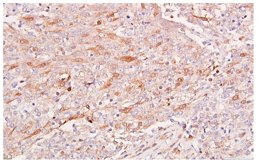
2                                    OPREAN et al: CARCINOMATOUS-LIKE MASTITIS AFTER NPC

in the head and neck (6). The incidence is higher in men than             By November 2015, the patient had undergone 3 cycles
in women with an M:F ratio of 2.75 (3,4). Epstein‑Barr virus          of docetaxel, cisplatin, and 5‑fluorouracil (TPF) induction
infection is also a factor in most neck and head cancer cases,        chemotherapy associated with prophylactic supportive granu‑
along with smoking and alcohol consumption (7‑9). Relative            locyte colony‑stimulating factor (GCSF) treatment, with PR
survival for NPC in Europe is 49% at 5 years, with no differ‑         after induction chemotherapy, followed by concomitant weekly
ences in survival between sexes (3). HNSCC has been proven            cisplatin (40 mg/m2) radio‑chemotherapy 66 Gray (Gy) on the
to distantly spread below the clavicles (10). The chance of           NP, 58 Gy on the bilateral cervical lymph nodes and 50 Gy on
dissemination is related to the cervical lymph node involve‑          the bilateral supraclavicular areas. Both treatment adherence
ment. For N0‑N2 tumors, the dissemination occurs in less than         and tolerance were satisfactory. The post therapeutic brain
10% of cases compared with 30% in N2‑N3 cases (10).                   CT scan, performed in March 2016, showed PR at the primary
    The lymphatic drainage from the head and neck occurs              NPC, and CR at the latero‑cervical lymph nodes.
through a superficial and deep system. The superficial group              On February 2017, the patient reported pain, edema,
of cervical lymph nodes includes occipital, postauricular,            erythema and swelling of the left breast. The physical exami‑
parotid, facial, submandibular, sub‑mental and superficial            nation noted left axillary lymphadenopathy and left‑sided
cervical nodes, which are situated alongside the external             mastitis. In order to explore the probability of a coexistent
jugular vein. The profound group of cervical lymph nodes lies         breast cancer (carcinomatous mastitis), an excisional biopsy
along the internal jugular vein. All of the lymphatic vessels of      was performed on the left axillary lymph nodes, which
the head and neck empty into the deep group, either directly or       excluded a neoplastic process. The patient received local and
via the superficial group of nodes (11). The lymphatic system,        oral treatment with anti‑inflammatory drugs (300 mg of keto‑
within the axilla region, normally flows from the distal portion      profen per day) and antibiotics (amoxycillin/clavulanic acid
of the upper limb and the chest wall along the axillary vein          1,000 mg/62.5 mg, twice a day) for 10 days, after which the
toward the subclavian venous system (12).                             mastitis went into slight remission for the next 4 months.
    Like most other locations of HNSCC, the lymphatic                     In June 2017, the inflammation of the left breast reoccurred
drainage of the nasopharynx (NP) is predominantly directed            along with the worsening of the previous symptoms. Chest
to the cervical lymph nodes (13,14). Lymph node metastases            CT scan confirmed left axillary (of 4.5 cm) and supraclavicular
are present in 80% of NPC cases, and in 50% of the cases,             lymph node enlargement (of 3.5 cm), and left mastitis. Left
these are bilateral. The worst aspect of cancer is its ability        axillary lymph node excisional biopsy was performed after
to spread or metastasize (15). Patients with lymph node               10 days of systemic antibiotic and anti‑inflammatory treat‑
metastases have a more favorable prognosis than those with            ment. The pathology report revealed another inflammatory
visceral metastases (16). It is known that NPC tumors are             reaction of the lymph node, without tumor infiltration.
associated with a high incidence of distant metastases (17,18).           In September 2017, due to the persistence of the clinical
Pulmonary metastases represent the most common localiza‑              signs of mastitis, despite oral antibiotic and anti‑inflammatory
tion, accounting for 66% of total distant metastases. Other           treatment, the patient attended our clinic looking for a second
metastatic sites include bone (22%) and liver (10%) (18‑20).          medical opinion. Clinical examination revealed severe mastitis,
Axillary lymph node metastasis is an uncommon event                   lymphedema of the left arm and lymphadenopathy in the left
in HNSCC. There are very few references in the literature             axilla and supraclavicular region (Fig. 1). A new subcutaneous
regarding axillary metastases from HNSCC (21). Only five              lesion on the left axilla and thorax was discovered. The total
reports have been published in the literature, including              body CT scan showed a progressive aspect of mastitis and
10 patients (22). None of these cases included a co‑occurrence        an increase in the axillary and supraclavicular lymph nodes
of mastitis. Axillary metastases are mentioned in only 2 of           (Figs. 2 and 3).
8 published autopsy studies (10). Upon autopsy reports,                   The primary NPC tumor and latero‑cervical lymph nodes
between 2 to 9% of patients with HNSCC tumors were                    were in CR. One subcutaneous nodule from the axillary left
found to have axillary node metastases, but it is assumed that        region was excised and sent for pathological examination. The
the incidence is higher, as impalpable lymph nodes are not            pathology report described large islands of tumor cells with
routinely examined during autopsy, except in breast cancer            prominent nucleoli and admixed inflammatory cells (Fig. 4).
cases and in some malignant melanomas (23,24).                        IHC staining was done on paraffin‑embedded tissue, with the
                                                                      following antibodies: Trans‑acting T cell‑specific transcrip‑
Case report                                                           tion factor (GATA‑3), mammaglobin, epithelial membrane
                                                                      antigen (EMA), cytokeratin (CK) AE1/AE3, CK5, high‑molec‑
We present the case of a 40‑year‑old woman, a stay‑at‑home            ular‑weight cytokeratin (HMWCK) (34BE12), CK7, p63,
mother of two children (aged 5 and 8), who lives in the rural area,   neutral peptidase 24.11 (NEP) (CD10), S100, smooth muscle
with no family history of neoplasm or any other comorbidities.        actin (SMA), p16, chromogranin A (CgA), synaptophysin
The onset of the disease was in April 2015, when our patient          (Syn). Serial sections, 3‑µm thick, were constructed from one
started noticing a nasopharyngeal discomfort, nasal obstruc‑          selected paraffin‑block with the representative tumor area, and
tion, rhinorrhea and epistaxis. The ear‑nose‑throat (ENT)             then mounted on silanized slides, so as to prevent detachment
exam revealed a lesion of 2.3/2 cm in the NP, for which a             during antigen unmasking procedures. Tissue sections were
biopsy was completed. Clinically, the patient presented bilat‑        deparaffined in xylene and rehydrated. For unmasking the
eral cervical lymphadenopathy with diameters of up to 3.5 cm.         antigens, we used heat induced epitope retrieval (HIER) by
In May 2015, the diagnosis was confirmed: Stage III, T1N2M0           boiling the slides for 20 min at 98˚C in target retrieval solution
poorly differentiated NSCC of the NP.                                 pH 6.0 or 9.0. Endogenous peroxidase activity was blocked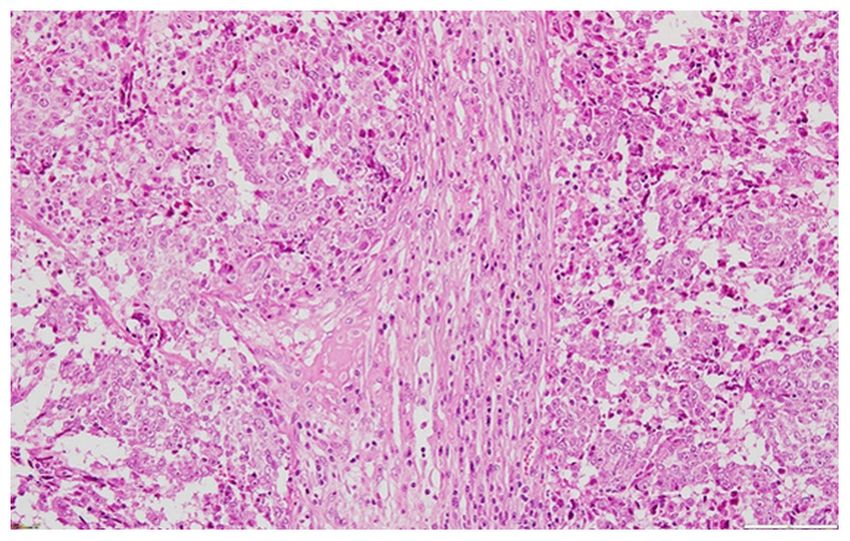
EXPERIMENTAL AND THERAPEUTIC MEDICINE 22: 1026, 2021                                                   3

                                                                      Figure 3. Left breast mastitis and axillary lymph nodes on CT (September
                    Figure 1. Left breast mastitis.                   2017). CT, computed tomography.

                                                                      Figure 4. Large islands of tumor cells with prominent nucleoli and admixed
                                                                      inflammatory cells. H&E, x 200. H&E, hematoxylin and eosin.

                                                                      Barr virus (EBV) were investigated. Early antigen (EA)
Figure 2. Left breast mastitis on CT (September 2017). CT, computed   immunoglobulin (Ig) G was positive (>150 U/ml); antibodies
tomography.                                                           against Epstein‑Barr nuclear antigen (EBNA) IgG were
                                                                      highly positive (>600 U/ml), virus capsid antigen (VCA)
                                                                      IgM antibodies were negative (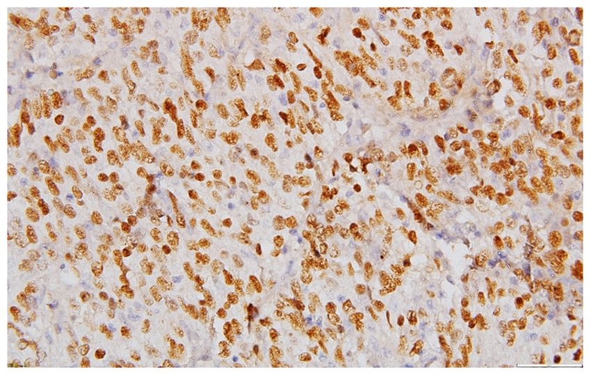
4 OPREAN et al: CARCINOMATOUS-LIKE MASTITIS AFTER NPC
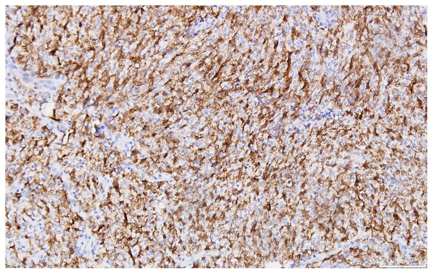
Figure 5. Extensive expression for p63 in the tumor cells. Anti‑p63, DAB,
counterstain with hematoxylin x 400. DAB, diaminobenzidine.
Figure 7. Remissive mastitis of the left breast after 4 cycles of chemotherapy
(January 2018).
Figure 6. Less extensive reactivity for HMWCK (clone 34ßE12) in the tumor
cells. Anti‑HMWCK, DAB, counterstain with hematoxylin x400. HMWCK,
high‑molecular‑weight cytokeratin; DAB, diaminobenzidine.
symptoms started to alleviate, and after a total of six weeks,
they completely disappeared.
The patient started chemotherapy with cisplatin (75 mg/m2)
and docetaxel (75 mg/m2) every 3 weeks, with GCSF support.
After 4 cycles, in January 2018, the edema of the left breast
and arm was in remission (Fig. 7). Clinically, the left axil‑
lary lymph nodes decreased and the supraclavicular ones
disappeared. The patient had a mild cough, no fever, but an Figure 8. Mild diffuse edema of the left breast (September 2018).
unpleasant fetid breath. Chest CT scan confirmed two pulmo‑
nary abscesses, PR of the left axillary lymph nodes and CR
of the left supraclavicular nodes. Chemotherapy was post‑
poned for two months, due to this severe lung infection. After axillary lymph node metastasis recurred. The clinical exam
remission, the same chemotherapy schedule was restarted and showed left breast mild mastitis and a mobile 3.3 cm axillary
continued until cycle 11. lymphadenopathy, which was programmed for excision.
In September 2018, full‑body CT‑scan showed persistence The retractile scar‑tissue was removed surgically, with a
of CR at primary NPC and loco‑regional lymph nodes, a mild thorough dissection of the fibrous axillary tissue that resulted
diffuse edema of the left breast and a decrease in the left axil‑ from scarring and radiotherapy, while isolating the lymphade‑
lary lymph nodes (at 1.8 cm) (Figs. 8 and 9). Afterwards, our nopathy block of the vascular pedicle and making a Z‑plasty,
patient underwent a surgical excision of the left axillary lymph in order to eliminate the adhesions. The pathology examina‑
nodes. The pathological report revealed the presence of 4 lymph tion, including an extended IHC panel which found positive
nodes between 0.5 and 2 cm, all of them metastatic NSCC, reaction for p63, CK5 (Fig. 10), CK AE1/AE3, focal reactivity
most probably originating in the NP. Our patient received for p16 (Fig. 11), cytoplasmic and nuclear and lack of reactivity
adjuvant radiotherapy of 50 Gy/25 fractions on the axilla, until for SMA, GATA‑3, Syn, chromogranin A (CgA), CK7, EMA,
January 2019. The treatment was well‑tolerated. After 9 months S100, CD10, confirmed metastasis of a poorly differentiated
of disease‑free interval (DFI), left breast mastitis and a left NSCC.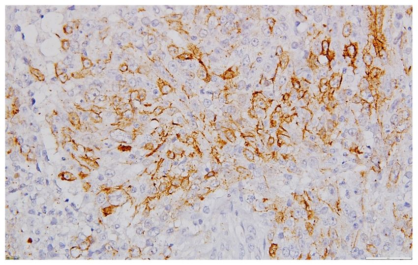
EXPERIMENTAL AND THERAPEUTIC MEDICINE 22: 1026, 2021                                                         5

                                                                               Figure 11. Focal reactivity for p16. Anti‑p16, DAB, counterstain with
                                                                               hematoxylin, x400. DAB, diaminobenzidine.

Figure 9. Left mastitis in remission and decrease of the left axillary lymph
nodes (September 2018).

                                                                               Figure 12. Local remission of left breast edema and persistent lymphedema
                                                                               of the left arm (April 2020).

                                                                               especially in patients with massive metastases or delayed
Figure 10. Diffuse expression of CK5 in the tumor cells. Anti‑CK5, DAB,
                                                                               metastases at a low level of the head and neck (25). This article
counterstain with hematoxylin, x 200. CK, cytokeratin; DAB, diaminoben‑        presents the case of a young patient, initially diagnosed and
zidine.                                                                        treated for NPC, who presented, in the evolution of the disease,
                                                                               axillary lymph node metastasis and carcinomatous‑like
                                                                               mastitis. At the time of the axillary recurrence, the primary
                                                                               NPC had CR. Due to the clinical impression of the inflamma‑
    The paraffin block from this last biopsy sample was used                   tory breast carcinoma, we began by trying to diagnose a breast
to test genomic alteration. The result showed NOTCH1 muta‑                     cancer, which was disproved following the results of the IHC
tion‑V1578 deletion. The patient was invited to be included in                 and pathological examinations. Previous literature reports
a phase I/II clinical trial abroad, but she refused. The pre‑ther‑             suggest that the involvement of axillary sites may be related
apeutic total body CT scan evaluation did not highlight any                    to previous neck surgery, following a change in lymphatic
other metastasis. Primary NPC tumor and loco‑regional lymph                    drainage (11). Our patient had not undergone prior head and
nodes remained in CR. The patient restarted chemotherapy                       neck surgery.
with cisplatin and docetaxel in January 2020, with the same                        The pathological aspects corresponded to a metastasis of
dosages and following the same schedule, as an ‘adjuvant’                      a poorly differentiated/undifferentiated tumor, which is diffi‑
treatment. On April 2020, the patient was undergoing chemo‑                    cult to diagnose based on routine stained slides. The positive
therapy treatment, and the clinical symptoms were in complete                  reaction for EMA and CK established the epithelial nature of
remission, but the upper left limb edema persisted (Fig. 12).                  the proliferation. Given the clinical aspect of inflammatory
                                                                               carcinoma combined with axillary lymphadenopathy, the IHC
Discussion                                                                     investigation, performed over two stages (2017 and 2019), was
                                                                               focused on removing the possibility of a metastasis from a
Axillary lymph node involvement is rare in nasopharyngeal                      second primary metachronous breast carcinoma.
carcinoma (NPC) (21). Yet, they are a frequent site of regional                    The absence of CK7, mammaglobin and GATA3
metastatic disease from breast carcinoma (25). The recogni‑                    reactivity, excluded the majority of most common breast
tion of axillary metastasis in patients with HNCC is crucial,                  carcinoma subtypes. The diffuse positivity for p63, CK5 and a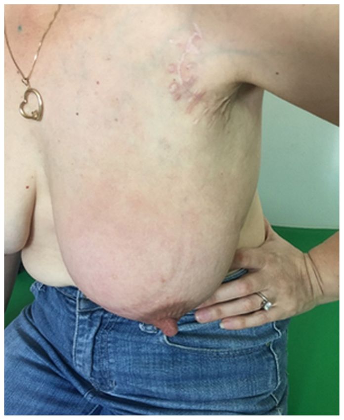
6                                  OPREAN et al: CARCINOMATOUS-LIKE MASTITIS AFTER NPC

heterogeneous one for HMWCK suggested a metastasis from                This particular case required a multidisciplinary
a squamous or myoepithelial carcinoma, both of which are           management, where the oncology, plastic surgery, pathology
considered to be extremely rare subtypes of breast carcinoma.      and psychiatric medical specialists acted together in order
The absence of S100 and SMA reactivity excluded a breast           to establish the correct diagnosis and adopt the appropriate
lesion with myoepithelial differentiation. Knowing that p63        therapeutic pathway. Currently, neoplastic patients benefit
and HMWCK are positive in metaplastic breast carcinomas            from personalized therapy, with the ultimate target of offering
with spindle cells and/or squamous differentiation (26), the       the patient a good quality of life.
possibility that the axillary lesion represented a metastasis
from these entities could not be completely ruled out. But         Acknowledgements
considering what Wargotz and Norris (1990) affirmed, namely
that before diagnosing a primary squamous cell carcinoma of        The authors would like to thank the patient for allowing them
the breast it is necessary to exclude a metastatic squamous cell   to publish her case and to all the healthcare professionals who
carcinoma to the breast, the scenario of a metastasis from the     helped care for this patient.
previous diagnosed NPC was favored (27).
    The main arguments for NPC metastasis against a                Funding
metastatic metaplastic breast carcinoma with spindle cell
and/or squamous differentiation included: An initial diagnosis     No funding was received.
of NPC; the important bilateral latero‑cervical lymphadenop‑
athy, staged cN2; the absence of a primary breast malignancy       Availability of data and materials
documented by imaging and excisional biopsies; the IHC profile
of the axillary metastasis which overlaps with the nasopha‑        Further information regarding this case report is available
ryngeal lesion (conducted in another pathology department);        from the corresponding author on reasonable request.
the identification, by next generation sequencing (NGS), of
NOTCH1 V1578 del and missense mutation of p53, when the            Authors' contributions
NOTCH1 mutation is the second most common mutated gene
(after TP53) in HNSCC (28); the positive serology for EBV,         CMO, NAS and AS were in charge of patient management,
being well‑known that nonkeratinizing NPC is associated with       establishing the chemotherapy treatment and follow‑up of the
EBV in almost all cases (29).                                      treatment response, and clinical evolution of the patient. ALCD
    Unfortunately, the in situ hybridization for EBER, a           and CSS were in charge of the laboratory diagnosis: Pathology,
helpful investigation for establishing the NP as the primary       IHC and genomic testing. DG performed the two diagnostic
site for a meta‑virus capsid antigen static undifferentiated or    interventions (biopsy) and the surgical axillary excision. ACB
poorly differentiated squamous cell carcinoma, could not be        and IAR performed the psychiatric evaluation, established a
performed.                                                         treatment plan and offered psychological support. All authors
    Emerging from the main genomic alteration identified,          read and approved the final manuscript for publication.
the NOTCH pathway, targeted therapy appeared to be a
logical therapeutic option for this case. A theory regarding       Ethics approval and consent to participate
the favorable evolution of our patient may be due to, partly,
the NOTCH1 mutation, since, more often than not, this              For this case‑report we obtained the patient's informed
mutation acts as a tumor‑suppressor gene (30). The timely          consent and her permission to use the images that were taken
recovery surgery of the axillary metastasis after exclusion        during her periodical check‑ups. An ethical approval from the
of other distant metastasis may improve the survival of these      hospital was not required.
patients (25). HNSCC is one of the most aggressive malignan‑
cies (31). However, our patient had a survival of almost 5 years   Patient consent for publication
from the diagnosis of the primary tumor, and of about 3 years
from the diagnosis of recurrence. For this patient, the surgical   Written informed consent was obtained from the patient
removal of the axillary lymph node was completed twice, for        regarding the publication of this case report and any accom‑
two local recurrences. We aimed to improve local control           panying images. A copy of the written consent is available for
by adding radiotherapy of the axilla, after the first complete     review.
surgical excision. Chemotherapy was administered twice, in
a ‘neo‑adjuvant’ setting, first due to the initially inoperable    Competing interests
recurrence, and second, in an ‘adjuvant’ setting, after the last
surgery for the recurrence. In the absence of clear evidence       The authors declare no competing interests.
on the efficacy of adjuvant therapy in this clinical situation,
adjuvant chemotherapy was used for the second recurrence,          References
which, although surgically removed, did not dismiss the possi‑
bility of systemic micro‑metastasis, that cannot be identified      1. Kuo DY, Chang MH, Wang SY, Hsieh PY and Shueng PW:
                                                                       Unusual axillary metastasis of recurrent nasopharyngeal cancer:
by standard diagnostic methods. The correct duration of this           A case report. Medicine (Baltimore) 96: e6854, 2017.
present ‘adjuvant’ chemotherapy remains an open question.           2. Mahdavifar N, Ghoncheh M, Mohammadian‑Hafshejani A,
                                                                       Khosravi B and Salehiniya H: Epidemiology and inequality
The role of local radiotherapy and systemic chemotherapy               in the incidence and mortality of nasopharynx cancer in Asia.
remains to be established in the future.                               Osong Public Health Res Perspect 7: 360‑372, 2016.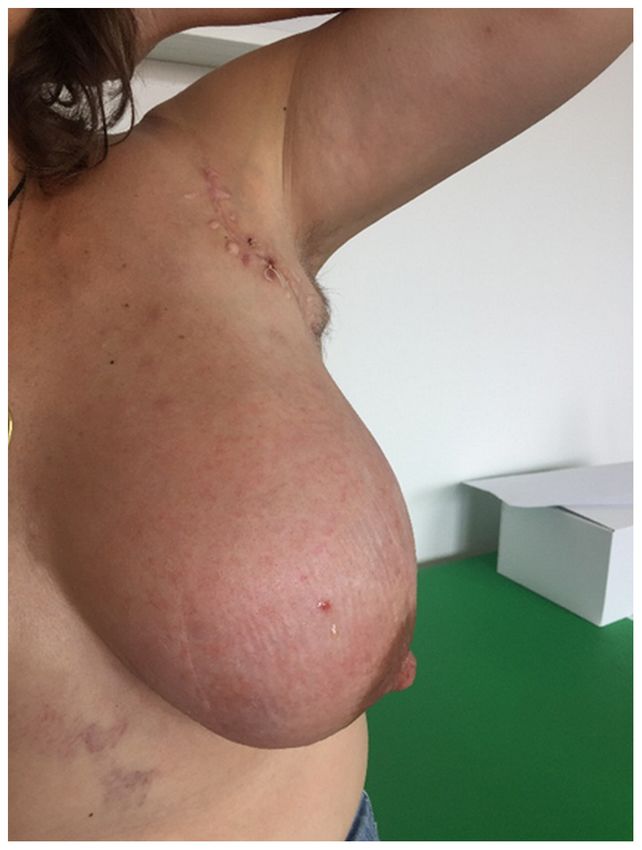
EXPERIMENTAL AND THERAPEUTIC MEDICINE 22: 1026, 2021                                                  7

 3. Bossi P, Chan AT, Licitra L, Trama A, Orlandi E, Hui EP,            18. Tomao F, Miele E, Spinelli GP, Caprio G, Ranieri E, Mingazzini P,
    Halámková J, Mattheis S, Baujat B, Hardillo J, et al:                   Zullo A and Tomao S: Axillary and subcutaneous breast metas‑
    Nasopharyngeal carcinoma: ESMO‑EURACAN clinical prac‑                   tases from rhinopharyngeal carcinoma: A case report and
    tice guidelines for diagnosis, treatment and follow‑up †. Ann           literature review. Anticancer Res 28: 419‑424, 2008.
    Oncol 32: 452‑465, 2021.                                            19. Chan AT, Teo PM and Johnson PJ: Nasopharyngeal carcinoma.
 4. Parkin DM, Bray F, Ferlay J and Pisani P: Global cancer statis‑         Ann Oncol 13: 1007‑1015, 2002.
    tics, 2002. CA Cancer J Clin 55: 74‑108, 2005.                      20. Ahmad A and Stefani S: Distant metastases of nasopharyngeal
 5. Chang ET and Adami HO: The enigmatic epidemiology of naso‑              carcinoma: A study of 256 male patients. J Surg Oncol 33:
    pharyngeal carcinoma. Cancer Epidemiol Biomarkers Prev 15:              194‑197, 1986.
    1765‑1777, 2006.                                                    21. Koch WM: Axillary nodal metastases in head and neck cancer.
 6. Zhou X, Cui J, Macias V, Kajdacsy‑Balla AA, Ye H, Wang J and            Head Neck 21: 269‑272, 1999.
    Rao PN: The progress on genetic analysis of nasopharyngeal          22. Wormald R, Sheahan P and Timon C: A case of head and neck
    carcinoma. Comp Funct Genomics 2007: 57513, 2007.                       cancer metastasizing to the axillary lymph nodes. Ear Nose
 7. Fernandes Q, Merhi M, Raza A, Inchakalody VP, Abdelouahab N,            Throat J 89: E24‑E26, 2010.
    Zar Gul AR, Uddin S and Dermime S: Role of Epstein‑Barr virus       23. Islam S, Cole CV, Hoffman GR and Brennan PA: Bilateral
    in the pathogenesis of head and neck cancers and its potential as       axillary metastasis from a primary ethmoidal squamous cell
    an immunotherapeutic target. Front Oncol 8: 257, 2018.                  carcinoma. J Laryngol Otol 120: 353‑355, 2006.
 8. Pezzuto F, Buonaguro L, Caponigro F, Ionna F, Starita N,            24. Kowalski LP: Noncervical lymph node metastasis from head and
    Annunziata C, Buonaguro FM and Tornesello ML: Update on                 neck cancer. ORL J Otorhinolaryngol Relat Spec 63: 252‑255,
    head and neck cancer: Current knowledge on epidemiology, risk           2001.
    factors, molecular features and novel therapies. Oncology 89:       25. Oo AL, Yamaguchi S, Iwaki H and Amagasa T: Axillary nodal
    125‑136, 2015.                                                          metastasis from oral and maxillofacial cancers: A report of
 9. Sturgis EM, Wei Q and Spitz MR: Descriptive epidemiology and            3 cases. J Oral Maxillofac Surg 62: 1019‑1024, 2004.
    risk factors for head and neck cancer. Semin Oncol 31: 726‑733,     26. Koker MM and Kleer CG: p63 expression in breast cancer: A
    2004.                                                                   highly sensitive and specific marker of metaplastic carcinoma.
10. Nelson WR and Sisk M: Axillary metastases from carcinoma of             Am J Surg Pathol 28: 1506‑1512, 2004.
    the larynx: A 25‑year survival. Head Neck 16: 83‑87, 1994.          27. Wargotz ES and Norris HJ: Metaplastic carcinomas of the
11. McKenzie BJ and Loock JW: Axillary nodal metastasis at                  breast. IV. Squamous cell carcinoma of ductal origin. Cancer 65:
    primary presentation of an oropharyngeal primary carcinoma:             272‑276, 1990.
    A case report and review of the literature. J Med Case Rep 3:       28. Fukusumi T and Califano JA: The notch pathway in head and
    7230, 2009.                                                             neck squamous cell carcinoma. J Dent Res 97: 645‑653, 2018.
12. Rayatt SS, Dancey AL, Fagan J and Srivastava S: Axillary            29. El‑Naggar AK, Chan JKC, Grandis JR, Takata T and
    metastases from recurrent oral carcinoma. Br J Oral Maxillofac          Slootweg PJ (eds): WHO Classification of Head and Neck
    Surg 42: 264‑266, 2004.                                                 Tumours. Vol. 9. 4th edition. International Agency for Research
13. Ho FC, Tham IW, Earnest A, Lee KM and Lu JJ: Patterns of                on Cancer. IARC, Lyon, 2017.
    regional lymph node metastasis of nasopharyngeal carcinoma:         30. Agrawal N, Frederick MJ, Pickering CR, Bettegowda C,
    A meta‑analysis of clinical evidence. BMC Cancer 12: 98, 2012.          Chang K, Li RJ, Fakhry C, Xie TX, Zhang J, Wang J, et al: Exome
14. Brennan B: Nasopharyngeal carcinoma. Orphanet J Rare Dis 1:             sequencing of head and neck squamous cell carcinoma reveals
    23, 2006.                                                               inactivating mutations in NOTCH1. Science 333: 1154‑1157,
15. Dangore‑Khasbage S: Local metastasis in head and neck                   2011.
    cancer‑an overview. Contemporary Issues in Head and                 31. Solomon I, Voiculescu V, Caruntu C, Lupu M, Popa A, Ilie M,
    Neck Cancer Management. IntechOpen, pp152‑167, 2015.                    Albulescu R, Caruntu A, Tanase C, Constantin C, et al:
    https://doi.org/10.5772/60072. Accessed 8th July, 2015.                 Neuroendocrine factors and head and neck squamous cell carci‑
16. Alavi S, Namazie A, Sercarz JA, Wang MB and Blackwell KE:               noma: An affair to remember. Dis Markers 2018: 9787831, 2018.
    Distant lymphatic metastasis from head and neck cancer. Ann
    Otol Rhinol Laryngol 108: 860‑863, 1999.
17. Chen MY, Jiang R, Guo L, Zou X, Liu Q, Sun R, Qiu F, Xia ZJ,
    Huang HQ, Zhang L, et al: Locoregional radiotherapy in patients                      This work is licensed under a Creative Commons
    with distant metastases of nasopharyngeal carcinoma at diag‑                         Attribution-NonCommercial-NoDerivatives 4.0
    nosis. Chin J Cancer 32: 604‑613, 2013.                                              International (CC BY-NC-ND 4.0) License.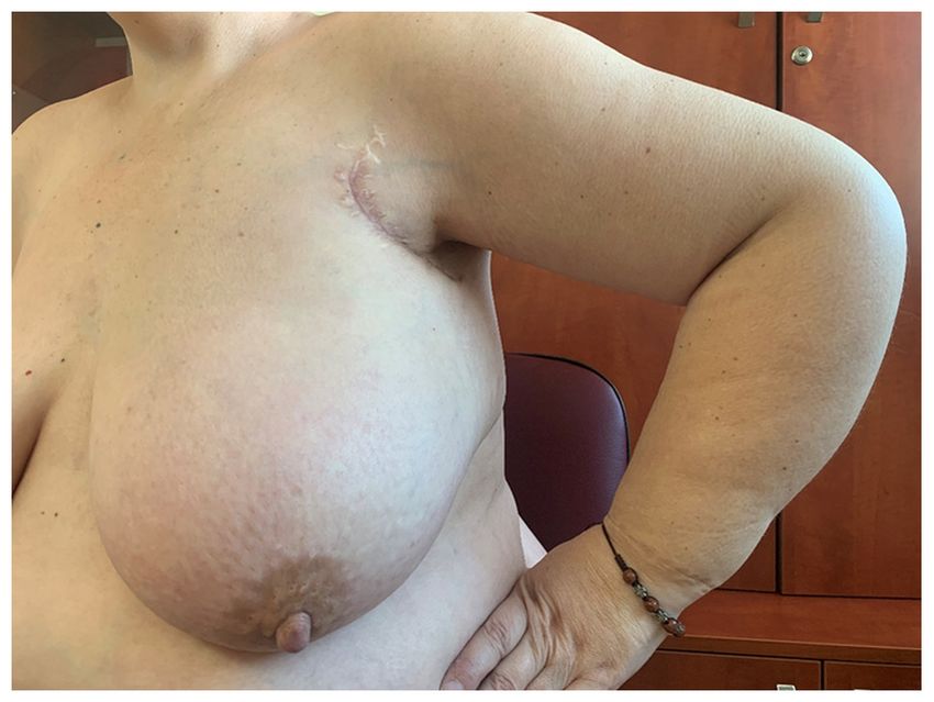
You can also read