Cerebellar Stroke in a COVID-19 Infected Patient. A Case Report
←
→
Page content transcription
If your browser does not render page correctly, please read the page content below
The Journal of Critical Care Medicine 2021;7(2):130-135 CASE REPORT
DOI: 10.2478/jccm-2021-0004
Cerebellar Stroke in a COVID-19 Infected
Patient. A Case Report
Khang Ning Loo1*, You Jiang Tan2, Kaavya Narasimhalu2, Krishan Kumar Sharma3, Dorinda
Chee Yee Chew4, Hei Man Wong5, Yvonne Fu Zi Chan5, Ken Cheah Hooi Lee6
1 Department of Internal Medicine, Singapore General Hospital, Singapore, Singapore
2 Department of Neurology, National Neuroscience Institute, Singapore General Hospital, Singapore, Singapore
3 Department of Neurosurgery, National Neuroscience Institute, Singapore General Hospital, Singapore, Singapore
4 Department of Diagnostic Radiology, Singapore General Hospital, Singapore, Singapore
5 Department of Infectious Disease, Singapore General Hospital, Singapore, Singapore
6 Department of Respiratory and Critical Care Medicine, Singapore General Hospital, Singapore, Singapore
Abstract
Background: Recent studies have reported that COVID-19 infected patients with stroke, who were often in the older
age group, had a higher incidence of vascular risk factors, and more severe infection related respiratory symptoms.
These observations provided little evidence to suggest that COVID-19 infection is a potential causative factor for
stroke. This report describes a young patient with a cerebellar stroke secondary to COVID-19 infection. Case presen-
tation: A 45-year old male presented at a hospital, reporting a two-day history of headache, vertigo, persistent vomit-
ing, and unsteady gait. Physical examination revealed gaze-evoked nystagmus on extraocular movement testing, left-
sided dysmetria and dysdiadochokinesia. He was diagnosed with a left cerebellar stroke. An external ventricular drain
was inserted, and sub-occipital craniectomy was performed to manage the effects of elevated intracranial pressure
due to the extent of oedema secondary to the infarct. He also underwent screening for the COVID-19 infection, which
was positive on SARS-COV-2 polymerase chain reaction testing of his endotracheal aspirate. Blood and cerebrospinal
fluid samples were negative. After the surgery, the patient developed atrial fibrillation and had prolonged vomiting
symptoms, but these resolved eventually with symptomatic treatment. He was started on aspirin and statin therapy,
but anticoagulation was withheld due to bleeding concerns. The external ventricular drain was removed nine days af-
ter the surgery. He continued with active rehabilitation. Conclusions: Young patients with COVID-19 infection may be
more susceptible to stroke, even in the absence of risk factors. Standard treatment with aspirin and statins remains
essential in the management of COVID-19 related stroke. Anticoagulation for secondary prevention in those with
atrial fibrillation should not be routine and has to be carefully evaluated for its benefits compared to the potential
harms of increased bleeding associated with COVID-19 infection.
Keywords: COVID-19, SARS-CoV-2, cerebellar stroke, atrial fibrillation
Received: 11 July 2020 / Accepted: 10 January 2021
Background vascular risk factors or more severe respiratory symp-
toms from COVID-19 infection [2, 3]. Such observa-
Neurological manifestations in COVID-19 infection tions suggest that the same risk factors for stroke may
have been well documented in recent observational be associated with more severe COVID-19 infection,
studies, ranging from mild symptoms of dizziness to but provide little evidence to support the infection as a
more severe stroke and seizures complications. An ear-
direct causative factor for stroke.
ly study reported on over a third of COVID-19 patients
who had neurological symptoms. Dizziness and head- On the other hand, young patients with no risk fac-
ache were the commonest at 16.8% and 13.1% respec- tors, presenting with large vessel ischemic stroke would
tively, while stroke occurred less frequently in 2.8% of raise a higher suspicion for COVID-19 infection as a
patients based on that study [1]. Subsequent case series contributory factor to stroke development once other
reported that COVID-19 patients with stroke were of- predisposing factors have been excluded [4]. Similar
ten in the older age group, had a higher incidence of findings of large artery cerebral infarctions were previ-
* Correspondence to: Khang Ning Loo, Singapore General Hospital, Singapore, Singapore. E-mail: loo.khang.ning@singhealth.com.sgAvailable online at: www.jccm.ro The Journal of Critical Care Medicine 2021;7(2) • 131
ously reported in patients with Severe Acute Respira- diminished, but the contractions of the facial muscles
tory Syndrome (SARS) virus infection [5]. This case were mostly symmetrical. His tongue was central and
report is another example of a young patient without palatal elevation was normal. Examination of the limbs
significant risk factors presenting with stroke as the revealed normal tone, power, reflexes and sensation.
primary manifestation of COVID-19 infection. There were left-sided dysmetria and dysdiadochokine-
sia. Examination of the heart, lungs and abdomen were
unremarkable. The National Institutes of Health Stroke
Case Presentation
Scale (NIHSS) score on admission was 2. Computed
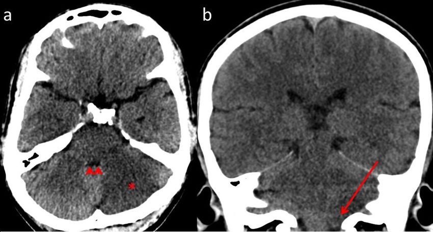
A 45-year-old Bangladeshi male presented at the Emer- tomography (CT) of the brain, performed on admis-
gency Department of Singapore General Hospital, Sin- sion, showed hypodensity with a loss of grey-white
gapore, giving a two-day history of moderately severe differentiation in the left cerebellar hemisphere, with
throbbing headache, associated with vertigo, multiple the left middle cerebellar peduncle’s involvement and
vomiting episodes aggravated by head movements, and left hemipons. There was a mass effect on the fourth
unsteady gait. He negated having any visual symptoms, ventricle and crowding of the left foramen magnum re-
dysarthria, dysphagia, anosmia, and ageusia. He had gion (Figure 1). CT angiography of the major vessels
no fever or respiratory symptoms. He was a smoker, re- from the level of the aortic arch to the Circle of Wil-
porting that this was limited to one cigarette a day. No lis was performed to evaluate for abnormalities of the
other significant medical history was reported. vertebral and carotid arteries, with no abnormalities
Upon admission, his vital signs were stable, and detected. The patient was diagnosed with a left cerebel-
his Glasgow Coma Scale (GCS) was 15. He had mild lar infarct.
anisocoria with a 2mm left pupil and 3mm right pu- The following investigations were also performed on
pil. Extraocular movement testing revealed hypermet- admission. A full blood count showed an elevated white
ric saccades in all directions, gaze-evoked nystagmus, blood cell count of 11.97x10⁹/L with mild lymphope-
with a jerky, slow pursuit. The left nasolabial fold was nia (0.97x10⁹/L). The haemoglobin and platelet counts
Fig. 1. Axial (a) and Coronal (b) images of an unenhanced CT brain demonstrating an area of hypodensity with loss of
grey-white differentiation in the left cerebellar hemisphere (*), extending to involve the left middle cerebellar peduncle,
in keeping with an established infarct. There is associated mass effect with mild effacement of the fourth ventricle
(arrowheads) and mild left inferior cerebellar tonsillar descent with crowding of the foramen magnum’s left side (long
arrow).132 • The Journal of Critical Care Medicine 2021;7(2) Available online at: www.jccm.ro
were normal. C-reactive protein level was 3.5mg/L. D- A cardiology opinion was sought, and the arrhyth-
dimer level was 0.33mg/L Fibrinogen Equivalent Units mia was attributed to dysautonomia from brainstem
(FEU). The glycated haemoglobin (HbA1c) measured involvement of the stroke.
6%, and fasting lipid levels were normal. A chest radio- Endotracheal (ET) aspirates were collected on Day
graph was reported as normal. 2 and Day 3, post-admission; blood and cerebrospinal
The patient was admitted to the high dependency fluid (CSF) samples were collected on Day 4, post-ad-
ward of the hospital for close monitoring on the day of mission, for SARS-CoV-2 PCR testing.
admission. Negative pressure ventilation was started as The ET aspirate tested positive for SARS-CoV-2 vi-
he had a positive contact history with suspected COV- rus, but results were negative for the blood and CSF
ID-19 patients, in his worker dormitory. Oro-naso- samples.
pharyngeal swabs for SARS-CoV-2 polymerase chain
The patient had persistent cyclic vomiting even after
reaction (PCR) testing were not performed to avoid
the craniectomy. These were sudden episodes with no
gagging that might cause a further rise in his intracra-
associated symptoms of headache or abdominal disten-
nial pressure. Blood samples were obtained for SARS-
sion. Gastric residual volume was minimal. The symp-
CoV-2 IgG serology, which tested positive. The hospital
toms were alleviated with intravenous ondansetron
neurosurgeon reviewed the patient. He advised intra-
(Intas Pharmaceuticals Ltd, Ahmedabad, India) 4mg
venous mannitol 20%, (B.Braun Medical Industries
eight hourly and intravenous metoclopramide (Yung
Sdn Bhd, Penang, Malaysia) 100mL, six-hourly, start-
Shin Pharmaceutical Ind. Co. Ltd, Taichung, Taiwan)
ing on the day of admission, and continued until Day
10mg, eight hourly, starting on Day 5 post-admission.
3 post-admission, and 3% sodium chloride solution to
The symptoms resolved after nine days.
target a sodium level of 140 to 145mmol/L and serum
osmolality up to 320 mmol/kg. Two days after the craniectomy, the patient became
drowsier again, recording a GCS=9, E2V1 (intubated)
On the Day 2 post-admission, the patient became
M6, and had weakness in his left upper and lower limbs.
drowsier registering a GCS 13 (E3V4M6). After a neu-
The repeated brain-CT showed no change in the size
rosurgical consult, he underwent a right frontal exter-
of the infarct or evidence of haemorrhagic conversion,
nal ventricular drain (EVD) insertion and a sub-occip-
but there was a slight increase in both lateral ventricles’
ital craniectomy, as an emergency treatment.
size. The EVD level was lowered from 10 to 5cmH2O;
Intraoperatively the left cerebellum appeared swol- the sodium level target was raised to between 145 and
len, herniated and infarcted. Some infarcted left cer- 150mmol/L to reduce cerebral oedema. His condition
ebellar tissue had to be removed to reduce the hernia- continued to improve and was eventually extubated af-
tion. Postoperatively the patient remained intubated ter two days. The EVD was safely removed on day nine
and was sent to the intensive care unit. post-craniectomy.
The initial EVD level was set at 15cmH2O above The following investigations were performed on Day
the tragus. He received pressure-controlled ventilation 8 post-admission to evaluate potential causes of stroke.
with these initial settings: driving pressure 17cmH2O, Transthoracic echocardiography (TTE) showed an av-
PEEP 5cmH2O, FiO2 30%. His PaCO2 remained in erage left ventricle ejection fraction of 55 to 60%, with
the range between 35 to 42 mmHg, and pH was nor- no regional wall motion abnormality. The left atrium
mal throughout. When he was more awake, these were (LA) size was normal, with an LA volume of 19.56ml/
quickly weaned to spontaneous ventilation with pres- m2. No valvular abnormalities or intracardiac thrombi
sure support of 8cmH2O, PEEP 5cmH2O, FiO2 25%. were detected. A thyroid function test, done on first-day
The patient’s post-operative recovery was compli- post-admission was normal. Extensive investigations
cated by hypotension requiring thirty hours of intra- for thrombophilia were performed on the sixth-week
venous noradrenaline (Laboratoire Aguettant, Lyon, post-admission. These included: Anti-cardiolipin IgM
France), 0.02-0.15 mcg/kg/min from Day 2 to Day 4, & IgG antibodies negative, Lupus anticoagulant nega-
post-admission, to maintain adequate cerebral perfu- tive, Protein C 125%, Protein S 87%, Antithrombin III
sion pressure of at least 60mmHg. There was also a 111%, Homocysteine 6.7 micromol/L. Autoimmune
new onset of atrial fibrillation with intermittent sinus markers, including Antinuclear antibody (ANA), Anti
pauses, the longest lasting twenty-two seconds, which Myeloperoxidase (MPO) antibody, Anti Proteinase 3
spontaneously reverted to sinus rhythm. (PR3) antibody, and Anti-double-stranded DNA (ds-Available online at: www.jccm.ro The Journal of Critical Care Medicine 2021;7(2) • 133
DNA) antibody, were all negative. pressure and cardiovascular homeostasis. In particular,
Based on these results, the patient was deemed un- Angiotensin II (ATII) has been implicated in lung in-
likely to have any underlying thrombophilia or autoim- jury, leading to severe respiratory failure in COVID-19
mune diseases contributing to stroke. The patient was patients [9]. Although the BBB insulates the brain from
started on long term oral aspirin (Reckitt Benckiser circulating angiotensin effects, the brain has a local
Healthcare Ltd, Hull, United Kingdom) 100mg daily independent RAS with all the “classical” RAS compo-
and atorvastatin (manufactured by Pfizer Pharmaceu- nents and can synthesise angiotensin. Like lung injury,
ticals LLC, Vega Baja, Puerto Rico) 40mg daily for sec- it is postulated that ischemic stroke results from the ef-
ondary prevention starting on Day 10 post-admission. fects of ATII, causing vasoconstriction of cerebral ves-
At the time of writing, he remained well and was sels and increasing inflammation and oxidative stress
actively participating in rehabilitation before eventual in the brain parenchyma. Under normal circumstances,
discharge to a community recovery facility. the activation of an alternative axis comprising ACE2-
mediated cleavage of ATII to angiotensin (1-7) binds
to the Mas receptor, would counter the effects of ATII.
Discussion This results in vasodilation, anti-inflammatory and an-
COVID-19 related stroke is thought to be due to mul- tioxidant responses. The ACE2-AT (1-7)-Mas pathway
tiple factors including coagulopathy and endothe- has also demonstrated further benefits of improved
lial dysfunction, features that are increasingly seen as angiogenesis, anti-thrombotic activity, and increased
hallmarks of the COVID-19 infection. Coagulopathy stability of atherosclerotic plaques. These protective
is seen mainly in patients with more severe infection effects against cerebral ischemia are reduced in COV-
[6, 7]. However, the prothrombin time, D-dimer and ID-19 infected patients due to the downregulation of
platelet levels were normal in our patient. The present ACE2 receptors [10].
patient’s symptoms would suggest an aetiology with The protracted vomiting in our patient was initially
the central nervous system’s more direct involvement attributed to the cerebellar stroke’s effects, including a
rather than complications of a severe systemic infec- raised intracranial pressure and compression on the
tion. A more plausible explanation should include the fourth ventricle where the area-postrema, i.e. the vom-
virus’ ability to target its effects on the brain without iting centre, is located. However, it would not explain
affecting the rest of the body. why the vomiting persisted even after decompressive
The neurotropism of coronaviruses has been well craniectomy. An old experimental model on coronavi-
recognised. A recent literature review on the expres- rus infection in pigs has suggested that vomiting may
sion of angiotensin-converting enzyme 2 (ACE2) re- be induced by a viral infection of the neurons in a few
ceptors in the cerebral vasculature, neurons, astrocytes target-tissues, including the brainstem. The vomiting
and oligodendrocytes, as well as localisation of ACE2 centre receives impulses from infected neurons and
receptors in the olfactory bulb and other areas of the triggers vomiting once sufficient afferent stimuli have
brain, provided the basis for the possible mechanisms been reached. This may explain the persistent vomiting
of neuro-invasion and stroke development secondary in our patient [11].
to COVID-19 infection. These include how the virus Anticoagulation therapy is essential for secondary
crosses the blood-brain barrier (BBB) via retrograde prevention of stroke in patients with atrial fibrillation.
neuronal transport along the sensory and olfactory The risk of recurrent stroke in our patient was 2.2% per
nerves, or through the infection of macrophages and year based on a CHA2DS2-Vasc score of 2; whereas the
host cells that pass through the BBB. Infection of the HAS-BLED score of 1 indicated risk of 1.02 bleeds per
vascular endothelium also causes inflammation and 100 patient-years. This should be carefully considered
vascular injury, increasing the BBB permeability and in our patient due to large infarct with mass effect in the
facilitating the virus’s entry [8]. posterior fossa, of which a haemorrhagic event would
The pathogenesis of stroke following neuro-invasion be disastrous. Neuroimaging studies have reported
of the virus should be explained with an understand- more haemorrhagic strokes in COVID-19 infected
ing of the renin-angiotensin system (RAS). The RAS is patients who received therapeutic or prophylactic an-
a complex system with important physiological func- ticoagulation [12, 13]. It is important to recognise that
tions, including fluid and electrolyte balance, blood post-stroke cardiac arrhythmias can occur. Two pro-134 • The Journal of Critical Care Medicine 2021;7(2) Available online at: www.jccm.ro
spective studies reported 25.1% and 29.5% incidence required mechanical ventilation [19, 20]. Moreover,
of cardiac arrhythmias, respectively in the first 48 to previous trials on steroids’ utility in acute stroke have
72 hours after a stroke. The incidence of slow atrial fi- demonstrated no evidence of significant benefits [21].
brillation was 4.8% and 2.7%; and the sinoatrial block Our patient had a primary neurological insult rather
was 1.6% and 0.6% respectively [14, 15]. Also, it has than respiratory compromise secondary to COVID-19
been reported that patients with lone atrial fibrillation, infection. Hence none of these additional therapies was
without any cardiovascular risk factors, and with nor- offered.
mal left atrial volume, did not have an increased risk of
stroke compared to the general population in a 30-year
Conclusion
follow up period [16]. Our patient had an average base-
line electrocardiogram and a TTE which did not show We have reported a COVID-19 related cerebellar
any left atrial enlargement or valvular abnormalities. stroke in a young patient. Absence of typical charac-
The atrial fibrillation started after surgery and reverted teristics and risk factors described in the literature in
to normal sinus rhythm after a day, with no further epi- our patient’s case gives greater credence to COVID-19
sodes during his stay. The overall assessment suggested infection as an independent risk factor for stroke. As-
that atrial fibrillation was a consequence rather than pirin and statins are the mainstays of treatment, while
the cause of the stroke. anticoagulation requires careful consideration due to
Furthermore, extensive investigations were done to the increased risk of intracranial bleed associated with
rule out underlying thrombophilic conditions, as evi- COVID-19 infection. Future studies should explore the
dent by negative antiphospholipid antibodies, autoim- role of additional therapies, including antiviral treat-
mune markers, and typical coagulation factors. After ment for COVID-19 related stroke.
careful consideration and discussion between neurolo-
gists, a decision was made to withhold anticoagulation
Conflicts of interests
due to the more significant concern of haemorrhagic
complications due to the large infarct size and tight The authors declare that they have no conflicts of in-
posterior fossa requiring decompression craniectomy terests.
initially. As we gained more knowledge of this disease,
we felt that coagulopathy associated with COVID-19
Consent for publication
infection varies between patients due to disease sever-
ity. Hence, the criteria for selecting patients who may Consent obtained from the patient for case report pub-
benefit from anticoagulation in the acute setting and lication.
the duration of anticoagulation in the longer term re-
mains uncertain. We would recommend a close follow-
up for these patients after they have recovered from
Reference
COVID-19 infection until more literature is available 1. Mao L, Jin H, Wang M, et al. Neurologic Manifestations of
on the disease’s long-term consequences. The patient Hospitalised Patients With Coronavirus Disease 2019 in Wuhan,
received aspirin and atorvastatin treatment. In addition China. JAMA Neurol. 2020;77(6):683-690.
to treatment for stroke, statin therapy’s potential bene- 2. Beyrouti R, Adams ME, Benjamin L, et al. Characteristics of
fits in COVID-19 infection have been widely discussed ischaemic stroke associated with COVID-19. J Neurol Neurosurg
Psychiatry. 2020;91(8):889-891..
in recent literature, including its anti-inflammatory,
3. Li Y, Wang M, Zhou Y, et al. Acute Cerebrovascular Disease
anti-thrombotic and immunomodulatory properties
Following COVID-19: A single-centre, Retrospective,
[17, 18]. An ongoing trial is being conducted to evalu- Observational Study. SSRN Electronic Journal. 2020;5.
ate aspirin’s potential protective effects in COVID-19
4. Oxley TJ, Mocco J, Majidi S, et al. Large-Vessel Stroke as a
patients (ClinicalTrials.gov Identifier: NCT04365309). Presenting Feature of Covid-19 in the Young. N Engl J Med.
COVID-19 specific therapies such as remdesivir and 2020;382(20):e60.
dexamethasone in COVID-19 related stroke remains 5. Umapathi T, Kor AC, Venketasubramanian N, et al. Large artery
uncertain because indications for their use based on ischaemic stroke in severe acute respiratory syndrome (SARS). J
published studies include patients with lower respirato- Neurol. 2004;251(10):1227-31.
ry tract infection who had poor oxygen saturation and 6. Helms J, Tacquard C, Severac F, et al. High risk of thrombosisAvailable online at: www.jccm.ro The Journal of Critical Care Medicine 2021;7(2) • 135
in patients with severe SARS-CoV-2 infection: a multicenter 14. Fernández-Menéndez S, García-Santiago R, Vega-Primo A,
prospective cohort study. Intensive Care Med. 2020;46(6):1089- et al. Cardiac arrhythmias in stroke unit patients. Evaluation
98. of the cardiac monitoring data. Neurología (English Edition).
7. Tang N, Li D, Wang X, et al. Abnormal coagulation parameters 2016;31(5):289-95.
are associated with poor prognosis in patients with novel 15. Kallmunzer B, Breuer L, Kahl N, et al. Serious cardiac arrhythmias
coronavirus pneumonia. J Thromb Haemost. 2020;18(4):844-7. after stroke: incidence, time course, and predictors--a
8. Zubair AS, McAlpine LS, Gardin T, et al. Neuropathogenesis and systematic, prospective analysis. Stroke. 2012;43(11):2892-7.
Neurologic Manifestations of the Coronaviruses in the Age of 16. Osranek M, Bursi F, Bailey KR, et al. Left atrial volume predicts
Coronavirus Disease 2019. JAMA Neurology. 2020;77(8):1018- cardiovascular events in patients originally diagnosed with
1027. lone atrial fibrillation: three-decade follow-up. Eur Heart J.
9. Imai Y, Kuba K, Rao S, et al. Angiotensin-converting 2005;26(23):2556-61.
enzyme 2 protects from severe acute lung failure. Nature. 17. Castiglione V, Chiriaco M, Emdin M, et al. Statin therapy in
2005;436(7047):112-6. COVID-19 infection. Eur Heart J Cardiovasc Pharmacother.
10. Divani AA, Andalib S, Di Napoli M, et al. Coronavirus Disease 2020;6(4):258-259.
2019 and Stroke: Clinical Manifestations and Pathophysiological 18. Lee KCH, Sewa DW, Phua GC. Potential role of statins in
Insights. J Stroke Cerebrovasc Dis. 2020;29(8): 104941 COVID-19. Int J Infect Dis. 2020; 96: 615–617.
11. Andries K, Pensaert M. Vomiting and wasting disease, a 19. Beigel JH, Tomashek KM, Dodd LE, et al. Remdesivir for the
coronavirus infection of pigs. Adv Exp Med Biol. 1981;142:399- Treatment of Covid-19 - Final Report. N Engl J Med. 2020;
408. 383(19):1813-1826.
12. Dogra S, Jain R, Cao M, et al. Hemorrhagic stroke and 20. Group RC, Horby P, Lim WS, et al. Dexamethasone in
anticoagulation in COVID-19. J Stroke Cerebrovasc Hospitalised Patients with Covid-19- Preliminary Report. N Engl
Dis.2020;29(8):104984. J Med. 2020;2021436.
13. Jain R. Evolving Neuroimaging Findings during COVID-19. AJNR 21. Sandercock PA, Soane T. Corticosteroids for acute ischaemic
Am J Neuroradiol. 2020 Aug;41(8):1355-1356. stroke. Cochrane Database Syst Rev. 2011(9):CD000064.You can also read