CASE REPORT: A RARE TRUNCATING VARIANT OF THE CFHR5 GENE IN IGA NEPHROPATHY - SERVAL
←
→
Page content transcription
If your browser does not render page correctly, please read the page content below
CASE REPORT
                                                                                                                                                    published: 20 May 2021
                                                                                                                                           doi: 10.3389/fgene.2021.529236

                                              Case Report: A Rare Truncating
                                              Variant of the CFHR5 Gene in IgA
                                              Nephropathy
                                              Gabriella Guzzo 1,2,3*, Salima Sadallah 2*, Heidi Fodstad 4 , Jean-Pierre Venetz 1 ,
                                              Samuel Rotman 5 , Daniel Teta 3 , Thierry Gauthier 6 , Giuseppe Pantaleo 2 ,
                                              Andrea Superti-Furga 4 and Manuel Pascual 1
                                              1
                                                Organ Transplant Center, Lausanne University Hospital, University of Lausanne, Lausanne, Switzerland, 2 Service of
                                              Immunology and Allergy, Lausanne University Hospital, University of Lausanne, Lausanne, Switzerland, 3 Service of
                                              Nephrology, Valais Hospital, Sion, Switzerland, 4 Division of Genetic Medicine, Lausanne University Hospital, University of
                                              Lausanne, Lausanne, Switzerland, 5 Service of Clinical Pathology, Lausanne University Hospital, University of Lausanne,
                                              Lausanne, Switzerland, 6 Hôpital Riviera Chablais, Vevey, Switzerland

                          Edited by:
           Tarunveer Singh Ahluwalia,
                                              IgA nephropathy (IgAN) is the most common primary glomerulonephritis worldwide.
  Steno Diabetes Center Copenhagen
                   (SDCC), Denmark            Despite appropriate therapy, 20–40% of affected-patients evolve toward end-stage
                          Reviewed by:        kidney disease (ESKD). Mesangial IgA deposits are the hallmark of IgAN, and
                          Pedro Dorado,       complement deposition (C3) seems to differentiate latent IgA mesangial deposits from
     University of Extremadura, Spain
              Nancy Monroy-Jaramillo,
                                              active IgAN. Atypical hemolytic uremic syndrome (aHUS), another disease in which
    National Institute of Neurology and       complement plays an important role, is caused by inherited or acquired deregulation
                  Neurosurgery, Mexico
                                              of the alternative pathway (AP) of complement. A subgroup of IgAN shows thrombotic
                   *Correspondence:
                                              microangiopathy (TMA) lesions in kidney biopsies, the histological characteristic of
                        Gabriella Guzzo
              gabriella.guzzo@chuv.ch         aHUS. Genetic variants of complement Factor H (CFH), known to be present in aHUS,
                       Salima Sadallah        have been associated with rapidly progressive forms of IgAN and a clinical pattern
              salima.sadallah@chuv.ch
                                              of aHUS. Genome-wide association studies (GWAS) have confirmed that the 1q32
                  Specialty section:          region, encoding for CFH and its related proteins, is an IgAN susceptibility locus. A
        This article was submitted to         30 year-old man was admitted for seizures and malignant hypertension. The kidney
      Human and Medical Genomics,
              a section of the journal
                                              biopsy showed IgAN associated with features of TMA. Despite five plasma exchanges,
                 Frontiers in Genetics        the patient remained dialysis-dependent, and ESKD was diagnosed. Functional and
             Received: 03 April 2020          genetic complement analysis were performed. A monoallelic protein-truncating, likely
             Accepted: 22 April 2021          loss-of-function variant was identified in the CFHR5 gene. Eculizumab is the treatment
             Published: 20 May 2021
                                              of aHUS. As it has been successfully used in a few cases of rapidly progressive IgAN, it
                             Citation:
    Guzzo G, Sadallah S, Fodstad H,           was decided to administer eculizumab over a period of 12 months in addition to the usual
        Venetz J-P, Rotman S, Teta D,         immunosuppression for renal transplantation. After a follow-up of 3 years, there was no
               Gauthier T, Pantaleo G,
Superti-Furga A and Pascual M (2021)
                                              clinical disease recurrence. Systematic biologic and genetic screening of complement in
      Case Report: A Rare Truncating          individuals with IgAN might be useful to better delineate the role of the AP of complement
    Variant of the CFHR5 Gene in IgA          in renal disease progression, and this may have therapeutic implications.
                         Nephropathy.
            Front. Genet. 12:529236.          Keywords: IgA nephropathy, complement, CFHR5, CFH, eculizumab, atypical hemolytic uremic syndrome,
    doi: 10.3389/fgene.2021.529236            thrombotic microangiopathy (TA-TMA)

Frontiers in Genetics | www.frontiersin.org                                           1                                              May 2021 | Volume 12 | Article 529236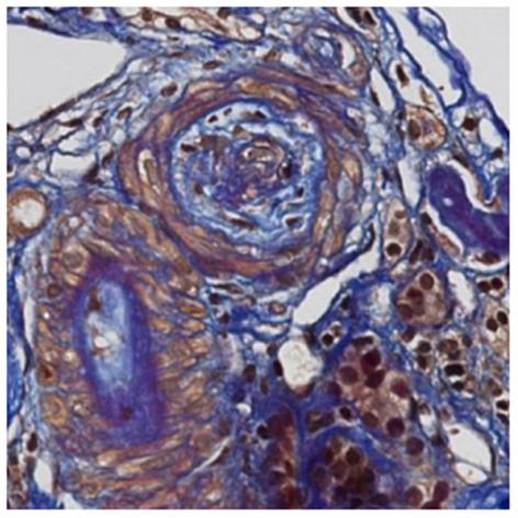
Guzzo et al.                                                                                                              CFHR5 in IgAN With TMA

INTRODUCTION                                                                CFHRs, which can compete with CFH and deregulate the AP
                                                                            of complement.
IgA nephropathy (IgAN) is the most common cause of end-                        Higher plasma CFHR5 protein levels (Zhu et al., 2018) and
stage kidney disease (ESKD) among primary glomerulonephritis                increased CFHR5 glomerular deposition have been associated
(Donadio and Grande, 2002). Even though IgAN was first                      with progressive IgAN (Medjeral-Thomas et al., 2017), but no
described more than half a century ago by Berger et al.                     genetic analysis was performed in both studies. A severe form
the pathogenesis remains incompletely elucidated (Berger and                of IgAN was associated with CFH mutations and a clinical
Hinglais, 1968).                                                            pattern of atypical hemolytic uremic syndrome (aHUS), and
    The hallmark of IgAN is the mesangial deposition of IgA1                eculizumab was successfully employed in the treatment of this
immunoglobulins with an aberrant glycosylation (Moldoveanu                  patient (Nakamura et al., 2018).
et al., 2007). While over 50% of patients with IgAN have increased             We report a case of rapidly progressive IgAN associated with
IgA1 serum level compared to healthy controls, this alone is not            a clinical pattern of aHUS and thrombotic microangiopathy
sufficient to cause the disease. The mesangial accumulation of              (TMA) features on kidney biopsy, leading to ESKD. Genetic
IgA1 has been described as the first step of a “4-hit process,”             complement screening identified CFH-H3, MCP-H2, CFHR1∗ B
which finally leads to complement activation (Suzuki et al., 2011).         polymorphisms, known to constitute moderate risk factors for
The second hit is the occurrence of IgG or IgA1 autoantibodies              aHUS, together with a rare deleterious variant in the CFHR5
directed against the antigenic galactose deficient IgA1 (Gd-IgA1).          gene. Treatment with eculizumab successfully prevented clinical
Formation of circulating immune complexes (CIC) containing                  recurrence of the disease, with a follow-up of 3 years after
Gd-IgA1 then occur (third hit) and their mesangial deposition               renal transplantation.
(fourth hit) triggers cells hyperplasia and hypertrophy, release
of inflammatory cytokines and chemokines with complement
activation. The frequent post-transplant disease recurrence                 CASE REPORT
confirmed the CIC mediated nature of IgAN (Ortiz et al., 2012).
                                                                            A 30 year-old Asian man, born in Sri-Lanka, was referred to
Conversely, IgA deposits are rapidly cleared from kidneys with
                                                                            our hospital for a kidney pre-transplant evaluation. At the age of
donor-derived IgAN when transplanted in recipients without
                                                                            16, he presented a unique episode of gross hematuria and lower
IgAN as cause of ESKD (Sanfilippo et al., 1982; Ponticelli et al.,
                                                                            limb oedema, which was not furtherly investigated. Of note, the
2001).
                                                                            patient showed no family history of TMA, IgA nephropathy or
    The co-localization of C3 and IgA deposits in 90% of IgAN
                                                                            other kidney diseases. At the age of 28, he was admitted to
kidney biopsies indicates complement activation by Gd-IgA1.
                                                                            the hospital for recurrent generalized seizures and oliguria, as
Therefore, complement plays a key role in the pathogenesis
                                                                            a manifestation of malignant hypertension. Laboratory analysis
of IgAN (Maillard et al., 2015). IgA mesangial deposition
                                                                            revealed hemolytic anemia (hemoglobin 71 g/L; schistocytes 9‰,
mostly occurs in active kidney disease (Suzuki et al., 2003),
                                                                            LDH 348 U/L, haptoglobin < 0.10 g/L), thrombocytopenia (70
and C3 co-deposition may be considered as a biomarker
                                                                            × 103 /µL) and severe kidney failure (creatinine 2,013 µmol/l)
of nephritogenic IgA1 deposition, where local and systemic
                                                                            with proteinuria (2.26 g/mmol from urine protein/creatinine
evidence of complement activation are prognostic marker of
                                                                            ratio). Kidney biopsy showed diffuse glomerular sclerosis (90%),
IgAN (Hastings et al., 2013).
                                                                            interstitial fibrosis (90%) and tubular atrophy, associated with
    Clinical and pathological manifestations of IgAN can
                                                                            severe vascular lesions characteristic of TMA (Figures 1, 2).
vary widely, with a 20–40% subgroup evolving to ESKD.
                                                                            Immunofluorescence was positive for abundant mesangial IgA
The identification of clinical, biological and/or histological
                                                                            (Figure 3) and C3 deposits, with focal IgA staining in some
prognostic markers at diagnosis could thus help tailoring
                                                                            arterioles. In addition, deposits of C5b-9, were present with
the best treatment option to the given patient’s risk profile
                                                                            IgA and C3 deposits, within the mesangium and the wall of
(Barbour et al., 2019).
                                                                            small arterioles.
    Complement in IgAN is activated by both the alternative
                                                                               IgAN associated with TMA was diagnosed. Five plasma
pathway (AP) and the lectin pathway (LP), as demonstrated
                                                                            exchanges were performed without improvement of renal
by the variable immunohistochemical positivity for properdin,
                                                                            function, and the patient remained dialysis-dependent.
complement factor H (CFH), complement FH related proteins
(CFHRs), C4d, mannose binding lectin (MBL), MBL-associated
serine proteases (MASPs), L-ficolin and C5b-9 (Maillard et al.,             COMPLEMENT ASSAYS AND GENETIC
2015). The classical pathway of complement does not seem to                 ANALYSIS
contribute to the pathogenic process, indeed C1q deposits are
rarely found and only confined in sclerotic areas (Lee et al., 2013).       One year after starting dialysis, no abnormality was detected in
    An interesting question is whether dysfunctional regulation of          the complement screening (C4, CH50, CFH, Factor I, CD46,
complement activation may have a significant role in the rapid              or anti-CFH autoantibodies). At the time of pre-transplant
progression to ESKD in some forms of IgAN (Tortajada et al.,                evaluation, he had persistently low platelet counts and low C3
2019). Two genome-wide association studies (GWAS) identified                level (0.66 g/L; reference range > 0.75 g/L).
and confirmed 1q32 as a locus of IgAN susceptibility (Gharavi                  Given the suspicion of aHUS, functional evaluation of
et al., 2011; Kiryluk et al., 2012). The 1q32 region codes for CFH,         complement and molecular genetic analysis were performed.
the major regulator of complement in soluble phase, and five                We used an in-house genetic panel that we routinely apply at

Frontiers in Genetics | www.frontiersin.org                             2                                     May 2021 | Volume 12 | Article 529236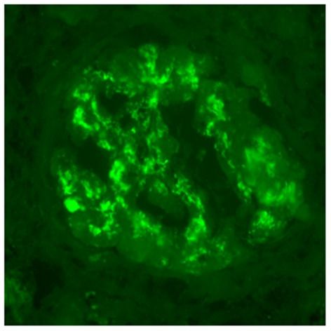
Guzzo et al. CFHR5 in IgAN With TMA
with a slightly increased risk for aHUS (Abarrategui-Garrido
et al., 2009). More significantly, he was found to be heterozygous
for a very rare CFHR5 variant (Chr.1: 196953197-TC>T;
NM_030787.4, c.361delC, p.Gln121Lysfs∗ 10, rs778029757),
which leads to a frameshift/premature stop codon [dbSNP
Database, 2021; Genome Aggregation Database (gnomAD),
2021]. It predicts the synthesis of a CFHR5 protein with a
normal amino acid sequence from the N-terminus up to the
SCR 2 domain, followed by a short stretch of 10 missense
amino acids and then terminating abruptly, leading to the
absence of two-thirds of the protein. Such proteins are frequently
FIGURE 1 | Light microscopy. Early stage of thrombotic microangiopathy: this
unstable. Alternatively, a stop codon occurring in the first
artery shows oedematous intima and few myointimal cells corresponding to third of the mRNA might likely lead to nonsense-mediated
“mucoid intimal hyperplasia” (FAOG, 400x). mRNA decay (NMD). In both cases, haploinsufficiency would
be the consequence. However, in absence of cellular studies,
it cannot be formally excluded that the variant may induce
a dominant negative effect on CFHR5 function (see section
Discussion, below).
The variant is represented in the gnomAD database [Genome
Aggregation Database (gnomAD), 2021] with 12 alleles on a
total of ∼250,000 (allelic frequency 0.000048, or 0.0048%; one
individual in ∼11,000) and all carriers are of South Asian
origin (our patient is from Sri Lanka). It has not been seen
at homozygosity and has not been reported in association
with IgAN so far. In spite of its deleterious effect on the
protein, its classification, according to the ACMG guidelines, is
between variant of unknown significance or likely benign variant
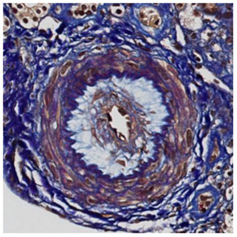
FIGURE 2 | Light microscopy. Later changes of thrombotic microangiopathy: (because of the presence of 12 heterozygous presumably healthy
the artery contains fibro-oedema with few collagen fibers within intima revealed individuals in gnomAD). However, this does not exclude its role
in blue with trichome FAOG (FAOG, 400x).
as a disease predisposing factor (see below).
PATIENT OUTCOMES
After a pre-transplant work-up, the patient received a kidney
allograft from a living unrelated donor. The day of transplant,
complement analysis revealed a low CH50 (59%; normal value
> 70%) and C3 (0.64 g/L). AP50, MBL, CFH, circulating C5b-9
were normal and there were no CFH autoantibodies.
Crossmatch assays were negative by both complement
dependent cytotoxicity (CDC) and flow cytometry techniques.
HLA match was of 2/6 antigens (A24, DQ5). Induction
and maintenance immunosuppression consisted of basiliximab
FIGURE 3 | Immunofluorescence microscopy. IgA deposits are observed
(20 mg at days 0 and 4), steroids, mycophenolate mofetil
within mesangium and glomerular membranes (400x).
(MMF) and tacrolimus. Considering the possible risk of aHUS
recurrence associated with the multiple complement genetic
our institutions to individuals with aHUS suspected to have a abnormalities found, prophylactic eculizumab was administered
genetic origin. This panel, that includes a NGS [Next-Generation peri- and post–transplant. Initially he received a weekly dose of
Sequencing using a TruSight One Expanded Sequencing Panel eculizumab (600 mg, D1 and D8), followed by one intravenous
(Illumina, USA)] analysis of ADAMTS13, C3, CD46, CFB, administration approximately every 3 weeks (900 mg), to
CFD, CFH, CFHR1, CFHR2, CFHR3, CFHR4, CFHR5, CFI, maintain a CH50 < 10%. No acute rejection occurred.
CFP, HOXA2, MMACHC, THBD, has been assembled by our Eculizumab was discontinued after 1 year post-transplant.
nephrologists, pediatric nephrologists and geneticists and is quite So far, an excellent allograft function has been observed,
similar to that used in diagnostic laboratories worldwide. with no clinical evidence of IgA recurrence after transplantation
The patient was found to be a carrier of the haplotypes and the patient is currently doing well with a serum creatinine
CFH-H3 and MCP-H2 and the CFHR1∗ B polymorphism of 92 µmol/l (eGFR by CKD-EPI of 92 ml/min/1.73 m2 ) and
(homozygous), which are common and have been associated no proteinuria.
Frontiers in Genetics | www.frontiersin.org 3 May 2021 | Volume 12 | Article 529236Guzzo et al. CFHR5 in IgAN With TMA
DISCUSSION population) and cannot be considered pathogenic per se. In
contrast, the CFHR5 variant identified in our case is very rare (see
Over the years, evidence has accumulated on the pathogenic role above). By analogy to the variant observed by Vernon et al. it can
of complement in IgAN. Already in 1994, Stad et al. showed that be predicted that this variant results in partial CFHR5 deficiency.
complement depletion induced by cobra venom factor abolished The circumstantial evidence of the role of similar CFHR5 variants
glomerular inflammation, proteinuria and C3 deposition in a rat in different complement-mediated nephropathies suggests its
model of IgAN (Stad et al., 1994). pathogenic potential. However, this variant can be present,
Furthermore, hereditary or acquired deregulation of the AP although very rarely, among healthy individuals. To summarize
of C may exacerbate the prognosis of IgAN (Gharavi et al., the evidence from genetic epidemiology, our observation seems
2011; Kiryluk et al., 2012; Zhai et al., 2016; Medjeral-Thomas to support and extend previous reports on the role of genetic
et al., 2017; Nakamura et al., 2018; Zhu et al., 2018; Tortajada variants of complement in the rapid progression of some cases
et al., 2019). The 1q32 gene locus has been associated with IgAN. of IgAN. However, the rare observation of these variants in
It codes for CFH and the five CFH-related (CFHR) proteins healthy individuals indicates that no single variant so far seems
(Gharavi et al., 2011; Kiryluk et al., 2012). These proteins have to be sufficient to induce renal disease per se at the heterozygous
a high degree of gene sequence identity, particularly in the C- state. The variants may increase the predisposition to develop the
terminal regions, which together with the gene locus proximity glomerulopathy or they could exacerbate the course of the disease
favor non-homologous recombination and gene rearrangements. (Kaartinen et al., 2019). The disease itself is likely triggered by
Abnormal CFHRs may compete with CFH, interfering with its other factors. The fact that CFHR5 variants have been observed
inhibitory activity and potentially causing deregulation of the in association with different types of nephropathies (namely,
AP of complement. A study conducted in 500 IgAN patients C3G, persistent post-infectious GN, HUS, and –as in this case-
confirmed a possible role of CHFR5 role in IgAN, with higher IgAN) further supports the concept that they may contribute to
soluble CFHR5 levels being associated with an increased risk of glomerular disease rather than triggering it.
IgAN progression (Zhai et al., 2016; Zhu et al., 2018). Moreover, In view of the potential pathogenic consequences of the
progressive IgAN has been associated with an increased patient’s genetic profile, we administered eculizumab up to 1
glomerular CFHR5 deposition (Medjeral-Thomas et al., 2017). year post-transplant in order to prevent recurrence of the IgAN.
In 2010, a monoallelic (heterozygous) variant characterized Eculizumab, a humanized monoclonal antibody directed against
by a two-exon duplication in the CFHR5 gene was described C5, is the treatment of choice of aHUS, and has been employed
in patients of Cypriot origin with familial glomerulonephritis in some cases of rapidly progressive IgAN associated with aHUS
(Gale et al., 2010). This “CFHR5 nephropathy,” which is and CFH variant. In our patient, it was discontinued after 1 year
morphologically a C3 glomerulopathy (C3G), shares a similar post-transplant, because the genetic risk profile of the patient did
clinical course with IgAN, with persistent microscopic hematuria not justify this long-term costly treatment and because its role in
and progressive ESKD, but without the pathognomonic IgA avoiding the recurrence of IgAN has not been established.
mesangial deposits. This Cypriot CFHR5 variant predicts an In summary, we report a case of IgAN associated with CFH-
abnormally large protein with duplicated dimerization domains. H3, MCP-H2, CFHR1∗ B polymorphisms, known to constitute
The pathogenesis is thought to be secondary to the hetero- moderate risk factors for aHUS, together with a very rare
dimerization competing with the AP inhibitory activity of CFH and deleterious CFHR5 gene variant. This finding prompted
(Goicoechea de Jorge et al., 2013). However, as Gale and Maxwell us to use eculizumab, in parallel with the usual induction and
have commented, the link between CFHR5 variants and C3G maintenance transplant treatment. Over 3 years post-transplant
is not fully understood. The two most likely possibilities are a the patient does not show clinical recurrence of IgAN. Biological
reduction in the effective concentration of active CFHR5 protein, and genetic screening of complement is currently part of the
or that the mutant CFHR5 protein acts in a dominant negative aHUS diagnostic process. We suggest that it should also be
manner (Gale and Maxwell, 2013). considered in rapidly progressive IgAN cases, to determine more
In 2012, Vernon et al. have reported on a 7-year old girl precisely the prevalence of genetic and/or acquired complement
with “persistent post-infectious glomerulonephritis” who had a abnormality, as this may have therapeutic implications in the
monoallelic (heterozygous) CFHR5 variant (Vernon et al., 2012) future (Harris et al., 2018).
(p.Glu163Argfs∗ 34), that is very similar to the one observed in
our patient (p.Gln121Lysfs∗ 10). That variant, which in gnomAD ETHICS STATEMENT
is ∼40 times more frequent than the variant of our patient, was
associated with reduced serum CFHR5 levels. Interestingly, the Written informed consent was obtained from the patient for the
variant was also present in her sister and mother, who were publication of any potentially identifiable images or data included
clinically healthy and who had a normal level of the CFHR5 in this article.
protein (Vernon et al., 2012).
The subject of our study carries a CFHR1∗ B allele, that AUTHOR CONTRIBUTIONS
determines a slightly higher risk of developing aHUS compared
with the CFHR1∗ A allele (Nakamura et al., 2018). However, All authors listed have made a substantial, direct and intellectual
this haplotype is extremely common (∼30–40% of the general contribution to the work, and approved it for publication.
Frontiers in Genetics | www.frontiersin.org 4 May 2021 | Volume 12 | Article 529236Guzzo et al. CFHR5 in IgAN With TMA
REFERENCES Moldoveanu, Z., Wyatt, R. J., Lee, J., Tomana, M., Julian, B. A., Mestecky,
J., et al. (2007). Patients with IgA nephropathy have increased serum
Abarrategui-Garrido, C., Martinez-Barricarte, R., Lopez-Trascasa, M., Rodriguez galactose-deficient IgA1 levels. Kidney Int. 71, 1148–1154. doi: 10.1038/sj.ki.
de Cordoba, S., and Sanchez-Corral, P. (2009). Characterization of complement 5002185
factor H-related (CFHR) proteins in plasma reveals novel genetic variations Nakamura, H., Anayama, M., Makino, M., Makino, Y., Tamura, K., and Nagasawa,
of CFHR1 associated with atypical haemolytic uremic syndrome. Blood 114, M. (2018). Atypical hemolytic uremic syndrome associated with complement
4261–4271. doi: 10.1182/blood-2009-05-223834 factor H Mutation and IgA nephropathy: a case report successfully treated with
Barbour, S. J., Coppo, R., Zhang, H., Liu, Z. H., Suzuki, Y., Matsuzaki, K., et al. eculizumab. Nephron 138, 324–327. doi: 10.1159/000485194
(2019). Evaluating a new international risk-prediction tool in IgA nephropathy. Ortiz, F., Gelpi, R., Koskinen, P., Manonelles, A., Räisänen-Sokolowski, A.,
JAMA Intern. Med. 179, 942–952. doi: 10.1001/jamainternmed.2019.0600 Carrera, M., et al. (2012). IgA nephropathy recurs early in the graft when
Berger, J., and Hinglais, N. (1968). Les depots intercapillaires d’IgA-IgG. J. Urol. assessed by protocol biopsy. Nephrol. Dial. Transplant. 27, 2553–2558.
Nephrol. 74, 694–695. doi: 10.1093/ndt/gfr664
dbSNP Database (2021). Available online at: https://www.ncbi.nlm.nih.gov/snp/ Ponticelli, C., Traversi, L., Feliciani, A., Cesana, B. M., Banfi, G.,
rs778029757 (accessed April 22, 2021). and Tarantino, A. (2001). Kidney transplantation in patients with
Donadio, J. V., and Grande, J. P. (2002). IgA nephropathy. N. Engl. J. Med. 347, IgA mesangial glomerulonephritis. Kidney Int. 60, 1948–1954.
738–748. doi: 10.1056/NEJMra020109 doi: 10.1046/j.1523-1755.2001.00006.x
Gale, D. P., de Jorge, E. G., Cook, H. T., Martinez-Barricarte, R., Hadjisavvas, Sanfilippo, F., Croker, B. P., and Bollinger, R. R. (1982). Fate of four cadaveric
A., McLean, A. G., et al. (2010). Identification of a mutation in complement donor renal allografts with mesangial IgA deposits. Transplantation 33,
factor H-related protein 5 in patients of Cypriot origin with glomerulonephritis. 370–376. doi: 10.1097/00007890-198204000-00006
Lancet 376, 794–801. doi: 10.1016/S0140-6736(10)60670-8 Stad, R. K., van Gijlswijk-Janssen, D. J., van Es, L. A., and Daha, M. R. (1994).
Gale, D. P., and Maxwell, P. H. (2013). C3 glomerulonephritis and CFHR5 Complement depletion abolishes IgA-mediated glomerular inflammation in
nephropathy. Nephrol. Dial. Transplant. 28, 282–288. doi: 10.1093/ndt/gfs441 rats. Exp. Nephrol. 2, 182–189.
Genome Aggregation Database (gnomAD) (2021). Available online at: https:// Suzuki, H., Kiryluk, K., Novak, J., Moldoveanu, Z., Herr, A. B., Renfrow, M. B.,
gnomad.broadinstitute.org/variant/1-196953197-TC-T?dataset=gnomad_r2_ et al. (2011). The pathophysiology of IgA nephropathy. J. Am. Soc. Nephrol. 22,
1 (accessed April 22, 2021). 1795–1803. doi: 10.1681/ASN.2011050464
Gharavi, A. G., Kiryluk, K., Choi, M., Li, Y., Hou, P., Xie, J., et al. (2011). Genome- Suzuki, K., Honda, K., Tanabe, K., Toma, H., Nihei, H., and Yamaguchi,
wide association study identifies susceptibility loci for IgA nephropathy. Nat. I. (2003). Incidence of latent mesangial IgA deposition in renal allograft
Genet. 43, 321–327. doi: 10.1038/ng.787 donors in Japan. Kidney Int. 63, 2286–2294. doi: 10.1046/j.1523-1755.63.
Goicoechea de Jorge, E., Caesar, J. J., Malik, T. H., Patel, M., Colledge, M., 6s.2.x
Johnson, S., et al. (2013). Dimerization of complement factor H-related proteins Tortajada, A., Gutierrez, E., Pickering, M. C., Praga Terente, M., and Medjeral-
modulates complement activation in vivo. Proc. Natl. Acad. Sci. U.S.A. 110, Thomas, N. (2019). The role of complement in IgA nephropathy. Mol.
4685–4690. doi: 10.1073/pnas.1219260110 Immunol. 114, 123–132. doi: 10.1016/j.molimm.2019.07.017
Harris, C. L., Pouw, R. B., Kavanagh, D., Sun, R., and Ricklin, D. (2018). Vernon, K. A., Goicoechea de Jorge, E., Hall, A. E., Fremeaux-Bacchi, V.,
Developments in anti-complement therapy; from disease to clinical trial. Mol. Aitman, T. J., Cook, H. T., et al. (2012). Acute presentation and persistent
Immunol. 102, 89–119. doi: 10.1016/j.molimm.2018.06.008 glomerulonephritis following streptococcal infection in a patient with
Hastings, M. C., Moldoveanu, Z., Suzuki, H., Berthoux, F., Julian, B. A., Sanders, J. heterozygous complement factor H-related protein 5 deficiency. Am. J. Kidney
T., et al. (2013). Biomarkers in IgA nephropathy: relationship to pathogenetic Dis. 60, 121–125. doi: 10.1053/j.ajkd.2012.02.329
hits. Expert Opion. Med. Diagn. 7, 615–627. doi: 10.1517/17530059.2013.856878 Zhai, Y. L., Meng, S. J., Zhu, L., Shi, S. F., Wang, S. X., Liu, L. J., et al. (2016).
Kaartinen, K., Safa, A., Kotha, S., Ratti, G., and Meri, S. (2019). Complement Rare variants in the complement factor H-related protein 5 gene contribute to
dysregulation in glomerulonephritis. Semin. Immunol. 45:101331. genetic susceptibility to IgA nephropathy. J. Am. Soc. Nephrol. 27, 2894–2905.
doi: 10.1016/j.smim.2019.101331 doi: 10.1681/ASN.2015010012
Kiryluk, K., Li, Y., Sanna-Cherchi, S., Rohanizadegan, M., Suzuki, H., Eitner, Zhu, L., Guo, W. Y., Shi, S. F., Liu, L. J., Lv, J. C., Medjeral-Thomas, N. R., et al.
F., et al. (2012). Geographic differences in genetic susceptibility to IgA (2018). Circulating complement factor H-related protein 5 levels contribute
nephropathy: GWAS replication study and geospatial risk analysis. PLoS Genet. to development and progression of IgA nephropathy. Kidney Int. 94, 150–158.
8:e1002765. doi: 10.1371/journal.pgen.1002765 doi: 10.1016/j.kint.2018.02.023
Lee, H. J., Choi, S. Y., Jeong, K. H., Sung, J. Y., Moon, S. K., Moon, J. Y., et al. (2013).
Association of C1q deposition with renal outcomes in IgA nephropathy. Clin. Conflict of Interest: The authors declare that the research was conducted in the
Nephrol. 80, 98–104. doi: 10.5414/CN107854 absence of any commercial or financial relationships that could be construed as a
Maillard, N., Wyatt, R. J., Julian, B. A., Kiryluk, K., Gharavi, A., Fremeaux- potential conflict of interest.
Bacchi, V., et al. (2015). Current understanding of the role of
complement in IgA nephropathy. J. Am. Soc. Nephrol. 26, 1503–1512. Copyright © 2021 Guzzo, Sadallah, Fodstad, Venetz, Rotman, Teta, Gauthier,
doi: 10.1681/ASN.2014101000 Pantaleo, Superti-Furga and Pascual. This is an open-access article distributed
Medjeral-Thomas, N. R., Troldborg, A., Constantinou, N., Lomax-Browne, H. J., under the terms of the Creative Commons Attribution License (CC BY). The use,
Hansen A. G., Willicombe, M., et al. (2017). Progressive IgA nephropathy distribution or reproduction in other forums is permitted, provided the original
is associated with low circulating mannan-binding lectin-associated serine author(s) and the copyright owner(s) are credited and that the original publication
protease-3 (MASP-3) and increased glomerular factor-H related protein-5 in this journal is cited, in accordance with accepted academic practice. No use,
(FHR5) deposition. Kidney Int Rep. 29, 426–438. doi: 10.1016/j.ekir.2017.11.015 distribution or reproduction is permitted which does not comply with these terms.
Frontiers in Genetics | www.frontiersin.org 5 May 2021 | Volume 12 | Article 529236Guzzo et al. CFHR5 in IgAN With TMA NOMENCLATURE aHUS: atypical haemolytic uremic syndrome AP: alternative pathway CIC: circulating immune complexes CFH: complement Factor H CFHR5: CFH-related 5 IgAN: IgA nephropathy CFHRs: complement FH related proteins (CFHRs) ESKD: end-stage kidney disease Gd-IgA1: galactose-deficient IgA1 GWAS: genome-wide association studies LP: lectin pathway TMA: thrombotic microangiopathy Frontiers in Genetics | www.frontiersin.org 6 May 2021 | Volume 12 | Article 529236
You can also read