Case report - Nexus Academic Publishers
←
→
Page content transcription
If your browser does not render page correctly, please read the page content below
Advances in Animal and Veterinary Sciences

    Case Report

Lethal Adverse Consequence of an Anticoccidial Therapy with Sulfa
Drugs in Inland Bearded Dragon (Pogona vitticeps)
Lai O.R. 1, Tinelli A.1*, Gelli D. 2, Escudero E. 3, Crescenzo G. 1
1
 Department of Veterinary Medicine, University of Bari “Aldo Moro”, s.p. Casamassima km 3, Valenzano 70010,
Bari, Italy; 2Department of Animal Medicine, Productions and Health, University of Padua, Legnaro 35020, Padua,
Italy; 3Department of Pharmacology, Faculty of Veterinary Medicine, University of Murcia, 30100 Murcia, Spain.

 Abstract | Coccidia have been recognized as a salient disease-causing parasite of captive reptile species, and this is
 linked with high mortality rate in youthful subjects. In this paper authors report the mortal effect of an anticoccid-
 ial therapy with sulfamides in a youthful bearded dragon (Pogona vitticeps) prescribed by a reptiles’ non-experienced
 practitioner. A formulation containing diaveridine 0.5% and sulfadimetoxine 0.5% was given orally at the dosage of
 71.4 mg/kg for both drugs, q 24 h for 7 days. No particular advisement was given to the owner about the exigency to
 integrate fluids assumption during therapy to avoid possible injurious effect of sulfa drugs on urinary system. During
 therapy, the animal became malnourished and lethargic, so it was referred to authors. Despite the therapy instituted
 on the histopathological examination of renal samples showed large multifocal areas of coagulative necrosis, and tu-
 bular lumina plugged with cell casts, crystals, calcium salts, erythrocytes leukocytes, and amorphous precipitates. Liver
 samples showed diffuse fatty change and necrosis of hepatocytes, and lungs presented hyperaemic areas. The final rec-
 ommendation for practitioners interested in exotic and reptile medicine practice is therefore to not device therapeutic
 protocols, but rather to refer to specially designed literature or more competent colleagues.

 Keywords | Sulfonamides; Pogona vitticeps; Coccidia; Histopathology.

Received | October 08, 2020; Accepted | October 19, 2020; Published | December 05, 2020
*Correspondence | Antonella Tinelli, Department of Veterinary Medicine, University of Bari “Aldo Moro”, s.p. Casamassima km 3, Valenzano 70010, Bari, Italy;
Email: antonella.tinelli@uniba.it
Citation | Lai OR, Tinelli A, Gelli D, Escudero E, Crescenzo G (2021). Lethal adverse consequence of an anticoccidial therapy with sulfa drugs in inland bearded
dragon (pogona vitticeps). Adv. Anim. Vet. Sci. 9(1): 21-25.
DOI | http://dx.doi.org/10.17582/journal.aavs/2021/9.1.21.25
ISSN (Online) | 2307-8316; ISSN (Print) | 2309-3331

Copyright © 2021 Tinellii et al. This is an open access article distributed under the Creative Commons Attribution License, which permits unrestricted use,
distribution, and reproduction in any medium, provided the original work is properly cited.

Introduction                                                    The inland bearded dragon (Pogona vitticeps) is a diurnal,
                                                                oviparous, omnivorous, agamid lizard native to inland Aus-

T     he pet reptile trade is a multimillionaire business, and tralia. Due to its placid nature, ease of adaptation to captive
      the popularity of reptiles as pets has resulted in a need conditions and relative hardiness, it has rapidly become
for veterinarians with preparation in their medical man- popular in captive collections and in pet business (Eliman,
agement. It involves knowledge of husbandry and medical 1997). The admittance of this species in captivity has seen
needs of approximately 7,500 vertebrate species, highly di- the concomitant need for control of the species’ parasites.
versified, having biological and medical peculiarities that The commonest endoparasites detected in bearded drag-
differ both between and within major groups. Therefore, ons are coccidia. Only two coccidia parasitizing P. vitticeps
reptiles medicine requests veterinarians experienced in have been described, Isospora amphiboluri and Eimeria pog-
caring for these exotic species to continually gather knowl- onae. I. amphiboluri is a parasite with strict host specificity
edge concerning both their proper husbandry and medical for the Australian agamid lizard from the genus Pogona,
and/or surgical casualties (de la Navarre, 2006; Jacobson et reported in wild and captive populations (Walden, 2009).
al., 2006).                                                     In the life cycle of the endogenous stage, the parasite ap-

January 2021 | Volume 9 | Issue 1 | Page 21                                                                                                                NE
                                                                                                                                                           Academic
                                                                                                                                                                      US
                                                                                                                                                                      Publishers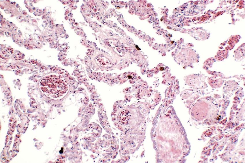
Advances in Animal and Veterinary Sciences
pears to be confined to the intestinal epithelial cells. On ly base. No advisement was given to the owner about the
the other hand, Eimeria pogonae has recently been reclassi- necessity to check for the hydration status of the bearded
fied into the correct genus Choleoeimeria, which is restrict- dragon, or the necessity to integrate fluids assumption dur-
ed located to the gallbladder epithelium of reptiles, mainly ing therapy because of the possible negative effect of sulfa
lizards; there it causes epithelial anomalies in gallbladders,drugs on urinary system. During the therapeutic cycle, the
with chronic inflammation induced by group of oocysts ac- animal became progressively anorectic and lethargic, and
companied with precipitates of tissue debris and gallstones at the end of the therapy it was referred to the authors. At
(Szczepaniak et al., 2009). Oocysts of both species can be admission, the bearded dragon was 23 cm length and 70 g
observed in fecal samples. weight, apathetic, moderately dyspnoic and seriously de-
hydrated, with pale dried mucous membranes and sunken
Isosporosis of young bearded dragons is commonly associ- eyes. A thermostatic cage was used for the hospitalization,
ated with high mortalities (>15%), while in adult individ- with heating sets (40°C in the basking site and 29°C in the
uals I. amphiboluri seem to be a low pathogenic parasite, cold side) to warranty thermoregulation. It was not possi-
even if it cause reduced fecundity and declining health in ble to collect samples for blood work because of the small
older dragons (Kim et al., 2002; Walden & Mitchell, 2012; size and the emaciated status of the animal, so supportive
Jańczak et al., 2014). In a study it was found that more than therapy was started considering the possibility of sulfa ne-
23% of adult bearded dragons in breeding colonies shed phrotoxicosis on an anamnestic basis. Via an intraosseous
this organism, and this is presumably underestimated be- catheter, placed in the tibial plateau anterior to the joint
cause it was based on a single faecal specimen examination capsule after local anaesthesia with lidocaine (Perry and
(Walden, 2009). I. amphiboluri has been known for over 40 Mitchell, 2019), fluid therapy was administered. Fluids
years, however no study has been conducted to assess the (1-part dextrose 5%, 1 part electrolytic rehydrating solu-
effectiveness of the various therapeutic protocols available tion, 1 part saline, 1 part sterile water) at 3 ml/day were
for coccidia in reptiles. administered using a small syringe infusion pump. At 24 h
since the admission, after a moderated correction of dehy-
Sulfonamides drugs have been a longstanding staple as a dration, assisted feeding (diluted Carnivore Care, Oxbow
treatment tool for coccidia ever since the 1940s. Since then, Enterprises, Inc.) at 1-2% b.w. was started, divided in 3
numerous drugs have been developed for use against coc- daily rations. After initial acceptance, tree day after admis-
cidia, but the sulfonamides persist to be one of the prima- sion the bearded dragon started regurgitation, so feeding
ry therapeutics applied to most species, including reptiles, was discontinued. In the same night the animal died.
they remain the most often approved therapy in reptiles,
with sulfadiazine, sulfamethazine and sulfadimethoxine In order to determine death causes, necropsy was per-
as the standard prescription (Walden, 2009). Nonetheless, formed according to the owner. At a gross examination
anatomical, biological, behavioral, and biochemical differ- (Figure 1), the abdominal cavity presented a moderate
ences in different reptile species should always be consid- amount of sero-sanguineous fluid. Fat pads were normal-
ered, in order to avoid therapeutic failures or serious, even ly developed. The liver was slightly enlarged with rounded
fatal, occurrences. margins, had a pale-yellow appearance, both on the cap-
sule and cut surface, and was crumbly. Kidneys were dis-
In this respect, the present paper reports a mortal effect of coloured and evenly moderately enlarged, while lungs ap-
a non-referenced therapy with sulfa drugs for coccidia in a peared hyperaemic. Stomach and intestines looked normal.
youthful bearded dragon, prescribed by a reptiles’ non-ex- Samples of liver, kidney and lung were collected and fixed
perienced practitioner. in 10% neutral buffered formalin (NBF), processed rou-
tinely and embedded in paraffin wax using an automatic
Case Description tissue processor. Serial 5-μm sections from all specimens
A youthful bearded dragon (Pogona vitticeps) was referred were cut with a 2030 Biocut microtome (Reichert-Jung,
to authors because of anorexia and lethargic status. A rep- Germany and were mounted on glass slides (Super-Frost,
tiles’ non-experienced vet practitioner had previously vis- Menzel-Gläser, Braunschweig, Germany). The sections
ited the bearded dragon, prescribing a non-accreditated were then stained with standard Haematoxylin and Eosin
therapy subsequently to a standard faecal flotation evidenc- (H&E) and examined under a light microscope for detec-
ing coccidia infection. Orally, a formulation (Disulfa, For- tion of histopathological alterations.
menvet s.r.l.) licensed in Italy for respiratory and enteric
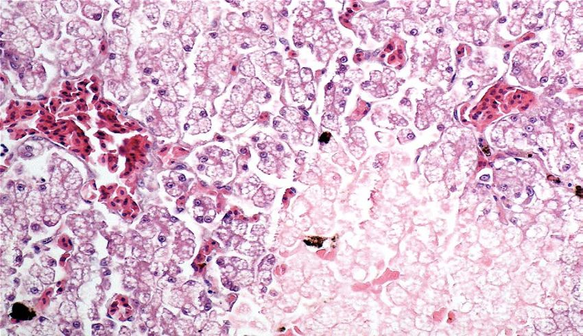
diseases of cage birds, was administered at the dose of 1 ml Renal samples (Figure 2) showed multifocal areas of coag-
q 24 h for 7 days. The formulation is based on diaveridine ulative necrosis, associated with marked marginal hyper-
0.5% and sulfadimetoxine 0.5%, corresponding to an actual aemia. The renal pattern showed interstitial nephritis, with
dosage schedule of 71.4 mg/kg for both drugs on a dai- presence of acidophilic mononuclear and polymorphonu-
clear cells. The examination of liver samples (Figure 3)
January 2021 | Volume 9 | Issue 1 | Page 22 NE
Academic
US
Publishers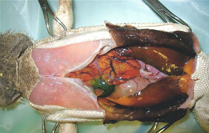
Advances in Animal and Veterinary Sciences

Figure 1: Necropsy examination of bearded dragon
showing a moderate amount of sero-sanguineous fluid          Figure 4: Lungs showing areas of extensive hyperaemia (H
in abdominal cavity; a slightly enlarged liver with pale     & E × 10, bar 100 μm).
yellow appearance; a moderately enlarged and discoloured
kidneys; and congested and hyperaemic lungs.                 showed cellular swelling and diffuse fatty change of hepat-
                                                             ocytes, associated with small scattered groups of necrot-
                                                             ic hepatocytes. Sinusoids were enlarged and congested.
                                                             Lungs showed areas of diffuse hyperaemia (Figure 4).

                                                             Discussion

                                                      Coccidia infections are a common finding in captive rep-
                                                      tiles, with most of these infections being perpetuated be-
                                                      cause of poor hygiene of enclosures. Coccidia infections
                                                      are typically self-limiting; however, autoinfection appears
                                                      common in captive reptiles and can represent a serious
                                                      risk for juvenile animals. Isospora amphiboluri may result in
                                                      enteritis in heavy infections. Heavy numbers can also be
Figure 2: Kidney showing large multifocal areas of associated with concurrent adenovirus (Atevirus, Adv) in-
coagulative necrosis, associated with marked marginal fections. This can lead to a wide range of clinical signs, in-
hyperemia, interstitial nephritis, with presence of cluding anorexia, lethargy, weight loss, diarrhea, tenesmus,
mononuclear and polymorphonuclear cells (H & E × 40, prolapses, and death if untreated. Treatment of coccidia is
bar 100 μm).                                          recommended for young animals or those with a moderate
                                                      to high parasite burden (Eatwell and Richardson, 2019).

                                                             Historically, sulfadimethoxine and trimethoprim-sul-
                                                             famethoxazole have been recommended as coccidiostats
                                                             for treating coccidia infections in reptiles. Unfortunately,
                                                             these recommendations were mainly based on empirical
                                                             data, especially for the posology, with little or no regard
                                                             for species peculiarities. To date, there have been no phar-
                                                             macokinetic studies performed to evaluate these drugs in
                                                             reptiles. Current dosing recommendations are varied, in-
                                                             cluding 15 to 25 mg/kg every 24 hours, 20 to 30 mg/kg
                                                             every 24 to 48 hours, and 30 mg/kg once a day for 2 days
Figure 3: Liver showing cellular swelling and diffuse        then 30 mg/kg every 48 hours. The practitioner should re-
microvacuolar fatty change of hepatocytes, associated with   member that these doses are anecdotal and consider the
scattered groups of necrotic hepatocytes; sinusoids were     general health status (mainly hydration status) of the pa-
enlarged and congested (H & E × 20, bar 100 μm).             tient before selecting a dose (Perry and Mitchell, 2019).

                                                             All sulfonamides work by interfering with the production
                                                             of folate through competitive replacement of para-amino
January 2021 | Volume 9 | Issue 1 | Page 23                                                                        NE
                                                                                                                   Academic
                                                                                                                              US
                                                                                                                              Publishers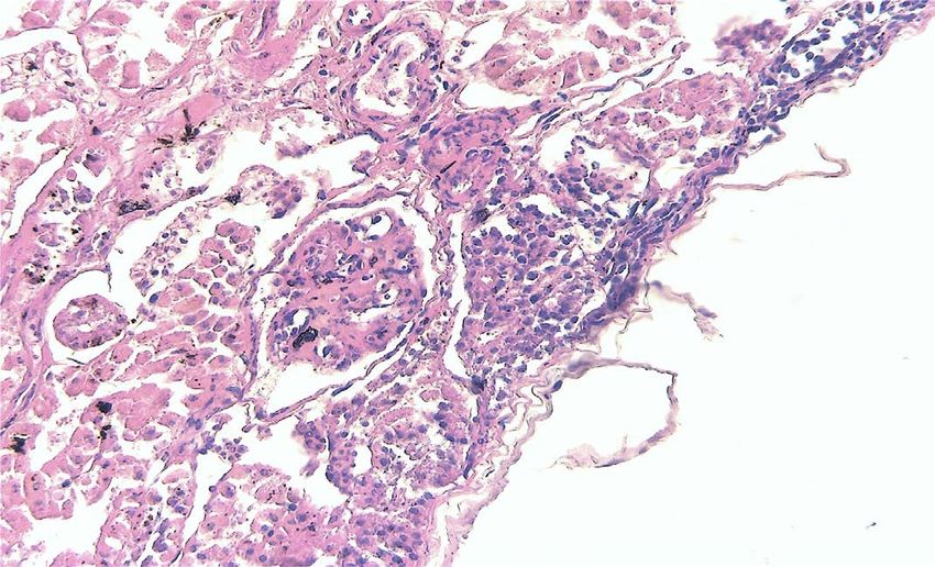
Advances in Animal and Veterinary Sciences
benzoic acid (PABA) in the structure of folate. The result        recommendations in exotic medicine literature (McAr-
of this competitive inhibition is a decreased amount of           thur, 2004; Perry and Mitchell, 2019); 2. the owner was not
RNA and DNA synthesis and a resulting interference with           instructed about carefully check the hydrating status and
metabolism, protein synthesis and growth of the parasite          water assumption in a desert species. The histopathological
(Bowman et al., 1999; Boothe, 2001) These effects are not         changes affecting the liver did not seem to be connected
evident in the host since vertebrates lack dihydropterate         to the coccidia infection; also, the gallbladder was free of
synthase and must ingest folate from plants and animal            debris/gallstones and not enlarged, occurrence that would
prey (Murray, 1996). Sulfonamides are primarily effective         have suggested the previous presence of C. pogonae. Gross
against the asexual stages of coccidian development (mero-        pathologic examination of reptiles that die with AdV in-
gony/schizogony), they operate as coccidiostatic drugs,           fection can involve only the liver, which may be enlarged
and often prolonged treatment is required (Bowman et al.,         and have petechiae or pale areas scattered throughout, with
1999). Potentiated sulfa drugs could be more fast effec-          hepatic necrosis histologically evident (Marschang, 2011).
tive. Trimethoprim, ormetoprim, pyrimethamine and dia-            Unfortunately, no virologic test was performed for Advs in
veridine are usually used in combination with long acting         the present case.
sulfonamides: they synergize their anticoccidial activity by
blocking the same metabolic pathway while exhibiting less         Hepatic alterations were evident, but regrettably the small
host toxicity (Menholm and Armstrong, 2001).                      size and the clinical status of the animal hampered the
                                                                  possibility to perform any hematologic and biochemical
Currently, there are several sulfonamides available, but          evaluation, that would have been an indispensable diag-
sulfadiazine, sulfamethazine and sulfadimethoxine remain          nostic tool, so no information on hepatic parameters is
mostly recommended as the standard therapy for many               available. The diversity of reptile species, their physiologic
species (Barnard and Upton, 1994). Sulfadimethoxine, one          features, and the effects of intrinsic and extrinsic factors
of the most widely used in reptiles, shares the same mode         present unique challenges for accurate interpretation of the
of action of other sulfonamides, but also the numerous            hemogram and biochemistry, but combining the clinical
disadvantages to using these compounds. The greatest in-          presentation with hematologic findings provides valuable
convenience is the necessity for careful monitoring of hy-        information in the diagnosis and monitoring of disease,
dration since sulfa drugs metabolites are excreted through        besides helping and guiding the clinician toward therapy
the kidneys. Sulfadimethoxine is acetylated to acetylsul-         and prognostic output (Stacy, 2011).
fadimethoxine in the liver of mammals and reptiles (Vree
et al., 1989), and the metabolite excreted through the kid-       Literature is lacking of data about safety of use of diave-
neys via both glomerular filtration and tubular secretion. It     ridine in reptiles, the low toxicity reported in mammals it
may crystallize in animals with concentrated urine causing        is likely to hypothesize less or no participation in the final
crystalluria and urinary tract obstruction in mammals. The        mortal toxic output.
increasing of the dose can potentially enhance harmful side
effects, and this possibility is still worse in desert species,   Conclusions
which highly concentrate urine to spare water.
                                                                  Based on little or no species-specific pharmacokinetic re-
Many desert species can behaviorally conserve water by            cords, drugs are used in reptile species. Most of the anti-
retreating to a humid underground burrow, but this may            biotic dosage procedure for these animals are empirical or
not be available in a cage with low relative humidity. In         deduced from other species. Moreover , the development
captivity, it is safest to assume all reptiles depend on en-      of a therapeutic treatment in reptile medicine depend by
vironmental water. Some reptiles are not adept at drink-          the broad range of anatomic, physiological and behavioural
ing from water bowls: many terrestrial species (included          peculiarities of the various species enclosed in the Reptilia
bearded dragons) seem to prefer to walk into shallow water        class, so cross-species deduction of drug dosage regimens
to drink or to have water dripped on their head (Boyer and        from birds, mammals or even between reptiles species can
Scott, 2019). Furthermore, the digestive system of beard-         be dangerous. The instruction for practitioners interested
ed dragons is used in conjunction with the urinary system         in exotic and reptile medicine practice is not to improvise
to prevent water loss: the urine flows into the cloaca and        therapeutic protocols, but rather to refer to specialised lit-
then it moves retrograde into the colon where the colon           erature or more qualified colleagues.
mucosa extracts more water and some salts before the con-
centrated urine is excreted with the faeces (Witten, 1993).       Acknowledgements
In the present case, the possibility of adverse effect of sulfa
drugs administration was announced by two concomitant             The authors would like to thank Mrs. Rosa Leone for her
occurrences: 1. the actual dose was higher respect to the         technical help during this work.

January 2021 | Volume 9 | Issue 1 | Page 24                                                                               NE
                                                                                                                          Academic
                                                                                                                                     US
                                                                                                                                     Publishers
Advances in Animal and Veterinary Sciences
Conflict of interest • Mader DR (2006). Reptile medicine and surgery. 2nd ed.,
Saunders Elsevier, St. Louis, Missouri, USA. pp. 631-639.
• Marschang RE (2011). Viruses infecting reptiles. Viruses.
The authors declare that they have no conflicts of interest. 3:2087-2126.
• McArthur S (2004). Feeding techniques and fluids. In: McArthur
authors contribution S, Wilkinson R, Meyer J (eds.) Medicine and surgery of
tortoises and turtles. Oxford: Blackwell Publishing Ltd,
2004; 257-264. https://doi.org/10.1002/9780470698877
All the authors equally contributed to the present study. • Mehlhorn H, Armstrong PM (2001). Encyclopaedic Reference
of Parasitology: Diseases, treatment, therapy. Sprinter Eds.,
References p. 141. https://doi.org/10.1007/3-540-29834-7
• Murray RK (1996). Harper’s biochemistry. 24th ed. Stamford,
Ct.: Appleton & Lange, p. 868.
• Barnard SM, Upton SJ (1994). A veterinary guide to the
• Perry S M, Mitchell MA (2019). Routes of Administration. In
parasites of reptiles. Malabar, Fla.: Krieger Pub. Co. v. p.
Mader’s reptile and amphibian medicine and surgery, 3rd
• Boothe DM (2001). Small animal clinical pharmacology and
edition, Elsevier inc., St. Louis, Missouri pp. 1130-1138.
therapeutics. Philadelphia: W.B. Saunders, p. 806.
https://doi.org/10.1016/B978-0-323-48253-0.00115-X
• Boyer TH and Scott PW (2019). Nutrition. In: Mader’s
• Perry SM, Mitchell MA (2019). Antibiotic Therapy. In Mader’s
reptile and amphibian medicine and surgery, 3rd edition,
reptile and amphibian medicine and surgery, 3rd edition,
Elsevier inc., St. Louis, Missouri, Pp. 201-223. https://doi.
Elsevier inc., St. Louis, Missouri pp. 1139-1154. https://doi.
org/10.1016/B978-0-323-48253-0.00027-1
org/10.1016/B978-0-323-48253-0.00116-1
• Bowman DD, Lynn RC, Georgi R (1999). Georgi’s parasitology
• Rachel E. Marschang (2019). Virology. In Mader’s reptile and
for veterinarians. 7th ed. Philadelphia, Penn.: W.B. Saunders,
amphibian medicine and surgery, 3rd edition, Elsevier inc.,
p. 414.
St. Louis, Missouri, Pp. 247-269. https://doi.org/10.1016/
• de la Navarre BJ (2006). Common procedures in reptiles and
B978-0-323-48253-0.00030-1
amphibians. Vet. Clin. North Am. Exot. Anim. Pract.
• Stacy NI, Alleman AR, Sayler KA (2011). Diagnostic
9(2):237-67, vi. https://doi.org/10.1016/j.cvex.2006.04.002
haematology of reptiles. Clin. Lab Med. 31(1):87-108.
• Eliman MM (1997). Hematology and plasma chemistry of
https://doi.org/10.1016/j.cll.2010.10.006
the Inland Bearded Dragon, Pogona vitticeps. Bulletin
• Szczepaniak KO, Tomczuk K, Lojszczyk-Szczepaniak A (2009).
of the Ass Reptile and Amphibian. Vet. 7(4). https://doi.
Reclassification of Eimeria pogonae Walden as Choleoeimeria
org/10.5818/1076-3139.7.4.10
pogonae comb. nov. (Apicomplexa: Eimeriidae). Parasitol.
• Eatwell K, Richardson J (2019). Gastroenterology—Small
Res. 115: 681–685 (2016). https://doi.org/10.1007/s00436-
Intestine, Exocrine Pancreas, and Large Intestine. In
015-4787-2
Mader’s reptile and amphibian medicine and surgery, 3rd
• Vree TB, Vree JB, Beneken Kolmer N, Hekster YA, Shimoda
edition, Elsevier inc., St. Louis, Missouri. Pp. 761-774.
M, Nouws JF, Yoshioka T, Hoji K. (1989). O-demethylation
https://doi.org/10.1016/B978-0-323-48253-0.00074-X
and N4-acetylation of sulphadimethoxine by the turtle
• Jacobson E, Heard D, Isaza R (2006). Future directions in
Pseudemys scripta elegans. Vet. Q. 11(3): 138-43. https://doi.
reptile medical education. J. Vet. Med. Educ. 33(3):373-81.
org/10.1080/01652176.1989.9694212
https://doi.org/10.3138/jvme.33.3.373
• Walden MR (2009). Characterizing the epidemiology of
• Jańczak, D, Barszcz, K, Toborek M (2014). Parasitic infestation
Isospora amphiboluri in captive bearded dragons (Pogona
of Isospora amphiboluri in bearded dragon (Pogona
vitticeps). Ph. D. thesis, Louisiana State University, Baton
vitticeps). Życie Weterynaryjne. 89(9): 778-780.
Rouge, Louisiana, USA etd-05292009-214931. https://doi.
• Kim DY, Mitchell MA, Bauer RW, Poston R, Cho DY (2002). An
org/10.1053/j.jepm.2012.06.008
outbreak of adenoviral infection in inland bearded dragons
• Walden M, Mitchell MA (2012). Evaluation of three treatment
(Pogona vitticeps) coinfected with dependovirus and coccidial
modalities against Isospora amphiboluri in inland bearded
protozoa (Isospora sp.). Vet. Diagn. Invest. 14(4):332-334.
dragons (Pogona vitticeps). J. Exotic. Pet. Med. 21(3):213-
https://doi.org/10.1177/104063870201400411
218.
• Long PL, editor. (1982). The Biology of the Coccidia. Baltimore:
• Witten G (1993). Fauna of Australia. In CG. Glasby, G.B. Ross,
University Park Press.
P.L. Beesley Editors, vol. 2, AGPS Canberra. p. 439.
January 2021 | Volume 9 | Issue 1 | Page 25 NE
Academic
US
PublishersYou can also read