Ultraviolet fluorescence discovered in New World flying squirrels (Glaucomys)
←
→
Page content transcription
If your browser does not render page correctly, please read the page content below
Journal of Mammalogy, xx(x):1–10, 2019
DOI:10.1093/jmammal/gyy177

                                                                                                                                            Downloaded from https://academic.oup.com/jmammal/advance-article-abstract/doi/10.1093/jmammal/gyy177/5299493 by ASM Member Access, Paula Spaeth on 24 January 2019
Ultraviolet fluorescence discovered in New World flying squirrels
(Glaucomys)
Allison M. Kohler, Erik R. Olson, Jonathan G. Martin, and Paula Spaeth Anich*
Department of Natural Resources, Northland College, 1411 Ellis Avenue, Ashland, WI 54806, USA (AMK, ERO, JGM, PSA)
Department of Wildlife and Fisheries Sciences, Texas A&M University, College Station, TX 77843, USA (AMK)
* Correspondent: panich@northland.edu

         Fluorescence of visible wavelengths under ultraviolet (UV) light has been previously detected in a wide range
         of birds, reptiles, and amphibians and a few marsupial mammals. Here, we report the discovery of vivid UV
         fluorescence of the pelage in Glaucomys, the New World flying squirrels. Fluorescence in varying intensities
         of pink was observed in females and males of all extant species (G. oregonensis, G. sabrinus, and G. volans)
         across all sampled geographic areas in North and Central America and a temporal range of 130 years. We
         observed fluorescence in museum specimens (n = 109) and wild individuals (n = 5) on both dorsal and ventral
         surfaces. Museum specimens of three co-occurring, diurnal sciurid species (Sciurus carolinensis, S. niger, and
         Tamiasciurus hudsonicus) were also examined but did not fluoresce. The ecological significance of this trait in
         the nocturnal–crepuscular flying squirrels warrants further investigation.

         Key words: crepuscular, fluorescence, flying squirrels, Glaucomys oregonensis, Glaucomys sabrinus, Glaucomys volans, museum
         specimens, nocturnal, Sciuridae, ultraviolet

Research into ultraviolet (UV) fluorescence in biological sub-         2007), green (Pine et al. 1985; Gruber and Sparks 2015; Taboada
stances has a long history. The phenomenon was first docu-             et al. 2017), blue (Taboada et al. 2017; Prötzel et al. 2018), and
mented in plants (Herschel 1845; Lloyd 1924; Goodwin and               pink (Meisner 1983; Pine et al. 1985; Weidensaul et al. 2011).
Kavanagh 1948) and has subsequently been detected in many              Despite the fact that fluorescence spans a wide range of taxa,
marine and terrestrial invertebrates (anthozoa—Miyawaki                tissue types, and color patterns, its ecological significance is
2002; arachnids—Gaffin et al. 2012; and lepidopterans—Olofs-           not clear. In some instances, compelling data suggest fluores-
son et al. 2010). Among the vertebrates, fish (Garcia and de           cence has important ecological functions such as influencing
Perera 2002; Michiels et al. 2008; Sparks et al. 2014), amphib-        mate choice (Arnold et al. 2002) or enhancing antipredator
ians (Nowogrodzki 2017; Taboada et al. 2017), reptiles (Gruber         defenses (Olofsson et al. 2010). In other cases, the ecological
and Sparks 2015; Prötzel et al. 2018), birds (Pearn et al. 2001;       significance of the trait is largely unknown (Weidensaul et al.
Weidensaul et al. 2011), and certain mammalian species within          2011; Gruber and Sparks 2015).
Didelphidae (Meisner 1983; Pine et al. 1985) fluoresce under              Within Mammalia, fluorescence has yet to be widely docu-
UV light. Although fluorescence occurs across a range of lin-          mented or studied. Externally visible UV fluorescence has only
eages, body sizes, and habitat types, it is manifested variably        been observed within Didelphidae. Meisner (1983) detected
across these scales.                                                   fluorescence of the skin and pelage of the Virginia opossum
   In vertebrates, fluorescent compounds have been found in            (Didelphis virginiana) under UV light. Pine et al. (1985) exam-
bone (Dooley and Moncrief 2012; Prötzel et al. 2018), plum-            ined museum skins of 31 didelphid species and reported UV
age (Pearn et al. 2001; Arnold et al. 2002; McGraw et al.              fluorescence in 23 species, without any notable patterns related
2007; Weidensaul et al. 2011), carapace and skin (Gruber and           to sex, age, or season. The fluorescing didelphids described by
Sparks 2015), and pelage (Meisner 1983; Pine et al. 1985).             Pine et al. (1985) inhabited a broad range of New World eco-
Furthermore, vertebrates fluoresce across a wide spectrum of           system and microhabitat types, but all had similar crepuscu-
visible colors including shades of red (Pine et al. 1985; Gruber       lar or nocturnal activity patterns. Diurnal eastern fox squirrels
and Sparks 2015), yellow (Arnold et al. 2002; McGraw et al.            (Sciurus niger) have a high incidence of UV fluorescence in

© 2019 American Society of Mammalogists, www.mammalogy.org

                                                                   1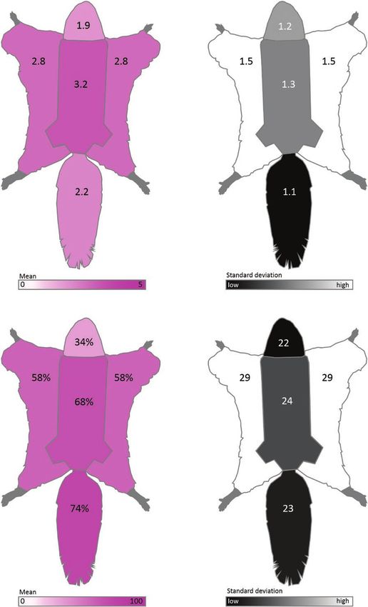
2                                                    JOURNAL OF MAMMALOGY

their crania and teeth (Dooley and Moncrief 2012), but this flu-       1). Glaucomys volans is found in eastern North American for-

                                                                                                                                              Downloaded from https://academic.oup.com/jmammal/advance-article-abstract/doi/10.1093/jmammal/gyy177/5299493 by ASM Member Access, Paula Spaeth on 24 January 2019
orescence has not been observed in any external features such          ests and highlands in Central America (Fig. 1). The omnivo-
as their skin or pelage.                                               rous, arboreal flying squirrels are unique among New World
   Here, we extend the list of visibly fluorescing mammals to          sciurids due to the presence of furred patagia extending from
include all three of the nocturnal–crepuscular New World fly-          their wrists to their ankles and furred, flattened tails that enable
ing squirrels (Glaucomys): Humboldt’s flying squirrel (G. ore-         them to glide from tree to tree (Dolan and Carter 1977; Wells-
gonensis), northern flying squirrel (G. sabrinus), and southern        Gosling and Heaney 1984). In visible light, their pelage is
flying squirrel (G. volans). The flying squirrels are small-bod-       camouflaged via counter-shading: dark on the dorsal surface
ied (50–150 g) and inhabit coniferous, hardwood, and mixed             (brown or gray) and light on the ventral surface (white or
forests from the northern United States and Canada to low-             cream). Sexual dimorphism of the pelage is minimal (Dolan
latitude highlands in Mexico, Guatemala, and Honduras (Fig.            and Carter 1977; Wells-Gosling and Heaney 1984).
1; Dolan and Carter 1977; Wells-Gosling and Heaney 1984).                 Glaucomys spp. are nocturnal–crepuscular and active year-
Glaucomys oregonensis, a newly described “cryptic species,”            round. Bimodal activity patterns with peaks of movement and
has a relatively small geographic distribution along the for-          foraging around sunset and sunrise are typical under cooler
ests of the Pacific coast and areas of overlap with G. sabrinus        conditions (Dolan and Carter 1977; Wells-Gosling and Heaney
in Washington, United States, and British Columbia, Canada             1984). The hours immediately after sunset and before sunrise
(Fig. 1; Arbogast et al. 2017). The geographic range of G. sab-        may be particularly significant for understanding UV fluores-
rinus includes northern forests across Canada and much of              cence. Under certain celestial and meteorological conditions,
the northern United States (where it overlaps with G. volans)          the light environments of dawn and dusk are dominated by
and sky islands in western and southwestern United States and          UV wavelengths (Johnsen et al. 2006; Cronin and Bok 2016).
Appalachia (another zone of co-occurrence with G. volans; Fig.         These UV wavelengths are also present during the daytime;

Fig. 1.—Geographic distribution of New World flying squirrels: Glaucomys oregonensis (black), G. sabrinus (light gray), and G. volans (dark
gray). Hatching indicates regions of overlap between species. We examined museum specimens from Guatemalan, Mexican, and United States
states and Canadian provinces highlighted with bold boundaries. Geographic ranges modified from Arbogast et al. (2017).
KOHLER ET AL.—UV FLUORESCENCE IN GLAUCOMYS 3
however, visible wavelengths are dominant. Thus, UV wave- specimens came from western, central, and eastern parts of
Downloaded from https://academic.oup.com/jmammal/advance-article-abstract/doi/10.1093/jmammal/gyy177/5299493 by ASM Member Access, Paula Spaeth on 24 January 2019
lengths are relatively more abundant during twilight and may the United States and Canada (Fig. 1; Table 1); and G. volans
therefore be crucial for animal perception and communication specimens spanned the United States, Mexico, and Guatemala
at dawn, dusk, and overnight (Johnsen et al. 2006; Cronin and (Fig. 1; Table 1). The specimens were collected between 1887
Bok 2016). and 2007 (Table 1; Supplementary Data SD1).
We studied the effects of UV wavelengths on the pelage We photographed the dorsal and ventral side of each speci-
characteristics of the nocturnal–crepuscular New World fly- men (Canon EOS 50D, Canon USA Inc., Melville, New York;
ing squirrels and compared them to three co-occurring diurnal Sigma 17–70 mm f2.8–4 DC Macro) under visible light (Canon
sciurids. Using a combination of opportunistic observations of Speedlite 430EX) and then separately under 395 nm UV light
living, wild Glaucomys individuals and a systematic examina- (LED UV flashlight, iLumen8 100 LED). We used a yellow filter
tion of museum specimens, we documented nearly ubiquitous (K&F Concept, Guangdong Sheng, China) for photographs taken
fluorescence in the three Glaucomys species. under UV light to absorb any residual light in the blue wave-
lengths. For each specimen, we parted a portion of fur on the dor-
sal surface to display the underfur. Images were captured using
Materials and Methods camera RAW files and post-processed with Adobe Lightroom
In May 2017, JGM made the initial opportunistic observation (ver. 3.6; Adobe, San Jose, California), where white balance was
of fluorescence in G. volans in Bayfield County, Wisconsin, adjusted from the neutral gray on the measurement scale refer-
United States, during an exploratory forest survey of early ences next to the specimens. Exposure was increased for dark
spring flora and lichens using a handheld LED UV flashlight images and the images presented here (Fig. 2) had dust and debris
(iLumen8 100 LED). JGM observed a bright pink fluorescence removed from the background. All other post-processing settings
of G. volans when exposed to UV light. Following this observa- were left untouched, including color saturation and intensity.
tion, we initiated our formal investigation into the prevalence of To rank the intensity of fluorescence for each photographed
fluorescence in Glaucomys and other North American sciurids. specimen, we used a qualitative visual fluorescence rank that
We examined skin specimens of the three New World flying ranged between 0 (no fluorescence) and 5 (vibrant fluorescence).
squirrels (G. oregonensis, G. sabrinus, and G. volans; n = 109; Each specimen was ranked for five body areas (dorsal surface,
Table 1; Supplementary Data SD1) and three diurnal sciurid ventral body, ventral chin and neck, ventral patagia [if appli-
species (Sciurus carolinensis, S. niger, and Tamiasciurus hud- cable], and ventral tail) by four independent observers (PSA,
sonicus; n = 26; Table 1; Supplementary Data SD1) at the Field AMK, JGM, ERO). Observers also estimated the percent cover
Museum of Natural History (FMNH, Chicago, Illinois) and the of fluorescence for each body area. Individual observer rankings
Science Museum of Minnesota (SMM, St. Paul, Minnesota). To were then averaged to yield a mean fluorescence score and per-
distinguish cryptic G. oregonensis and G. sabrinus specimens, cent fluorescence cover for each body area for each individual.
we relied on geographic range information from Arbogast We used generalized linear models to explore patterns of
et al. (2017) and only examined G. oregonensis specimens that fluorescence among flying squirrels. We examined the effects
were collected from areas that were allopatric to G. sabrinus of species and of seasonality by using month as a factor in our
(Arbogast et al. 2017; Fig. 1; Table 1). Glaucomys sabrinus models. In other taxa (e.g., Arnold et al. 2002), UV fluorescence
Table 1.—Sampling of North American sciurid specimens from museums (Field Museum of Natural History [FMNH] or Science Museum of
Minnesota [SMM]) with mean dorsal fluorescence scores (± SE) and mean ventral body fluorescence scores (± SE).
Species Sex n States or provinces Collection Repositories Dorsal Ventral body
dates fluorescence fluorescence
Glaucomys Female 3 Washington, Oregon, Unites States 1898–1935a FMNH, 1.11 ± 0.44 3.58 ± 0.52
oregonensis Male 3 SMM
Unknown 2
Glaucomys Female 37 British Columbia, Manitoba, Ontario, Canada; Idaho, 1894–1998 FMNH, 2.98 ± 0.17 3.21 ± 0.14
sabrinus Male 34 Massachusetts, Minnesota, Montana, Vermont, United States SMM
Unknown 4
Glaucomys Female 17 Chimaltenango, Guatemala; Chiapas, Mexico; Florida, Georgia, 1887–2007 FMNH, 2.83 ± 0.25 3.13 ± 0.26
volans Male 9 Illinois, Iowa, Michigan, Minnesota, New York, North Carolina, SMM
Ohio, South Carolina, Tennessee, West Virginia, United States
Sciurus Female 6 Minnesota, United States 1905–1992 SMM 0 0
carolinensis Male 4
Sciurus niger Female 3 Minnesota, United States 1968–2001 SMM 0 0
Male 2
Tamiasciurus Female 5 Alaska, Minnesota, Ohio, United States 1974–1999 SMM 0 0
hudsonicus Male 6
a
Collection year was not listed for all specimens.4 JOURNAL OF MAMMALOGY
Downloaded from https://academic.oup.com/jmammal/advance-article-abstract/doi/10.1093/jmammal/gyy177/5299493 by ASM Member Access, Paula Spaeth on 24 January 2019
Fig. 2.—Photographs of New World flying squirrels under visible light and 395 nm ultraviolet (UV) light. Panel 1: dorsal and ventral surfaces illu-
minated by visible light (a, b) and UV light (c, d) for Glaucomys sabrinus (FMNH 6482; dorsal fluorescence score = 2.3, chin and neck = 2.5, ventral
body = 4.8, tail = 1.9). Panel 2: dorsal and ventral surfaces illuminated by visible light (a, b) and UV light (c, d) for G. volans (FMNH 64181; dorsal
fluorescence score = 4.3, chin and neck = 2.4, ventral body = 4.0, tail = 2.3). Panel 3: dorsal and ventral surfaces illuminated by visible light (a, b)
and UV light (c, d) for G. oregonensis (FMNH 51510; dorsal fluorescence score = 3.5, chin and neck = 4.3, ventral body = 5.0, tail = 4.5). Panel 4:
variation in fluorescence on dorsal and ventral surfaces: (a) G. volans (FMNH 7726) fluoresced intensely on the dorsal surface (dorsal score = 3.6);
(b) G. oregonensis (FMNH 51509) fluoresced intensely on the ventral surface (ventral body score = 4.4); (c) G. sabrinus (FMNH 5905) showed
weak dorsal fluorescence (dorsal score = 0.5); (d) G. volans (FMNH 64183) showed weak ventral fluorescence (ventral body score = 1.3).KOHLER ET AL.—UV FLUORESCENCE IN GLAUCOMYS 5
Downloaded from https://academic.oup.com/jmammal/advance-article-abstract/doi/10.1093/jmammal/gyy177/5299493 by ASM Member Access, Paula Spaeth on 24 January 2019
Fig. 3.—Schematic diagrams summarizing qualitative ventral body fluorescence scores and the percent cover of fluorescence of New World fly-
ing squirrel museum specimens (Glaucomys spp.; n = 109). Top left: average fluorescence scores of each ventral region (body, chin and neck,
patagia, and tail) from 0 (no fluorescence) to 5 (vibrant fluorescence). Top right: SDs of fluorescence scores for each ventral body region. Bottom
left: average percent cover of fluorescence for each ventral region (body, chin and neck, patagia, and tail). Bottom right: SDs of percent cover of
fluorescence for each ventral body region.6 JOURNAL OF MAMMALOGY
appears to be related to sexual selection; therefore, we exam- Table 2.—Linear regression models predicting Glaucomys spp.
Downloaded from https://academic.oup.com/jmammal/advance-article-abstract/doi/10.1093/jmammal/gyy177/5299493 by ASM Member Access, Paula Spaeth on 24 January 2019
ined the influence of sex and the interactions between sex and ventral body fluorescence scores. ∆AICc = difference in Akaike’s
species on squirrel fluorescence scores. We also considered information criteria scores between focal model and top-ranked model;
latitude of collection as a factor to investigate whether the trait k = number of estimable parameters; ω = Akaike model weight; devi-
ance = measure of model fit.
varied systematically across the considerable latitudinal range
of the New World flying squirrels (Fig. 1). Collection local- Model ΔAICc k ω Deviance
ity was reported only at the state or province level for many Null 0 1 0.37 150.5
specimens, so we placed specimens in 10° latitude bins for our Month 1.72 2 0.16 150.3
analysis. All models were run in R (ver. 3.5.1—R Core Team Sexa 2.00 2 0.14 149.6
Latitude binb 2.03 2 0.13 150.3
2018) using the MuMIn package to summarize and rank models Year 2.05 2 0.13 149.9
according to second-order AICc (Akaike’s information criterion Speciesa 3.68 3 0.06 150.4
corrected for small sample size) scores. We also examined pat- Sex * species 7.85 6 0.01 145.6
terns of fluorescence among individuals’ body regions through
Categorical variable.
a
simple linear regressions using Statistical JMP (ver. 14—SAS
10° bins of latitude.
b
2018).
In addition to the museum specimens, we also made a num- body fluorescence (F1,1 = 225.33, P = 0.04); but sample sizes
ber of opportunistic observations of fluorescence in living, wild for this species were considerably lower. These positive rela-
Glaucomys in 2017 and 2018. These opportunistic observations tionships indicate that flying squirrels with vibrant fluorescence
of live Glaucomys allowed us to determine if this phenome- in one area are generally brightly fluorescent across their entire
non could also be observed in individuals within their natural bodies. In some cases, the pattern of fluorescence on the ventral
habitat. surface increased the contrast and visibility of the female mam-
mary glands (e.g., Fig. 2 panel 4b).
All opportunistic observations of living, wild Glaucomys
Results were made in Bayfield County, Wisconsin, between August
Pink UV fluorescence was seen in 108 of 109 Glaucomys and September of 2017 and 2018 (n = 5). All three locations
museum specimens and was not observed in any Sciurus or were in closed-canopy forests, with two observations occur-
Tamiasciurus specimens (Table 1; Figs. 2 and 3). Glaucomys ring in a forested residential area in Washburn, Wisconsin;
fluorescence was generally most pronounced on the ventral two observations in a mixed-forest area near a large, old white
body surface (Table 1; Figs. 2 and 3), an area that appears white pine, Pinus strobus (Laughlin et al. 2017); and the remaining
or cream-colored in visible light. Hence, we used the ventral observation occurred in a mixed-hardwood area on the Bayfield
body fluorescence score as the dependent variable in our linear Peninsula of Wisconsin. All individuals fluoresced similarly to
models. Of the resulting models, the intercept-only null model the museum specimens when exposed to UV light.
was the best fit to the data (Table 2). While there was substan-
tial variation among individuals, there were no meaningful
relationships between species, month, sex, year, or latitude and Discussion
the intensity of fluorescence (Table 2; Figs. 4 and 5). We report external UV fluorescence in a Eutherian mammal,
The percent cover of fluorescence was highest in the tail and Glaucomys spp. This trait was evident in nearly every individual
ventral body and lowest on the ventral chin and neck (Fig. 3). sampled, and fluorescence was seen in all species, sexes, and
Many specimens (n = 22) did not have exposed patagia; there- localities, although the intensity varied in idiosyncratic ways.
fore, we excluded the patagia scores from subsequent analyses. While it is possible that the UV fluorescence we observed is
The fluorescence scores of the dorsal surface, ventral chin and the result of an exogenous factor (e.g., consumption of fluores-
neck regions, and ventral tail showed positive associations with cent lichen) and not an intrinsic trait of the New World flying
ventral body score (Fig. 6), although the strengths of these cor- squirrels, it seems unlikely that a particular food item or other
relations varied. In G. sabrinus, all body regions for both sexes environmental factor could explain a pattern of pelage fluores-
were strongly positively correlated with ventral body intensity cence that spans species, seasons, decades, countries, and habi-
(female dorsal surface F1,33 = 6.98, P = 0.01; female chin and tat types to the extent that we document here.
neck F1,33 = 20.58, P < 0.0001; female ventral tail F1,33 = 38.35, We therefore suggest that the observed fluorescence is
P < 0.0001; male dorsal surface F1,32 = 25.01, P < 0.0001; an intrinsic, potentially ecologically significant trait of the
male chin and neck F1,32 = 41.59, P < 0.0001; male ventral tail Glaucomys flying squirrels. We posit four hypotheses to
F1,32 = 45.69, P < 0.0001). Glaucomys volans females showed explain the presence of fluorescence in Glaucomys: 1) the trait
significant correlation between all body regions with ventral is adaptive in the squirrels’ unique nocturnal–crepuscular light
body intensity (dorsal surface F1,14 = 20.29, P = 0.0005; chin environment; 2) fluorescence is especially important on land-
and neck F1,14 = 13.18, P = 0.003; ventral tail F1,14 = 13.88, scapes with snow cover; 3) fluorescence is used in intraspecific
P = 0.002), while males had significantly correlated chin and communication; and 4) fluorescence plays a role in antipreda-
neck (F1,7 = 10.77, P = 0.01). In G. oregonensis, only female tor behavior. These explanations are not necessarily mutually
chin and neck fluorescence was strongly correlated with ventral exclusive.KOHLER ET AL.—UV FLUORESCENCE IN GLAUCOMYS 7
Downloaded from https://academic.oup.com/jmammal/advance-article-abstract/doi/10.1093/jmammal/gyy177/5299493 by ASM Member Access, Paula Spaeth on 24 January 2019
Fig. 5.—Average fluorescence scores of the major body areas of mu-
seum specimens of Glaucomys oregonensis (black), G. sabrinus (light
Fig. 4.—Summary of the qualitative fluorescence scores for New gray), and G. volans (dark gray) by latitude. Fluorescence scores vary
World flying squirrel museum specimens by major body region. from 0 (no fluorescence) to 5 (vibrant fluorescence) and represent the
Fluorescence scores varied from 0 (no fluorescence) to 5 (vibrant fluo- average observer ranking for each body area.
rescence) and represent the average observer ranking for each body
area. active year-round at high latitudes (Dolan and Carter 1977;
Wells-Gosling and Heaney 1984). As a result, they perpetu-
Among New World squirrels, only Glaucomys are active ally inhabit low-light environments, in contrast to the other
at twilight and night (Dolan and Carter 1977; Wells-Gosling diurnal and often-hibernating New World sciurids. UV light is
and Heaney 1984). In addition, they do not hibernate and are important in these conditions (Johnsen et al. 2006; Cronin and8 JOURNAL OF MAMMALOGY
Downloaded from https://academic.oup.com/jmammal/advance-article-abstract/doi/10.1093/jmammal/gyy177/5299493 by ASM Member Access, Paula Spaeth on 24 January 2019
Fig. 6.—Relationships between the ventral body fluorescence scores and fluorescence scores of the dorsal body, chin and neck, and tail for
museum specimens of New World flying squirrels: Glaucomys oregonensis (left panel), G. sabrinus (center panel), and G. volans (right panel).
Fluorescence scores vary from 0 (no fluorescence) to 5 (vibrant fluorescence) and represent the average observer ranking for each body area.
Bok 2016), and prior research has shown that UV vision spe- that the vision, perception, and appearance of Glaucomys may
cifically appears to be important for nocturnal mammals (Zhao constitute a suite of adaptations to twilight and night environ-
et al. 2009). The eye lenses of one New World flying squirrel, ments that are relatively rich in UV wavelengths.
G. volans, have been examined and were found to be clear and Glaucomys’ fluorescence may be particularly adaptive dur-
capable of transmitting UV light, whereas the yellow lenses ing the winter season and in snowy conditions. Despite overall
of all examined diurnal sciurids filter UV light (Yolton et al. decreases in the intensity of sunlight, the amount of UV-B radi-
1974). It appears that vision may be stronger in low-light envi- ation may increase by a factor of two relative to summer levels
ronments in Glaucomys than the other squirrels. Combined with (Klein 1978). Furthermore, snow cover reflects UV radiation
the fluorescence we observed in the present study, this suggests that reaches the surface (McIntosh et al. 2011), amplifyingKOHLER ET AL.—UV FLUORESCENCE IN GLAUCOMYS 9
the importance of the UV wavelength band in the winter over Acknowledgments
Downloaded from https://academic.oup.com/jmammal/advance-article-abstract/doi/10.1093/jmammal/gyy177/5299493 by ASM Member Access, Paula Spaeth on 24 January 2019
much of the geographic distribution of the New World flying
We thank the Field Museum of Natural History, especially
squirrels.
L. Heaney, A. Ferguson, and L. Smith; the Science Museum of
Our third hypothesis is that fluorescence is beneficial to
Minnesota, especially R. Oehlenschlager; N. Anich, J. Burman,
flying squirrels as a means of intraspecific communication.
M. Laughlin, T. Pichler, T. Van Deelen, and R. Zifko. This
The prominent ventral fluorescence may allow Glaucomys to
research was funded in part by Northland College, a Parsonage
signal their movement patterns to one another as they glide
Fund awarded to AMK, the Risvedt Professorship awarded
through trees, flashing their ventral surfaces, and then con-
to JGM, the Sigurd Olson Professorship in Natural Sciences
cealing their fluorescent patches when latching onto or climb-
awarded to ERO, and a sabbatical leave awarded to PSA.
ing trees. Some fluorescent reef fish may communicate their
presence and activities to conspecifics in this way (Michiels
et al. 2008). If fluorescence is indeed used for intraspecific Supplementary Data
signaling, it may also be possible that the trait plays a role in
Supplementary data are available at Journal of Mammalogy
mate choice. In certain fish (Garcia and de Perera 2002) and
online.
bird species (Hunt et al. 1998; Arnold et al. 2002) UV fluo-
Supplementary Data SD1.—List of museum specimens pho-
rescence appears to indicate individual fitness and influences
tographed and analyzed for UV fluorescence.
mate choice—individuals with the brightest fluorescence have
a reproductive advantage (Hunt et al. 1998; Arnold et al. 2002;
Garcia and de Perera 2002). It is possible that fluorescence in Literature Cited
Glaucomys plays a role in intraspecific communication, pos- Arbogast, B. S., K. I. Schumacher, N. J. Kerhoulas,
sibly related to mate selection. A. L. Bidlack, J. A. Cook, and G. J. Kenagy. 2017. Genetic data
Finally, Glaucomys fluorescence may be ecologically sig- reveal a cryptic species of New World flying squirrel: Glaucomys
nificant for interspecific interactions, specifically predator oregonensis. Journal of Mammalogy 98:1027–1041.
avoidance. UV fluorescence has been observed in many organ- Arnold, K. E., I. P. F. Owens, and N. J. Marshall. 2002.
Fluorescent signaling in parrots. Science 295:92.
isms that share the twilight and nighttime landscape with fly-
Cronin, T. W., and M. J. Bok. 2016. Photoreception and vision in
ing squirrels. Co-occurring barn owls (Tyto alba), barred owls
the ultraviolet. Journal of Experimental Biology 291:2790–2801.
(Strix varia), and great horned owls (Bubo virginianus) are Dolan, P. G., and D. C. Carter. 1977. Glaucomys volans.
known predators of flying squirrels that also display bright Mammalian Species 78:1–6.
pink or magenta UV fluorescence on their ventral surfaces Dooley, A. C., Jr., and N. D. Moncrief. 2012. Fluorescence pro-
(Weidensaul et al. 2011). Several other co-occurring owls that vides evidence of congenital erythropoietic porphyria in 7000-year-
are not squirrel predators, including northern saw-whet owls old specimens of the eastern fox squirrel (Sciurus niger) from the
(Aegolius acadicus) and screech-owls (Megascops spp.), also Devil’s Den. Journal of Vertebrate Paleontology 32:495–497.
fluoresce pink ventrally. Flying squirrels using the same habi- Gaffin, D. D., L. A. Bumm, M. S. Taylor, N. V. Popokina, and
tats at the same times of night as the owls show the same colors S. Mann. 2012. Scorpion fluorescence and reaction to light.
and patterns of fluorescence. This may be a form of Batesian Animal Behaviour 83:429–436.
Garcia, C. M., and T. B. de Perera. 2002. Ultraviolet-based
mimicry, in which Glaucomys resemble predatory owls in order
female preferences in a viviparous fish. Behavioral Ecology and
to escape avian predation. In addition, some plants and lichens
Sociobiology 52:1–6.
contain UV fluorescent compounds (Lloyd 1924; Goodwin and Goodwin, R. H., and F. Kavanagh. 1948. Fluorescing substances in
Kavanagh 1948; Hale 1956; Klein 1978). In the same way that roots. Bulletin of the Torrey Botanical Club 75:1–17.
reef fishes exploit UV fluorescence (or UV reflectance) to blend Gruber, D. F., and J. S. Sparks. 2015. First observation of fluores-
in with the fluorescent corals and algae of their environments cence in marine turtles. American Museum Novitates 3845:1–8.
(Sparks et al. 2014), Glaucomys may appear camouflaged Hale, M. E., Jr. 1956. Fluorescence of lichen depsides and depsi-
against fluorescing lichen-covered trees. In short, fluorescent dones as a taxonomic criterion. Castanea 21:30–32.
flying squirrels may blend into to their UV-saturated, fluores- Herschel, J. F. W. 1845. On a case of superficial colour presented
cent environment. by a homogeneous liquid internally colourless. Philosophical
New World flying squirrels, like several species of New Transactions of the Royal Society of London 135:143–145.
Hunt, S., A. T. D. Bennett, I. C. Cuthill, and R. Griffiths.
World didelphids, demonstrate UV fluorescence of the pel-
1998. Blue tits are ultraviolet tits. Proceedings of the Royal Society
age. In both groups, the fluorescence can be seen in museum
of London, B. Biological Sciences 265:451–455.
specimens from different regions and habitats, after decades of Johnsen, S., et al. 2006. Crepuscular and nocturnal illumina-
preservation, and in both sexes. In the flying squirrels, we have tion and its effects on color perception by the nocturnal hawk-
also observed this trait in wild animals. These distantly related moth Deilephilia elpenor. Journal of Experimental Biology
mammals share a major ecological trait—nocturnal–crepuscu- 209:789–800.
lar activity patterns—that may be the key to understanding the Klein, R. M. 1978. Plants and near-ultraviolet radiation. Botanical
ecological significance of their fluorescence. The discovery of Review 44:1–127.
fluorescence in Glaucomys prompts many new questions about Laughlin, M. M., E. R. Olson, and J. G. Martin. 2017. Arboreal
how Eutherian mammals navigate the twilight and nocturnal camera trapping expands Hyla versicolor complex (Hylidae) can-
realm. opy use to new heights. Ecology 98:2221–2223.10 JOURNAL OF MAMMALOGY
Lloyd, F. E. 1924. The fluorescent colors of plants. Science Prötzel, D., M. Heß, M. D. Scherz, M. Schwager, A. van’t Padje,
Downloaded from https://academic.oup.com/jmammal/advance-article-abstract/doi/10.1093/jmammal/gyy177/5299493 by ASM Member Access, Paula Spaeth on 24 January 2019
59:241–248. and F. Glaw. 2018. Widespread bone-based fluorescence in cha-
McGraw, K. J., M. B. Toomey, P. M. Nolan, N. I. Morehouse, meleons. Scientific Reports 8:1–9.
M. Massaro, and P. Jouventin. 2007. A description of unique R Core Team. 2018. R: a language environment for statistical com-
fluorescent yellow pigments in penguin feathers. Pigment Cell puting. R Foundation, Vienna, Austria.
Research 20:301–304. SAS Institute Inc. 1989–2018. JMP, version 14. SAS Institute Inc.,
McIntosh, S. E., B. Guercio, G. C. Tabin, D. Leemon, and Cary, North Carolina.
T. Schimelpfenig. 2011. Ultraviolet keratitis among mountain- Sparks, J. S., et al. 2014. The covert world of fish biofluorescence: a
eers and outdoor recreationalists. Wilderness & Environmental phylogenetically widespread and phenotypically variable phenom-
Medicine 22:144–147. enon. PLoS One 9:e83259.
Meisner D. H. 1983. Psychedelic opossums: fluorescence of the skin Taboada, C., et al. 2017. Naturally occurring fluorescence in frogs.
and fur of Didelphis virginiana Kerr. The Ohio Journal of Science Proceedings of the National Academy of Sciences of the United
83:4. States of America 114:3672–3677.
Michiels, N. K., et al. 2008. Red fluorescence in reef fish: a novel Weidensaul, C. S., B. A. Colvin, D. F. Brinker, and J. S. Huy.
signalling mechanism? BMC Ecology 8:16. 2011. Use of ultraviolet light as an aid in age classification of owls.
Miyawaki, A. 2002. Green fluorescent protein-like proteins in reef Wilson Journal of Ornithology 123:373–377.
anthozoa animals. Cell Structure and Function 27:343–347. Wells-Gosling, N., and L. R. Heaney. 1984. Glaucomys sabrinus.
Nowogrodzki, A. 2017. First fluorescent frog found. Nature Mammalian Species 229:1–8.
543:297–297. Yolton, R. L., D. P. Yolton, J. Renz, and G. H. Jacobs. 1974.
Olofsson, M., A. Vallin, S. Jakobsson, and C. Wiklund. 2010. Preretinal absorbance in sciurid eyes. Journal of Mammalogy
Marginal eyespots on butterfly wings deflect bird attacks under low 55:14–20.
light intensities with UV wavelengths. PLoS One 5:e10798. Zhao, H., S. J. Rossiter, E. C. Teeling, C. Li, J. A. Cotton, and
Pearn, S. M., A. T. Bennett, and I. C. Cuthill. 2001. Ultraviolet S. Zhang. 2009. The evolution of color vision in nocturnal mam-
vision, fluorescence and mate choice in a parrot, the budgerigar mals. Proceedings of the National Academy of Sciences of the
Melopsittacus undulates. Proceedings of the Royal Society of United States of America 106:8980–8985.
London, B. Biological Sciences 268:2273–2279.
Pine, R. H., J. E. Rice, J. E. Bucher, D. J. Tank, Jr., and Submitted 30 November 2018. Accepted 20 December 2018.
A. M. Greenhall. 1985. Labile pigments and fluorescent pelage
in didelphid marsupials. Mammalia 49:249–256. Associate Editor was Edward Heske.You can also read