Histo-Morphological Comparison of the Tongue between Grainivorous and Insectivorous Birds
←
→
Page content transcription
If your browser does not render page correctly, please read the page content below
Int. J. Morphol.,
39(2):592-600, 2021.

                  Histo-Morphological Comparison of the Tongue
                  between Grainivorous and Insectivorous Birds

            Comparación Histo-Morfológica de la Lengua entre Aves Granívoras e Insectívoras

                         Nazema S. Abdel-Megeid1; Safwat Ali2; Mohamed Abdo3.4 & Samy F . Mahmoud5

ABDEL-MEGEID, N. S.; ALI, S.; ABDO, M. & MAHMOUD, S. F. Histo-morphological comparison of the tongue between grainivorous
and insectivorous birds. Int. J. Morphol., 39(2):592-600, 2021.

        SUMMARY: The study aimed to illustrate the influence of feeding habits in the anatomical feature and histological structure as
well as some histochemical observations on the tongue of two species of birds which differ in their classification, activity and habitat as;
The domestic pigeon, Grainivorous bird, and cattle egret, Insectivorous bird, using light and SEM studies. Results showed that the
tongue of two species was differing in size, shape and structure. The tongue of pigeon appeared short and triangular; while in cattle egret
was long lanceolate in shape with narrow tapering apex. Dorsal large conical lingual papillae presented between the body and the root of
both tongue of the studied birds. They were arranged in form of U-letter in pigeon and in form of wide V-letter in cattle egret. Histologically,
both dorsal and ventral lingual surfaces lined with keratinized stratified squamous epithelium, which decreased in the thickness and the
degree of keratinization toward the base. The dorsum of the apex of pigeon tongue showed desquamated epithelial cells with filiform
papillae, these papillae not observed in the egret¢s tongue. The tongue of cattle egret contained longitudinal tendinous tissue as intra-
lingual ligament appeared parallel and accompanied with skeletal muscle bundles and attached with entoglossal cartilage. It extended
longitudinally from the root to the body of the egret's tongue. The salivary glands presented in the propria submucosal layer of the dorsal
surface that extended laterally from apex to the root, while the ventral surface devoid from any glandular structures. The nature of lingual
salivary glands showed variations in their histochemical observation to Alcian blue stain and PAS technique. It has been summarized that
the morphological and histological variations of both tongues may be correlated to their feeding habits.

        KEY WORDS: Tongue; Birds; SEM; Histology; feeding habits; Salivary glands.

INTRODUCTION

        The pigeon considered the oldest world's                          is very important to detect the adaptation and perseverance
domesticated bird from about ten thousand years ago. They                 of vertebrates to their environmental habits (Darwish, 2012).
have an importance to humanity, particularly in the times of
war also considered important source of meat for people                           Variations in morphology in shape and size of the
(Blechman, 2007). The cattle egret spread in the hot areas                tongue allowing its functions as a special tool for obtaining,
with long expansions. Most people especially the farmers                  manipulation, swallowing and processing food. Many
called it Abo-quirdan, meaning “father of ticks”, and this                authors has been studied the morphology of the tongue in
name originate from the large number of ticks of birds which              vertebrates (Zweers, 1982). According to their lifestyle, birds
spreads in their breeding colonies. The egrets appeared                   have different feeding habits, with differences corresponding
mostly in Egypt, in Delta and Nile Valley, making nests                   to the shape of their beak and tongues. Most bird’s lives in
colony close to water bodies on trees and shrubs. They mostly             different environment such as the air, the land and the water,
appear together with animals to pick up insects as ticks and              many authors illustrated that the shape and structure of the
flies. The tongue has an important role in mechanism of food              tongue differs according to the type of food and method of
in vertebrates especially in birds. Therefore, this mechanism             food intake (Jackowiak et al., 2011; Al-Zahaby & Elsheikh,

1
  Cytology and Histology Department, Faculty of Veterinary Medicine, University of Sadat City, Sadat City 32897, Egypt.
2
  Department of Anatomy and Embryology, Faculty of Veterinary Medicine, Minia University, Egypt.
3
  Department of Animal Histology and Anatomy, School of Veterinary Medicine, Badr University in Cairo (BUC). Egypt.
4
  Department of Anatomy and Embryology, Faculty of Veterinary Medicine, University of Sadat City, Sadat City 32897, Egypt.
5
  Department of Biotechnology, College of Science, Taif University, P.O. Box 11099, Taif 21944, Saudi Arabia.

592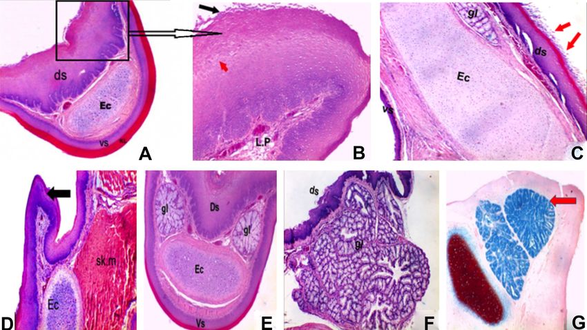
ABDEL-MEGEID, N. S.; ALI, S.; ABDO, M. & MAHMOUD, S. F. Histo-morphological comparison of the tongue between grainivorous and insectivorous birds. Int. J. Morphol., 39(2):592-600, 2021.

2014). The tongues of birds are adapted for manipulation,                                      7.4) the samples were post fixed in phosphate-buffered
collection, and swallowing of foods. Studies of                                                solutions (pH 7.4) of 1 % osmium tetroxide at 4 °C for 2
morphological and functional aspects of different species of                                   hours. These specimens were then washed in 0.1 M.
birds indicated a close relation to the histological structure                                 phosphate buffer solution several times before treating with
of the tongue with their feeding habits (Emura et al., 2009a;                                  3 N hydrochloric acid for 20 min at 60 °C to remove extra
Guimares et al., 2009; Abou-Zaid & Al-Jalaud, 2010;                                            cellular mucus from the lingual surface. The specimens were
Mahmoud et al., 2017).                                                                         then washed in phosphate buffer solution and dehydrated in
                                                                                               ascending graded ethanol series to the critical-point of drying
         The structure of mucosa of the tongue, type and                                       and gold coated. The specimens were then examined in a
distribution of lingual papillae and the degrees of                                            JEOL-JSM 5300 Scanning Electron Microscope at the
keratinization of the lingual epitheliumin relation to feeding                                 faculty of Medicine, Tanta University (Hayat, 2000).
habits were described by many authors as white tailed eagle
(Jackowiak & Godynicki, 2005), cormorant (Jackowiak et                                                 For light microscopy, different parts of the tongues
al., 2006), ostrich (Jackowiak & Ludwig, 2008), peregrine                                      were rapidly set in 10 % neutral-buffered formalin solution
falcon and common kestrel (Emura et al., 2008), spot-billed                                    for at least 48 hours, then dehydrated in ascending ethanol-
duck (Emura, 2009a), three species of herons (Emura,                                           xylene and embedded in paraffin wax. 5 µm serial sections
2009b), woodpecker (Emura et al.), common quail (Parchami                                      were cut transversely and stained with haematoxylin and
et al., 2010), domestic pigeon (Parchami & Dehkordi, 2011),                                    eosin, Masson's trichrome stain for detection of the
red jungle fowl (Kadhim et al., 2014), chukar partridge                                        demonstration of collagen fibers, Weigert's elastic tissue stain
(Erdogan et al., 2012), Muscovy duck (Igwebuike & Anagor,                                      for demonstration of elastic fibers and some special
2013), white-throated kingfisher and common buzzard (El-                                       histochemical stains as Alcian blue stain and Periodic acid
Beltagy, 2013), Black Francolin (Kadhim et al.), the common                                    Schiff techniques for detection the nature of lingual salivary
kingfisher (Al-Zahaby & Elsheikh), southern lapwing                                            glands secretion (Bancroft & Gamble, 2002).
(Erdogan & Perez, 2015). From the previous observations,
there are a correlation between shape, structure of the tongue
and nature of food, also the mechanism of food intake and                                      RESULTS
bird’s habits.

       This study aimed to illustrate the influence of the                                     Gross morphology. Neither the shape nor the dimensions
nature of food and the methods of feeding intake on the                                        of the tongue of the birds showed specific-sex differences.
macroscopic and microscopical observations of the tongue                                       The dorsum of the tongue of Pigeon and Egret were
between two different birds in feeding habits and type of                                      distinguished into three parts; apex, body and root. It is
food as the domestic pigeon (Grainivorous) and the egret                                       distinguished dorsally from its anterior half by a clear me-
(Insectivorous), using light microscopy and scanning                                           dian groove, except on its apical region and some parts along
electron microscope studies, to interpret the present result                                   its posterior part. This groove was dividing the lingual apex
to other previous studies in relation to avian feeding habits.                                 and the body of the tongue into two symmetrical halves.
                                                                                               The tongue of adult domestic pigeon appeared triangular,
                                                                                               small with narrow anterior part and wide fleshy posterior
MATERIAL AND METHOD                                                                            part (Fig. 1). While it was very long, lanceolate with narrow
                                                                                               and tapering apex in cattle egret (Fig. 2). Conical papillae
                                                                                               were arranged symmetrically in the marginal region between
        Eight tongues of both pigeons and egrets were                                          the body and the root, these papillae take the form of the
investigated. The birds were captured from El-Menofiya                                         letter (U) in pigeon while take (V) letter in egret. The lateral
Governorate, Egypt. The birds were observed prior to being                                     papillae were larger and thicker than the middle one in both
sacrificed, to ensure their health condition and for any clinical                              pigeon and egret (Figs. 1 and 2).
signs indicating infection. Only apparently healthy birds were
selected for this study; the tongues were quickly dissected                                    Scanning Electron Microscope Studies (SEM): SEM in
from the mouth cavity and processed as follows: For                                            the domestic pigeon revealed that, deep sulcus dividing the
scanning electron microscopy (SEM), different parts of                                         apex and body of the tongue into two symmetrical halves
tongue were rapidly fixed overnight in modified                                                (Fig. 3A). The surface of the dorsum of the anterior part of
glutraldehyde solutions (2 % paraformaldehyde and 2.5 %                                        the tongue was covered by a large number of irregular scaley
glutraldehyde containing 0.1 M phosphate-buffered solution,                                    protrusions of the deciduous epithelial growths. (Fig. 3B).
pH 7.4) at 4 °C. After rinsing in 0.1 M phosphate buffer (pH                                   Filliform papillae were compactly distributed along the
                                                                                                                                                                                 593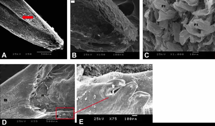
ABDEL-MEGEID, N. S.; ALI, S.; ABDO, M. & MAHMOUD, S. F. Histo-morphological comparison of the tongue between grainivorous and insectivorous birds. Int. J. Morphol., 39(2):592-600, 2021.

                                                                               Fig. 2. Photograph of the dorsal surface of the tongue of the egret showing
                                                                               parts of the tongue, apex (A); body (B); root (R), entoglossal bone (EB), the
                                                                               giant conical papillae (white spotted arrow ), and the median sulcus in the
                                                                               caudal part of the body (white arrow), hyoid bone (HB).

                                                      lingual body. They were long, slender with broad tip. The filliform papillae
                                                      were backwardly directed and situated around the median sulcus (Fig. 3C),
                                                      in the margin between the body and the base of the tongue, small sized
                                                      conical papillae were observed and they were numerous arranged
                                                      asymmetrically in the form of a letter (U). The lateral papillae were larger
                                                      and thicker than the middle one. The dorsum of the base of tongue showed
                                                      smooth appearance with no densely packed desquamated cells and no
Fig. 1. Photograph of the pigeon tongue showing parts lingual papillae are observed (Fig. 3D).
of the tongue, lingual apex (A), body (B) and the root
(R), the median groove (white arrow). Note, the               While in the cattle egret, SEM revealed that, the dorsum of the
arrangement of the - U- shaped conical papillae (black anterior lingual part from the apex to the end of the body of the tongue was
dotted arrow). Laryngeal mound (L), hyoid bone (HB). somewhat appeared smooth, devoid of any lingual papillae. The epithelium

Fig. 3. Scanning electron micrograph. of the dorsal surface of the pigeon tongue showing. a, Median groove dividing the tongue into two
symmetrical halves (arrow). b, the irregular scalyprotrusions and scales in tip of tongue (arrow). c, the filiform papillae (FI) backwardly directed
in the body of tongue. d, the arrangement of the lingual conical papillae (black arrow ), note the lateral papillae is more thicker than the middle one
(dark spotted arrow). e, higher magnification of Fig. (d) showing large lateral conical papilaae with some scaley protrusions on its surface.

594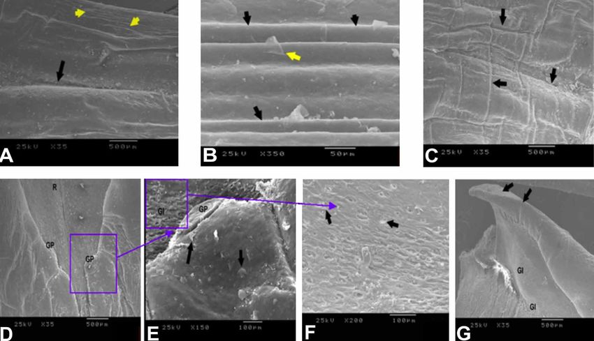
ABDEL-MEGEID, N. S.; ALI, S.; ABDO, M. & MAHMOUD, S. F. Histo-morphological comparison of the tongue between grainivorous and insectivorous birds. Int. J. Morphol., 39(2):592-600, 2021.

of this region was slightly undulated. Scanty number of irre-                                  4G). The surface of root also devoid of lingual papillae
gular scales was observed on the dorsal surface of the lingual                                 showing widely distributed rounded openings of the caudal
apex with characteristic median groove dividing the tongue                                     lingual salivary glands; the openings of the salivary glands
into two symmetrical halves (Fig. 4A). Longitudinal pellicae                                   were surrounded by capsule (Fig. 4F).
were observed at both sides of the anterior part of the lingual
body. These pellicae arranged at almost regular intervals, in                                  Light Microscope Studies. Histologically, the lining
addition few scales were frequently arranged over the surface                                  epithelium of the dorsal surface of the tongue in both pigeon
of the pellicae (Fig. 4B). On the caudal part of the lingual                                   and egret consisted of a stratified squamous keratinized
body, longitudinal and transverse ridges or pellicae also                                      epithelium. The keratinized layer was thicker in the ventral
noticed at both sides of the tongue around the median sulcus                                   and lateral surfaces especially in the tip of the lingual apex
with widely distributed tiny scales on their surfaces (Fig.                                    (Figs. 5A and 6A). The dorsal surface was thicker than the
4C). Giant papillae were located in a transverse row at the                                    ventral surface. The base of this epithelium was uneven,
end of the lingual body which completed by caudal                                              being thrown into shallow and narrow folds. The cells of
continuation of giant conical papillae, larger and thicker than                                both the basal and the deep intermediate layers were rounded
the middle one. (Fig. 4D). The middle papillae were dome-                                      or elliptical in shape and have large, central and spherical
shaped in outline and some scales frequently observed on                                       nuclei. From the deep intermediate layer to the superficial
their dorsal surface. Well-developed micro ridges were                                         layer, the cells and the nuclei gradually flattened and
widely distributed on the cell surface of the dorsum of the                                    desquamating cells were detectable on the upper surface
giant conical papillae (Fig. 4E). The lateral lingual papillae                                 (Figs. 5B and 6B). On the superficial layer of the dorsal
was larger inclined backwardly toward the pharynx (Fig.                                        surface of the body of the pigeon tongue, there were distinct

Fig. 4. Scanning electron micrograph of the dorsal surface of the cattle egret tongue showing. A, longitudinal groove on the
anterior part of the lingual body (dark arrow) and ridges on the lateral lingual surface (yellow arrow). B, Longitudinal
pellicae arranged at almost regular intervals, with few scales on their surface (black arrow) on the lingual body. C, longitudinal
and transverse ridges (pellicae) (black arrow) with compactly distributed scales and the central lingual sulcus. D, the
widely distributed openings of the caudal lingual salivary glands. Note, the giant conical papillae. (GP). on the root of the
tongue. E, Higher magnification of Fig.(D) showing the giant papillae (GP) of the egret tongue. Note that the giant papillae
are dome –shape in outline with characteristic scales on their surface. (Black arrow). F, opening of salivary glands on the
root of egret tongue. G, the giant conical lateral papillae directed caudally toward the pharynx (black arrows) and opening
of salivary glands (Gl) on the root).
                                                                                                                                                                                 595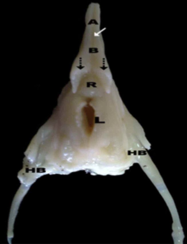
ABDEL-MEGEID, N. S.; ALI, S.; ABDO, M. & MAHMOUD, S. F. Histo-morphological comparison of the tongue between grainivorous and insectivorous birds. Int. J. Morphol., 39(2):592-600, 2021.

Fig. 5. Photomicrophotographs of the pigeon tongue showing. A, the thick dorsal surface (ds) and thin ventral surface (vs) with thick
keratinized layer( KL) and the entoglossal cartilage (EC) on the lingual apex of tongue, H & E × 40. B, desquamation of the epithelial
surface, lamina propria ( L.P) H & E × 100. C, superficial desquamated epithelial cell which represented as filliform papillae (black
arrows) on body of tongue. D, showing, the conical papillae, entoglossal cartilage and the attached skeletal muscle on the posterior part.
(H & E × 40). E, lingual mucus gland (gl), thick dorsal surface, thin keratinized ventral surface, entoglossal cartilage (Ec) H & E × 40.
F, root of tongue with caudal mucus salivary gland (gl) and non keratinized dorsal surface. G, positive alcianophilic substance in lingual
salivary gland. (AB PH 2,5 × 100).

protrusions of the desquamated epithelial cells which may                                      of the second half of the free part of the tongue till the root of
represented as filliform papillae (Fig. 5C). The keratinization                                the tongue. Therefore, the lingual salivary glands of both
was also highly developed at the giant conical papilla which                                   studied birds could be divided into two types: Anterior lingual
separating the body of the tongue from the base fixed part                                     glands at the anterior part of the tongue and posterior salivary
which inclined backwardly toward the pharynx (Figs. 5D and                                     glands at the root of the tongue, both of them were typically
6D). Gustatory lingual papillae were not found in the                                          mucous gland in the pigeon. The mucous secretory units
epithelium covering the tongue in both studied birds. The la-                                  composed of tall columnar cells with extensive vesicular
mina propria consisted of loose connective tissue containing                                   cytoplasm. These glands were surrounded by connective tissue
collagen fibers, thin elastic fibers and numerous blood vessels                                capsule with septa dividing the gland into lobules) (Figs. 5F
(Fig. 6C). There were muscles in the lamina propria of the                                     and 6G). They open on the epithelial surface through minute
dorsal surface of the tongue. The muscles were arranged thin                                   pores (Fig. 6F).
and striated in the form of circular in the lingual apex, but
oriented in the form of circular and longitudinal in different                                         The secretory cells of the lingual salivary glands of
direction in the body and root of the tongue.                                                  the pigeons, contained large amount of acid muco-
                                                                                               polysaccharide substances that showed strong positive reaction
        The skeleton of the tongue in pigeon was supported                                     to alcian blue stain while give negative reaction to PAS
by cartilage hyoid apparatus revealed on entoglossal bone as                                   techniques (Fig. 5G). The ventral surface of the tongue is
skeletal element of the tongue which extending from the lingual                                devoid of any glandular structure. while in the egret , the an-
root to lingual apex (Figs. 5C, D, E). While in cattle egret, an                               terior one appeared serous adenomere while mixed (sero
intra-lingual tendon was longitudinal fibrous structure parallel                               mucoid) in the posterior one. The alcianophilic substances
and accompanied with skeletal muscle fibers and attached with                                  appeared to be increased in amount in the cytoplasm of
entoglossal cartilage (Figs. 6E and F). The lingual salivary                                   secretory cells of the posterior salivary lingual glands and
glands of pigeons and egrets were located in the lamina propria                                showed strong positive reaction to alcian blue stain (Fig. 6H).
596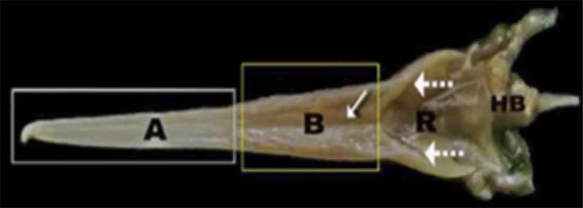
ABDEL-MEGEID, N. S.; ALI, S.; ABDO, M. & MAHMOUD, S. F. Histo-morphological comparison of the tongue between grainivorous and insectivorous birds. Int. J. Morphol., 39(2):592-600, 2021.

Fig. 6. Photomicrograph of the egret tongue. A, the dorsal and ventral surface in the lingual apex. B, distribution of elastic
fibers around the blood vessels. C, collagen fibers in lamina propria and surrounding the gland. D, the highly keratinized
lingual conical papillae. E, entglossal cartilage and the origin of the intra lingual tendon (red arrow). F, A higher magnification
of Fig. (E) showing the entoglossal tendon attached to the lingual skeletal muscle. G, lingual salivary glands opening in the
epithelium of the dorsal surface. H, positive alcian blue and PAS lingual salivary gland. (AB, PH2.5+PAS combination × 100).

DISCUSSION

Gross morphology. The present study was carried out to                                         (Erdogan, 2012). While the long cattle egret’s tongue of the
clarify the structural features of the tongue between two birds                                current study prolongs to fill almost the cavity of the lower
living in different environments feeding habits and food                                       beak and is terminating with sharp, tapered apex. This offers
sources. The structural characteristics of components of the                                   suitable eating possibilities for searching for small food items
avian digestive tract are largely determined by the kind of                                    such as insects in rubbish dumps and bodies of animals (Al-
diet consumed by the particular species. There are different                                   Zahaby, 2016). These description of the tongue is same si-
types of adaptation of the bird's tongue, such as for                                          milar to that of Chukar partridge (Erdogan et al.), which
accumulating food, handling food and swallowing                                                also feeds on insects in the ground pastures.
(Jackowiak & Godynicki).
                                                                                                      Data found from this study also showed that definite
        Previous studies in the avian tongue revealed that the                                 median sulcus divides the apex and body of the tongue into
morphology, structure of the epithelium linguae, supportive                                    two similar halves. These results resemble of those described
components also papillae localization are thoroughly related                                   on the tongue of white tailed eagle, grey heron (Abou-Zaid
with the nature of food, mode of feeding also the different                                    & Al-Jalaud) and domestic goose (Jackowiak et al., 2011),
habitats (Whittow, 2000). Results achieved from the current                                    however it is lacking on the tongue of chickens and ostrich,
study revealed that the tongue of domestic pigeon is a                                         besides these features show the adaptation of the tongue to
characteristic triangular organ with three distinct anatomical                                 aid swallowing grains as whole pieces in the esophagus
parts: apex, body and root. These morphological features                                       (Iwasaki, 2002). Also the current study showed that a main
resemble those of common quail (Parchami et al.), domestic                                     row of great conical papillae are situated symmetrically in
chicken (Homberger & Meyers, 1989) and chuker partridge                                        the form of the letter U in the marginal region between the

                                                                                                                                                                                 597
ABDEL-MEGEID, N. S.; ALI, S.; ABDO, M. & MAHMOUD, S. F. Histo-morphological comparison of the tongue between grainivorous and insectivorous birds. Int. J. Morphol., 39(2):592-600, 2021.
lingual body and the lingual root. These results are similar insects, which are soft enough for smooth swallowing. These
to those the documented by Parchami et al. and Parchami & giant papillae so-called “lingual spikes” by Kooloos (1986).
Dehkordi (2011), in common quail and domestic pigeon. In The distribution pattern of these papillae was the same in
the chucker partridge and common quail, the root of the the Mallard (Kooloos), in the chicken (Iwasaki & Kobayashi,
tongue have conical papillae with the pointed apex directed 1986), in the little tern (Iwasaki), in duck (Abdalla, 1994),
caudally arranged in the letter V; behind this row there is an in Middendroff’s Bean Goose (Iwasaki et al.). The presence
additional row composed of laterally sited large papillae of these giant papillae in these different species of birds may
(Erdogan et al.). While in the Middendroffs bean goose and be related to their phylogenic origin.
domestic goose there are giant conical papillae located
between the body and basal region also on the lateral sides Light microscope observation. In this current study, we
of the anterior region of the tongue. There are lingual hairy observed that mucosa of the dorsum of the lingual apex (tip)
papillae densely distributed and small numbers of large is lined with a thick keratinized stratified squamous
cylindrical papillae are arranged between these lingual hairs epithelium only on the lateral and ventral surface, whereas
(Iwasaki et al., 1997), that is not noted in the present study. the lingual body and root are lined with non- keratinized
On the other hand, Pasand et al. (2010) reported the giant stratified squamous epithelium, this result is similar to that
conical papillae in ostrich were not detected between the described through (Jackowiak & Godynicki) in white tailed
lingual body and root. Occurrence of these lingual papillae eagle and by Parchami & Dehkordi (2011) in domestic
has been reflected to be related to specific feeding habits of pigeon. Contrary to reports in chucker partridge, the dorsal
birds, the conical papillae found in the lingual body was surface of its tongue was lined with thick keratinized
helping in the transfer of swallowed food towards the stratified squamous epithelium Erdogan et al., whereas in
esophagus and preventing its regurgitation. the ostrich, the dorsal and ventral surfaces of the tongue were
lined by non-keratinized stratified squamous epithelium
Scanning Electron Microscope. The ultrastructural (Jackowiak & Ludwig).
observations in the present study revealed that the pigeon
tongue has large numbers of backwardly directed scales on The changes in the degree of keratinization of the
the lingual apex and slender filiform papillae with a broad lingual epithelium between different species look to be
tip on the rest of the lingual body. Moreover, no fungiform related to the differences in habitat. These differences clearly
papillae presents between the filiform papillae as mentioned appear in chickens´ live in habitat, being much drier than
in grey heron (Abou-Zaid & Al-Jalaud), domestic chick (El- that of the water fowls like Middendroff´s bean goose,
Beltagy) also King fisher (Al-Zahaby & Elsheikh). This domestic ducks and the little tern. In birds, the amount of
result was similar to some species of birds but the shape of keratinization of lingual epithelium looks to be a certain
the processes differing from each other, they were needle – degree, to reflect differences in their life style (Erdogan &
shaped processes in woodpecker (Emura et al.), lamellar Perez). In most birds, anterior tip of the tongue is directly
shaped in pigeon (Parchami & Dehkordo, 2011), carpet- contact with food and may avert injuries during feeding. In
shaped in the peregrine falcon and common kestrel (Emura this study, the lingual epithelium of pigeon is thick
et al.), thread-shaped in the owl species (Emura et al., 2009a), keratinized and has solid plates, as it is exposed to solid
spine-like in the penguins (Kobayashi et al., 1998), acicular grains and seeds during feeding. Lateral edges and ventral
processes in the European Magpie and the Common Raven surface of the tongue may be exposed to hard grains and
(Erdogan & Alan, 2012), also many processes can be seeds, during handling and when stored in the buccal cavity.
observed in the black kite (Emura). Therefore, epithelium covered with a thick keratinized layer.
This interpretation agreed with Al-Nefely (2015) in laughing
The microridges present on the root in the pigeon dove. Observations of the present study showed that the la-
and egret may develop the transport of food otherwise, seed mina propria is dense irregular connective tissue, which
or insects through the surface of the tongue and perform as contains adipose cells and numerous blood vessels. This
sites for preservation of the mucous produced by the lingual connective tissue supported by the strong layer of striated
salivary glands located on the base of tongue. The distribution muscle fibers, which are oriented in longitudinal and circu-
of the apical scales and in the present investigation, giant lar direction in the body and base of the tongue. In addition,
papillae were arranged at the final part of the lingual body the tongue contains hyaline cartilage, which extends from
in both specimens, being big and conical in shape in the the lingual apex to the lingual root and surrounded by lingual
pigeon and small with rounded profile in the egret. These muscle fibers. These observations are similar to that of
much larger giant papillae in the pigeon may help in pushing Pasand et al. in male ostrich and Parcham & Dehkordi (2011)
the dry seeds into the pharynx. However, these papillae are in domestic pigeon. Homberger and Meyers stated that in
smaller in the egret because the food consists of worms and birds, the tongue maintained by unpaired paraglossae
598ABDEL-MEGEID, N. S.; ALI, S.; ABDO, M. & MAHMOUD, S. F. Histo-morphological comparison of the tongue between grainivorous and insectivorous birds. Int. J. Morphol., 39(2):592-600, 2021.
extending through the lingual tip and articulates caudally ACKNOWLEDGMENTS
with the basihyoid apparatus.
In the present study, the tongue of domestic pigeon We appreciate and thank Taif University for the
and cattle egret supported by elements of the hyoid apparatus. financial support for Taif University Researchers Supporting
It spreads ventrally to the apex of the tongue as a narrow Project (TURSP-2020/138), Taif University, Taif, Saudi
point, paraglossal apex, then becomes flat and thicker in the Arabia.
tongue’s body called the corpus paraglossale. This last
cartilage remains caudally and bifurcates in the tongue’s root
as two paraglossalis caudalis ending on the each side of the ABDEL-MEGEID, N. S.; ALI, S.; ABDO, M. & MAHMOUD,
trachea as a hyoid bone. In cattle egret showing different S. F. Comparación histo-morfológica de la lengua entre aves
structure not observed in the pigeon hyoid apparatus, which granívoras e insectívoras. Int. J. Morphol., 39(2):592-600, 2021.
is the paraglossal apparatus enveloped with a definite
RESUMEN: La investigación tuvo como objetivo ilus-
perichondrium consisted of fibrous connective tissue and
trar mediante estudios de luz y SEM, la influencia de los hábitos
striated muscle fibers, assisting for moving of the tongue alimentarios en la característica anatómica y estructura histológica,
out of the mouth cavity. Likewise, Erdogan et al. stated that así como algunas observaciones histoquímicas en la lengua de
the hyaline paraglossal cartilage supporting the chukar dos especies de aves, que se diferencian en su clasificación, acti-
partridge’s tongue, which extends in the lingual root, body vidad y hábitat, como es la paloma doméstica, ave granívora y la
and apex. Igwebuike & Anagor also accepted the presence garcilla bueyera, ave insectívora. De acuerdo a los resultados la
of the paraglossal skeleton and associated striated muscle lengua de las dos especies difería en tamaño, forma y estructura.
fibers of the tongue in Muscovy duck. In brief, the tongue La lengua de las palomas tenía una forma corta y triangular; mien-
apparatus of the egret displays certain anatomical and tras que en la garceta bueyera era de forma larga lanceolada con
microscopical structures that are distinctive to this bird ápice estrecho y agusado. Grandes papilas dorsales linguales có-
nicas entre el cuerpo y la raíz de ambas lenguas estaban dispues-
species. This may be an adjustment to the method of food
tos en forma de letra U en paloma y en forma de letra V ancha en
intake, the type of food, lifestyles and bird’s habitat (Al- garza bueyera. Histológicamente, las superficies linguales dorsal
Zahaby). y ventral estaban revestidas con epitelio escamoso estratificado
queratinizado que disminuía en el grosor y el grado de
In this study, we showed that the lingual glands are queratinización hacia la base. El dorso del ápice de la lengua de
simply branched tubulo alveolar glands; they did not appear la paloma mostró células epiteliales descamadas, estas papilas no
in the lingual apex but appeared in the lingual body, which se observaron en la lengua de la garceta. Se observó tejido
open on the epithelial surface through minute pores. It tendinoso longitudinal en la lengua de la garceta bueyera, debido
increased in numbers through the basal region of the tongue. a que el ligamento intralingual aparecía en forma paralela y acom-
The lingual salivary glands prolonged from the apex of the pañado de haces de músculo esquelético y adherido con cartílago
entogloso. Se extendía longitudinalmente entre la raíz hasta el
tongue to both sides of the laryngeal cleft in the white-eared
cuerpo de la lengua de la garceta. Las glándulas salivales de la
bulbul (Parchami & Dehkordi, 2013). The lingual salivary lengua estaban incrustadas en la capa submucosa propia de la
glands are absent in the fundamental tongue of cormorants superficie dorsal que se extendía lateralmente desde el ápice has-
(Jackowiak et al., 2006). In this study, the lingual salivary ta la raíz, mientras que la superficie ventral carecía de estructuras
gland in both pigeon and egret take positive reaction to alcian glandulares. En las glándulas salivales linguales se observaron
blue stain while negative to PAS technique as observed on variaciones histoquímicas a la tinción con azul de Alcian y la
the tongue of the little egret. In contrast, in red jungle fowl, técnica PAS. En conclusión, las variaciones morfológicas e
Zebra finch, Black Francolin and common kestrel which their histológicas de ambas lenguas pueden estar correlacionadas con
salivary gland take strong positive to PAS reaction which sus hábitos alimentarios.
indicated to their content of neutral mucin (El-Beltagy;
PALABRAS CLAVE: Lengua; Aves; SEM; Histología;
Kadhim et al.). Saliva aids in softening ingested food to
Hábitos alimentarios; Glándulas salivales.
facilitate swallowing, keep the mucous membrane of the
upper digestive tract protected from injures of hard grains
(Parchami & Dehkordi, 2011). REFERENCES
In conclusion, results of the present study revealed
that the tongue of both studied birds were different from Abdalla, K. E. A comparative anatomical study of the tongue in chicken
pigeon and duck. Egypt. J. Anat., 17(4):221-37, 1994.
each other in their shape; electron microscope observations Abou-Zaid, D. F. A & Al-Jalaud, N. A. The structural adaptations of the
and histological analysis identified their differences in lingual apparatus of the grey heron, ardea cinerea. Egypt. J. Exp. Biol.
feeding habit and habitat. (Zool.), 6(2):307-17, 2010.
599ABDEL-MEGEID, N. S.; ALI, S.; ABDO, M. & MAHMOUD, S. F. Histo-morphological comparison of the tongue between grainivorous and insectivorous birds. Int. J. Morphol., 39(2):592-600, 2021.
Al-Nefely, F. A. Functional morphology of the tongue and lingual epithelium Jackowiak, H. & Ludwig, M. Light and scanning electron microscopic study
of the laughing dove in relation to feeding habit. Int. J. Res. Stud. Biosci., of the structure of the ostrich (Strutio camelus) tongue. Zoolog. Sci.,
3(2):14-9, 2015. 25(2):188-94, 2008.
Al-Zahaby, S. A. & Elsheikh, E. H. Ultramorphological and histological Jackowiak, H.; Andrzejewski, W. & Godynicki, S. Light and scanning
studies on the tongue of the common kingfisher in relation to its feeding electron microscopic study of the tongue in the cormorant
habit. J. Basic Appl. Zool., 67(3):91-9, 2014. Phalacrocorax carbo (Phalacrocoracidae, Aves). Zoolog. Sci.,
Al-Zahaby, S. A. Light and scanning electron microscopic features of the 23(2):161-7, 2006.
tongue in cattle egret. Microsc. Res. Tech., 79(7):595-603, 2016. Jackowiak, H.; Skieresz-Szewczyk, K.; Godynicki, S.; Iwasaki, S. & Meyer,
Bancroft, J. D. & Gamble, M. Theory and Practice of Histological W. Functional morphology of the tongue in the domestic goose (Anser
Techniques. 5th ed. London, Churchill Livingston, 2002. anser f. domestica). Anat. Rec. (Hoboken), 294(9):1574-84, 2011.
Blechman, A. Pigeons. The Fascinating Saga of the World's Most Revered Kadhim, K. K.; Mahdi, A. A. & Al-Timmemi, H. Histomorphological and
and Reviled Bird. St Lucia, Queensland, University of Queensland Press, Histochemical Study on the Tongue of Black Francolin (Francolinus
2007. francolinus). Int. J. Anim. Vet. Adv., 6(6):156-61, 2014.
Darwish, S. T. Comparative histological and ultrastructural study of the Kobayashi, K.; Kumakura, M.; Yoshimura, K.; Inatomi, M. & Asami, T.
tongue in Ptyodactylus guttatus and Stenodactylus petrii (Lacertilia, Fine structure of the tongue and lingual papillae of the penguin. Arch.
Gekkonidae). J. Am. Sci., 8(2):603-12, 2012. Histol. Cytol., 61(1):37-46, 1998.
El-Beltagy, A. M. Comparative studies on the tongue of white throated Kooloos, J. G. M. A conveyer belt model for pecking in the mallard. Anas
king fisher and common Bazard. Egypt. Acad. J. Biol. Sci., 4(1):1-14, Platyrhynchos. J. Zool., 36:47-87, 1986.
2013. Mahmoud, A. M.; Gadel-Rab, A. G. & Shawki, N. A. Effect of different
Emura, S. SEM studies on the lingual papillae and their connective tissue feeding behaviors on the roof of buccal cavity of two bird species.
cores of the black kite (Milvus migrans) (in Japanese). Med. Biol., Egypt. J. Zool., 67:175-90, 2017.
152:43-7, 2008. Parchami, A. & Dehkordi, R. A. Light and electron microscopic study of
Emura, S.; Okumura, T. & Chen, H. Scanning electron microscopic study the tongue in the White-eared bulbul (Pycnonotus leucotis). Iran. J.
of the tongue in the oriental scops owl (Otusscops). Okajimas Folia Vet. Res., 14(1):9-14, 2013.
Anat. Jpn., 86(1):1-6, 2009a. Parchami, A. & Dehkordi, R. A. Lingual structure in the domestic pigeon
Emura, S.; Okumura, T. & Chen, H. Scanning electron microscopic study (Columba livia domestica):a light and scanning electron microscopic
of the tongue in the peregrine falcon and common kestrel. Okajimas study. World Appl. Sci. J., 12(9):1517-22, 2011.
Folia Anat. Jpn., 85(1):11-5, 2008. Parchami, A.; Dehkordi, R. A. & Bahadoran, S. Fine structure of the dorsal
Emura, S.; Okumura, T. & Chen, H. Scanning electron microscopic study lingual epithelium of the common quail (Coturnix coturnix). World
of the tongue in the Japanese Pygmi Woodpecker (Dendrocopos kizuki). Appl. Sci. J., 10(10):1185-9, 2010.
Okajimas Folia Anat. Jpn., 86(1):31-5, 2009b. Pasand, A.; Tadjalli, M. &Mansouri, H. Microscopic study on the tongue
Erdogan, S. & Alan, A. Gross anatomical and scanning electron microscopic of male Ostrich. Eur. J. Biol. Sci., 2(2):24-31, 2010.
studies of the oropharyngeal cavity in the European magpie (Pica pica) Whittow, G. C. Sturkie’s Avian Physiology. NewYork, Academic Press, 2000.
and the common raven (Corvus corax). Microsc. Res. Tech., 75(3):379- Zweers, G. The feeding system of the pigeon (Columba livia L.). Adv. Anat.
87, 2012. Embryol. Cell Biol., 73:1-108, 1982.
Erdogan, S. & Perez, W. Anatomical and scanning electron microscopic
characteristics of the oropharyngeal cavity (tongue, palate and laryngeal
entrance) in the southern lapwing (Charadriidae: Vanellus chilensis,
Molina 1782). Acta Zool., 96(2):264-72, 2015.
Erdogan, S.; Sagsoz, H. & Akbalik, M. E. Anatomical and histological Corresponding author:
structure of the tongue and histochemical characteristics of the lingual Mohamed Abdo
salivary glands in the Chukar partridge (Alectoris chukar, Gray 1830). Department of Anatomy and Embryology
Br. Poult. Sci., 53(3):307-15, 2012. Faculty of Veterinary Medicine
Guimarães, J. P.; de Britto Mari, R.; de Carvalho, H. S. & Watanabe, I. S. University of Sadat City
Fine structure of the dorsal surface of ostrich's (Struthio camelus)
Sadat City 32897
tongue. Zoolog. Sci., 26(2):153-6, 2009.
Hayat, M. A. Principles and Techniques of Electron Microscopy: Biological
EGYPT
Applications. 4th ed. Cambridge, Cambridge University Press, 2000.
Homberger, D. G. & Meyers, R. A. Morphology of the lingual apparatus of
the domestic chicken, Gallus gallus, with special attention to the E-mail: Mohamed.abdo@vet.usc.edu.eg
structure of the fasciae. Am. J. Anat., 186(3):217-57, 1989.
Igwebuike, U. M. & Anagor, T. A. The morphology of the oropharynx and
tongue of the muscovy duck (Cairina moschata). Vet. Arhiv., 83(6):685- Received: 21-11-2020
93, 2013.
Iwasaki, S. & Kobayashi, K. Scanning and transmission electron microscopy
Accepted: 01-12-2020
studies on the lingual dorsal epithelium of chickens. Kaibogaku Zasshi,
61(2):83-96, 1986.
Iwasaki, S. Evolution of the structure and function of the vertebrate tongue.
J. Anat., 201(1):1-13, 2002.
Iwasaki, S.; Asami, T. & Chiba, A. Ultrastructural study of the keratinization
of the dorsal epithelium of the tongue of Middendorff's bean goose,
Anser fabalis middendorffii (Anseres, Antidae). Anat. Rec., 247(2):149-
63, 1997.
Jackowiak, H. & Godynicki, S. Light and scanning electron microscopic
study of the tongue in the white tailed eagle (Haliaeetus albicilla,
Accipitridae, Aves). Ann. Anat., 187(3):251-9, 2005.
600You can also read