Entomological investigation of Japanese encephalitis outbreak in Malkangiri district of Odisha state, India - Scielo.br
←
→
Page content transcription
If your browser does not render page correctly, please read the page content below
ORIGINAL ARTICLE Mem Inst Oswaldo Cruz, Rio de Janeiro, Vol. 113(6): e170499, 2018 1|7
Entomological investigation of Japanese encephalitis outbreak
in Malkangiri district of Odisha state, India
Sudhansu Sekhar Sahu/+, Smrutidhara Dash, Thankachy Sonia, Subramaniam Muthukumaravel,
Thirumal Sankari, Kasinathan Gunasekaran, Purushothaman Jambulingam
Vector Control Research Centre, Indian Council of Medical Research, Medical Complex, Indira Nagar, Puducherry, India
BACKGROUND A severe outbreak of Japanese encephalitis (JE) and acute encephalitis syndrome (AES) with high case fatality was
reported from Malkangiri district of Odisha state, India during September to November 2016 affecting 336 children with 103 deaths.
OBJECTIVES The purpose of this study was to investigate the outbreak in the light of entomological determinants.
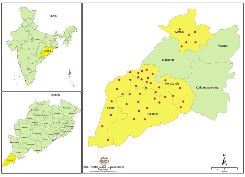
METHODS Entomological investigation was carried out in 48 villages from four mostly affected Community Health Centres (CHCs)
of Malkangiri district. Dusk collections of resting adults was done in villages from indoor and outdoor sites to record the density of
mosquito species, including the known JE vectors, feeding behaviour, parity, dusk index and infection status with JE virus (JEV).
FINDINGS The per man hour density and dusk index of JE vector species varied from 2.5 to 24.0 and 0.81 to 7.62, respectively in
study villages. A total of 1136 mosquitoes belonging to six vector species were subjected to PCR and one pool of Culex vishnui
was found to be positive for JEV.
CONCLUSION The JE transmission in Malkangiri district was confirmed. Thorough screening of human blood samples of JE/
AES suspected cases and JE vector mosquitoes for the presence of JEV during rainy season every year is recommended.
Key words: Japanese encephalitis - outbreak - entomological investigation - Malkangiri - Odisha - India
Japanese encephalitis (JE) is a common mosquito India only one outbreak of JE was reported from Ro-
borne flaviviral encephalitis and one of the leading urkela city of Sundergarh district in 1989 (Vajpayee et
forms of viral encephalitis covering a population of over al. 1991). Sporadic JE cases have been diagnosed from
three billion (Ghosh and Basu 2009). It is one of the most hospitalised children between 1992 and 1995 (Devi et al.
important forms of outbreak and sporadic encephalitis in 1996, Dash et al. 2001). Since then, there is no record of
the tropical regions of Asia, including Japan, China, Tai- JE infection in the state. Malkangiri district of Odisha
wan, Korea, Philippines, all of South eastern Asia, and state reported 9 deaths among children in 2009 due to
India (Solomon 1997). Japanese encephalitis virus (JEV) acute encephalitis syndrome (AES) (Nayak et al. 2016a).
is maintained in a zoonotic cycle, which can be both During September-November 2012, children with AES
enzootic and epizootic. This cycle involves pigs as the followed by 38 deaths were reported from Malkangiri
major reservoir/amplifying host, water birds as carriers district of Odisha by the state Health Department. In
and mosquitoes as vectors. The Culex vishnui subgroup 2014, eight deaths of children were reported due to AES
of mosquitoes consisting of Cx. tritaeniorhynchus, Cx. in seven villages of Korkunda and Kalimela Community
vishnui and Cx. pseudovishnui have been implicated as Health Centres (CHCs) of the district.
major vectors of JE in different parts of India (NVBDCP Malkangiri district has a long history of high falci-
2018). Outbreaks of JE are reported from many parts of parum malaria incidence (Anuse et al. 2015, Gunase-
India, and it is considered a major paediatric problem. karan et al. 2016). From 2010-2016, the annual parasite
The first recognition of JE based on serological surveys incidence (API, which is the number of clinical cases per
was in 1955, in Tamil Nadu, India (Namachivayam and 1000 people per year) in the district ranged from 18.5 to
Umayal 1982). A total of approximately 65 cases were 60.3 (Source: data from the Office of the Chief District
reported between 1955 and 1966 in Southern India (Car- Medical Officer, Koraput and Malkangiri). A severe out-
ey et al. 1968). Subsequent surveys carried out by the break of viral encephalitis was reported from 9th Sep-
National Institute of Virology of Pune indicated that tember to 2nd December 2016 in the Malkangiri district.
approximately half of the population in Southern India It was the most severe outbreak occurred in Odisha ever
has neutralising antibodies to the virus. Since 1955, since in the past; 336 children were affected with JE/
many major outbreaks in different parts of the country AES from 187 villages in seven CHCs. The outbreak
have been reported. From the state of Odisha in eastern first started with four cases of encephalitis admitted at
District Head Quarters Hospital (DHH), Malkangiri on
9th September 2016. Among the four admitted, two chil-
dren died on next day. A total of 579 children were ad-
mitted in the general fever ward of the DHH Malkangiri
doi: 10.1590/0074-02760170499
+ Corresponding author: sssahu1961@gmail.com
with the complaint of fever. Among them, 243 children
Received 16 November 2017 were cured and discharged next day of their admission.
Accepted 14 March 2018 Blood samples of the remaining 336 children were tested
online | memorias.ioc.fiocruz.br2|7 Outbreak investigation of JE in Odisha • Sudhansu Sekhar Sahu et al.
and among them 175 were found IgM positive for JE vi- Entomological investigation was carried out by In-
rus by ELISA. Out of the 103 deaths during the outbreak, dian Council of Medical Research-Vector Control Re-
37 deaths were due to JE and remaining 66 were due to search Centre (ICMR-VCRC), Puducherry during Sep-
AES. Among the 336 children suffered from either JE or tember 2016 to November 2016, covering 48 villages
AES, 233 children were cured and discharged from the selected randomly from four CHCs, viz., Korkunda,
hospital. For the first time, such a large number of deaths Mathili, Kalimela and Podia of the Malkangiri district
occurred due to JE/AES in Odisha state. (Fig. 1). The Korkunda CHC has a population of 143,867
In view of the severe outbreaks with life loss and on living in 29,667 households, Kalimela CHC with 127,819
the request of the Health department, Govt. of Odisha, population in 25,510 households, Mathili CHC with
an entomological investigation was carried out in the 103,046 population in 21,529 houses and Podia CHC
affected villages during the outbreak period to monitor with a population of 55,427 in 11,337 households. Aver-
the abundance of JE vectors, their infection status, risk age rainfall in the area in 2016 was 1471.75 mm and the
factors for transmission of JE and to suggest appropriate temperature ranged between 16.0 and 42.6ºC.
vector control intervention measures for implementation
Mosquito collections - For containment of the out-
by the state/district Health Department.
break, fogging (with 5% malathion and 5% cyphe-
MATERIALS AND METHODS nothrin) was initiated in some of the affected villages.
Children between the age group four months to 13 Therefore, the mosquito collections were done in the
years were affected in 187 villages in Malkangiri district. affected villages where fogging was not done and also
The common clinical presentations were fever, abdominal in villages where one round of fogging was completed.
pain, recurrent vomiting, lethargy, convulsion and loss of Density of mosquito species, including the known JE
consciousness. The main presenting features were sud- vectors and their blood meal source, parity, dusk index
den onset of convulsions followed by rapid progression and infection rate for JE virus were recorded.
to unconsciousness. Most of the affected children showed Dusk collections of resting adults indoors (six human
hypoglycaemia. No symptomatic adult case was admit- dwellings and three cattle sheds) spending five minutes in
ted during the outbreak period. Blood samples of the 336 each human dwelling and 10 min in each cattle shed and
children were tested by the IgM antibody capture ELISA outdoors (from sites such as bushes, plantations, stand-
(MAC ELISA) by DHH, Malkangiri. This technique was ing crops, root-intrices) spending 1 h in each village were
used for the detection of JE virus- specific IgM antibod- done using oral aspirators (hand catches); thereby spend-
ies using kits acquired from Indian Council of Medical ing a total of two man hours in each village. All the col-
Research-National Institute of Virology (ICMR-NIV), lected mosquitoes were identified to species level (Bar-
Pune, India. The kits were supplied by National Vector raud 1934). Per man hour density (PMHD) of known JE
Borne Disease Control Programme (NVBDCP), India. vector mosquitoes, i.e., number collected per man hour
For the first time, such a large number of deaths (n = 103) was calculated and recorded for each species. The known
occurred due to JE/AES in Odisha state. JE vector mosquitoes were dissected out and their ovari-
The seven affected CHCs in the district have 108 oles were examined for dilatations to determine the parity.
Gram Panchayats (GP) and 1,045 revenue villages nearly The proportion of parous was determined as the number
25% are inhabited by settlers and the remaining by tribes. of parous mosquitoes out of total dissected. The dusk in-
The district has a population of 641,385 (2014 census dex (DI) was estimated for measuring the density of rec-
conducted by health department) living in 109,483 house- ognised vectors of JEV, which incorporates proportion of
holds; population density was 106 inhabitants per km2. parous female mosquitoes with abundance. This index
The distance between CHC and different villages ranged was calculated by multiplying the PMHD and the propor-
from 1 to 35 km. Three seasons, summer (March-June), tion of parous females mosquitoes (Mani et al. 1991).
rainy (July-October) and cold (November-February), are Vector infection with JE virus - The unfed and fully
generally prevailing in the district. Malkangiri, lying gravid female mosquitoes, after dissection for parity,
just south of the Tropic of Cancer, has a tropical climate. was pooled and kept in sterile 1.5 mL eppendorf tube.
During the last five years, the district is warm almost A pool containing five mosquitoes (from same species
throughout the year with a maximum temperature of 42 with same gonotrophic condition or from same habitat or
to 47ºC in summer months. In winter, the temperature same type of collection or same village) was kept in each
is lower, ranging from 13 to 16ºC. The relative humidity tube and stored in liquid nitrogen. The mosquito samples
(RH) varies between 60 and 97% and generally higher in liquid nitrogen were transported to ICMR-VCRC lab-
during monsoon and post-monsoon months. The district oratory and stored at -80ºC for JE virus detection.
receives rainfall mostly from southwest monsoon during Ninety four pools containing 1136 mosquitoes were
July to October with an average rainfall of 1349.2 mm. subjected to real-time polymerase chain reaction (RT-PCR)
The actual rainfall recorded in the district from 2010 to for JE virus detection. A pool containing five mosquitoes
2015 was 1852.9 mm, 1103.9 mm, 1715.2 mm, 1783.3 was homogenised and RNA was extracted using TRI re-
mm, 1509.6 mm, and 1771.02, respectively. The month of agents method (Molecular Research Center, Inc. USA).
July is the wettest and the major rivers may get flooded. The whole mosquito tissue was used for RNA extraction
The district also receives a small rainfall from the retreat- by homogenisation. The extracted RNA from five pools
ing monsoon in the month of November. (Each PCR pool contains RNA from 25 mosquitoes) wasMem Inst Oswaldo Cruz, Rio de Janeiro, Vol. 113(6), 2018 3|7
Map showing study villages in Japanese encephalitis (JE) affected community health centres (CHCs) of Malkangiri district.
amplified using primers specific to capsid pre membrane the two surrounding wells. Gel plates were covered with
region (Liang 2007) of JE virus genome using transcriptor wet chamber petridish and allowed to stand overnight at
one step RT-PCR kit (Roche Applied Science, Penzberg, room temperature. Diffusion of the antigen and antisera
Germany). RNA from 25 mosquitoes in a pool for detec- took place through the agar so that the blood-meal ex-
tion of JE virus was standardised previously in ICMR- tract made contact with each of the antisera mid-way be-
VCRC laboratory. RT-PCR was performed along with tween wells. Homologous reactions left a white precipi-
negative and positive controls. After the amplification, 675 tate band in the agar gel. Tests were read and recorded
bp PCR amplicons were separated on 1.5% agarose gel and the following day. The proportion that fed on human and
results was documented using gel documentation system bovine was used to calculate human and bovine blood
(Gelstan Medicae, India). Based on the number of positive index (HBI and BBI) for each tested species.
pools for JE virus, infection rate was calculated.
Statistical analysis - Data were entered into Microsoft
Blood meal analysis - The mid gut of freshly fed JE Excel spreadsheet and statistical analyses were carried
vector mosquitoes obtained from resting collections was out using SPSS version 16.0. Pearson correlation test was
drawn and placed on a circle marked on Whatman No.1 done to find out if there was a linear association between
filter paper and squashed to make a round smear, and the mosquito diversity and JE incidence and between the
labelled. Each filter paper with mosquito blood meal PMHD of major JE vectors and JE incidence. For all sta-
smears was kept separately in a self-sealing cover and tistical tests, the level of significance was taken as p < 0.05.
stored at +4ºC until analysis was done for blood meal RESULTS
source using agar gel diffusion method (Crans 1969).
The reagents were from MP Biomedicals, (Solon, OH). Enzyme-linked immunosorbent assay (ELISA) test with
Before testing the blood meal, the reagents were tested human blood - Blood samples of the 336 children admitted
with positive controls to confirm the specificity of the in the DHH, Malkangiri were tested for the detection of JE
reagents. The mosquito blood-meal was extracted in virus-specific IgM antibodies and among them 175 were
0.5 mL saline. A portion of this unknown antigen was found IgM positive by ELISA. The test was performed by
placed in each of the two centre wells on a gel plate. Each the state Health Department. The JE prevalence among
mosquito blood meal was simultaneously tested for hu- the children admitted was found to be 52.1%. Out of the
man and bovine hosts by placing the antisera in each of 103 deaths during the outbreak, 37 deaths were due to JE4|7 Outbreak investigation of JE in Odisha • Sudhansu Sekhar Sahu et al.
and remaining 66 were due to AES. While the total (JE/ to 6.32 (Cx. whitmorei), respectively in the villages with
AES) case fatality rate in the current outbreak was 30.7%, fogging. The PMHD and dusk index of Cx. vishnui was
the case fatality rate due to JE was 21.1%. Among the 336 16.82 and 4.53, respectively (Table I). There was no cor-
children suffered from either JE or AES, 233 children were relation between the number of vector species and JE
cured and discharged from the hospital. incidences (r = -0.013, p = 0.926). Further, correlation
analysis indicated that the densities of Cx. vishnui (r =
Entomological evaluations - A total of 14 collections
-0.048, p = 0.735) and Cx. tritaeniorhynchus (r = -0.102,
were made spending 28 man-hours in 12 non-fogging vil-
p = 0.465) showed no correlation with JE incidences.
lages. In total, 2214 mosquitoes belonging to six known
JE vector species were collected. Among them, Cx. vish- JE virus detection - RT-PCR assay was performed
nui (30.4%) was predominant followed by Cx. whitmorei in 94 pools containing 1136 mosquitoes belonging to
(24.9%), Cx. tritaeniorhynchus (17.7%), Cx. bitaeniorhyn- the six vector species collected during the study period
chus (13.6%), Cx. gelidus (7.9%) and Cx. fuscocephalus and of which, one pool of Cx. vishnui was found to be
(5.6%). The PMHD of the JE vector species varied from positive for JE virus and the minimum infection rate was
4.4 to 24.0. The dusk index ranged from 1.39 (Cx. gelidus) found to be 0.88%.
to 7.62 (Cx. vishnui) among the vectors (Table I). Blood meal analysis - A total of 259 samples were
A total of 39 collections were made spending 78 analysed for the blood meal source and among them,
man-hours in 36 villages where one round of fogging two samples (HBI-0.008) were found to contain human
was completed and 4,564 culicine mosquitoes belong- blood and the remaining were bovine (BBI-0.992). The
ing to six species were collected. Out of the six vector species found positive for human blood were Cx. vishnui
species collected, the proportion of Cx. whitmorei, Cx. (HBI-0.014) and Cx. whitmorei (HBI- 0.021) (Table II).
vishnui, Cx. tritaeniorhynchus, Cx. bitaeniorhynchus,
Cx. fuscocephalus and Cx. gelidus was 34.5%, 28.7%, DISCUSSION
14.2%, 11.1%, 7.2% and 4.2%, respectively. The PMHD JE is a viral disease spread by mosquitoes to hu-
and dusk index of the six species varied from 2.5 (Cx. mans, from infected animals usually pigs and wading
gelidus) to 20.2 (Cx. whitmorei) and 0.81 (Cx. gelidus) birds. Areas with rice fields, where mosquitoes thrive
TABLE I
The per man hour density (PMHD), proportion parous (PP) and dusk index (DI)
of different Japanese encephalitis (JE) vectors in study villages
Non fogging Fogging
Sl. no Species Total collected PMHD PP DI Total collected PMHD PP DI
1 Culex bitaeniorhynchus 301 10.8 0.36 3.85 506 6.49 0.16 1.02
2 Cx. fuscocephalus 123 4.4 0.36 1.60 329 4.22 0.26 1.10
3 Cx. gelidus 175 6.3 0.22 1.39 193 2.47 0.33 0.81
4 Cx. tritaeniorhynchus 392 14.0 0.38 5.28 648 8.31 0.29 2.40
5 Cx. vishnui 672 24.0 0.32 7.62 1312 16.82 0.27 4.53
6 Cx. whitmorei 551 19.7 0.30 5.82 1576 20.21 0.31 6.32
TABLE II
Human blood index (HBI) of Japanese encephalitis (JE) vector mosquitoes collected from
JE/acute encephalitis syndrome (AES) outbreak villages
Sl. no Species Total tested Number positive to human HBI
1 Culex bitaeniorhynchus 58 0 0
2 Cx. fuscocephalus 11 0 0
3 Cx. gelidus 15 0 0
4 Cx. tritaeniorhynchus 57 0 0
5 Cx. vishnui 70 1 0.014
6 Cx. whitmorei 48 1 0.021
Total 259 2 0.008Mem Inst Oswaldo Cruz, Rio de Janeiro, Vol. 113(6), 2018 5|7
and where there is a lot of pig farming, are especially tions before and after fogging, man hour spent etc. could
risky (Bhowmik et al. 2012). JE has been known in India not be planned beforehand. Since, this was an outbreak
since 1952 (Work and Shah 1956). A total of 63 deaths condition, fogging was done profusely in affected vil-
occurred in Malkangiri from four outbreaks (JE/AES) lages. Hence, same villages could not be monitored for
happened during 2009 to 2014 (Nayak et al. 2016a). All change in density before and after one round of fogging.
the victims were children. But the recent outbreak was During earlier outbreaks occurred in Malkangiri dis-
reported to be the most severe one occurred in the state trict, the association of JE was confirmed as JE virus
till the end of 2016. The initial stages of the current out- IgM from serological samples and JE virus RNA from
break evidence pointed to JE because all the cases oc- cerebrospinal fluid (CSF) samples was detected (Dwibe-
curred during monsoon. JE is a disease principally of di et al. 2015, Nayak et al. 2016a) and was lacking the
agricultural areas, particularly in rice cultivation areas, supporting entomological confirmations from viral isola-
where vector mosquitoes proliferate in close association tion of JE vectors. In the current outbreak, the presence
with pigs, wading birds and ducks. The current study of JEV was confirmed in vector mosquitoes by PCR as-
was conducted in rainy and post rainy seasons. Paddy say. Further, Cx. vishnui was found to have fed on human
fields are the favourable breeding places during rainy blood and virus detection was confirmed from the same
seasons. Cx. vishnui subgroup of mosquitoes breed in species. Hence, Cx. vishnui could be involved in JE trans-
water with luxuriant vegetation, mainly in paddy fields mission in the current outbreak affected area. Earlier dif-
and their abundance may be related to their breeding in ferent studies showed that maximum detection of JEV
rice fields (Medhi et al. 2017). In the current study, all was obtained from the Culex vishnui subgroup (Chakra-
the JE/AES affected villages were surrounded by paddy varty et al. 1975, Banerjee et al. 1978, Mourya et al. 1989,
fields thereby, providing favourable breeding places for Dhanda et al. 1997, Gajanana et al. 1997, Mariappan et
the vector mosquitoes. However, persistence of a high al. 2014) and this subgroup mosquitoes were also shown
number of cases with similar symptoms for a prolonged to be capable of transmitting the JE virus in the labora-
period might be due to the existence of another causative tory (Soman et al. 1977). The current observations on the
agent. Majority of cases showed symptoms of vomiting, presence of JEV in Cx. vishnui is also in line with the ear-
dizziness and loss of appetite when they were admitted lier findings observed in Mayurbhanj district of Odisha
to the hospital. This led the doctors to suspect that they state (Nayak et al. 2016b). The evidence gathered such
were suffering from JE/AES, which is characterised by as availability of paddy fields, abundance of JE vectors,
inflammation of brain leading to death in some cases.
their dusk index, HBI and infection status in the current
The present outbreak of JE/AES persisted for three
investigation pointed towards JE virus aetiology.
months. The human (175 JEV positive out of 336 sam-
Based on the findings of this investigation, appro-
ples tested) and pig (18 JEV positive out of 239 samples
priate vector control measures were recommended to
tested) (Data source: Chief District Veterinary Office,
local general/health administration and they mounted a
Malkangiri) serological investigation confirmed the oc-
well-funded campaign to control the outbreak. Intense
currence of JEV infection in the Malkangiri district.
The current outbreak of viral encephalitis extended to vector control interventions such as fogging indoors and
Sukuma district (neighbouring district of Malkangiri) of outdoors with 5% malathion and 5% cyphenothrin, lar-
Chhattisgarh state where five AES cases were admitted vicidal (Bti) spray in temporary breeding habitats found
in DHH of Malkangiri and three of them died. Another inside the villages and LLINs/non-impregnated nets
neighbouring district, i.e., Koraput of Odisha, where 62 were distributed in the affected villages. Five rounds of
AES cases were reported during the same period and fogging were carried out in the affected villages up to
among them, 29 were found positive for JE virus on test- the mid December 2016. A total of 6200 odomos mos-
ing their serum samples. Among the 62 cases admitted quito repellent creams were distributed by the female
in Koraput DHH, four died due to JE and 10 due to AES. health workers to the children of the both affected and
Entomological investigations revealed the presence nearby affected villages to apply on their body during
of six vector species of JE in the study villages, includ- evening time for preventing from mosquito bites. As
ing the two major vector species viz., Cx. vishnui and Cx. JE vector mosquitoes are transmitting the disease from
tritaeniorhynchus, which constituted 44.6% of the total infected pigs to humans, measures were taken by the
JE vector mosquitoes. The PMHD and the DI of these two government and community to keep the pigs in pig pens
vector species were relatively higher in the affected vil- constructed in the isolated places 2.5 km away from the
lages where no fogging was done. Since, the same villages villages. In addition, the people in affected villages sold
could not be monitored for change in density before and nearly 15,000 pigs to neighbouring districts as a means
after one round of fogging, the differences in mosquito of eliminating the source of the virus. Bush cutting in
occurrences might not be related to fogging. The detec- the affected villages was carried out intensively. All the
tion of JEV in Cx. vishnui has important implications for low lying area where water accumulation occurred was
public health and for JE control agenda. This is the first filled with sand and soil. JE vaccines have been admin-
demonstration of detection of JEV from outbreak affected istered within a week after control of outbreak (second
villages of Malkangiri district in Odisha state. week of December 2016) to all the children (221,155) be-
The limitation of the study was that, the present study low 15 years for preventing the outbreak in future. An
being an entomological outbreak investigation of JE/AES, active programme of health education and social mobili-
the selection of study villages for entomological collec- sation has been mounted in affected areas.6|7 Outbreak investigation of JE in Odisha • Sudhansu Sekhar Sahu et al.
The geographic features of this district support the Carey DE, Myers RM, Pavri KM. Japanese encephalitis studies in
spread of JEV. Paddy fields of the affected villages are Vellore, South India. II. Antibody response of patients. Indian J
Med Res. 1968; 56(9): 1319-29.
located within 0.3 to 0.5 km from the human habitations.
During 2016, the district received 1471.75 mm rainfall Chakravarty SK, Sarkar JK, Chakravarty MS, Mukherjee MK, Mukher-
from July to the end of October. Water accumulations jee KK. The first epidemic of Japanese encephalitis studied in India-
in the paddy fields, which are good breeding grounds virological studied. Indian J Med Res. 1975; 63(1): 77-82.
for the JE vector mosquitoes, were more during the out- Crans WJ. An agar-gel diffusion method for identification of mos-
break period indicating a possible association between quito blood meal. Mosq News. 1969; 29(4): 563-6.
rice cultivation season and JE transmission. Mostly trib- Dash AP, Chhotray GP, Mahapatra N, Hazra RK. Retrospective anal-
al villages of the district rear the pigs as it is a source ysis of epidemiological investigation of Japanese encephalitis
of economy for them. In addition, high temperature and outbreak occurred in Rourkela, Orissa, India. Southeast Asian J
relative humidity prevailed in the district provided a suit- Trop Med Public Health. 2001; 32(1): 137-9.
able environment for JEV transmission. The district is Devi PS, Behera PL, Swain A. Japanese encephalitis in Orissa. Indian
also endemic for falciparum malaria since many decades Pediatr. 1996; 33(8): 702-3.
(Gunasekaran et al. 2016). As the rice agro eco-system
supports the breeding of the vectors of malaria (Sahu et Dhanda V, Thenmozhi V, Kumar NP, Hiriyan J, Arunachalam N, Bal-
asubramaniam A, et al. Virus isolation from wild-caught mosqui-
al. 2017) and JE (Dwibedi et al. 2015), integrated vec- toes during a Japanese encephalitis outbreak in Kerala in 1996.
tor management (IVM) is recommended to prevent the Indian J Med Res. 1997; 106(1): 4-6.
transmission of both the vector-borne diseases.
In summary, we confirmed the outbreak of JEV infec- Dwibedi B, Mohapatra N, Rathorea SK, Panda M, Patia SS, Sabata J,
et al. An outbreak of Japanese encephalitis after two decades in
tion during 2016 in Malkangiri district, as JE virus was Odisha, India. Indian J Med Res. 2015; 142(Suppl.): 30-2.
detected from Cx. vishnui collected from the outbreak
affected villages. The environmental conditions, vector Gajanana A, Rajendran R, Samuel PP, Thenmozhi V, Tsai TF, Kimu-
abundance, dusk index and HBI of JE vectors supported ra-Kuroda J, et al. Japanese encephalitis in South Arcot District,
Tamil Nadu: a three-year longitudinal study of vector abundance
the occurrence of JE transmission in the district. Based and infection frequency. J Med Entomol. 1997; 34(6): 651-9.
on these findings, the appropriate vector control mea-
sures were recommended and the district administration Ghosh D, Basu A. Japanese encephalitis-a pathological and clinical
implemented intense vector control activities as per the perspective. PLoS Negl Trop Dis. 2009; 3(9): e437.
recommendations. The district has environmental risk for Gunasekaran K, Sahu SS, Vijayakumar T, Subramanian S, Yadav RS,
acquiring JE infection. This report of occurrence of JE Pigeon O, et al. An experimental hut evaluation of Olyset Plus, a
in Malkangiri district points towards the need for public long lasting insecticidal net treated with a mixture of permethrin
health vigilance and thorough screening of JE vectors and piperonyl butoxide, against Anopheles fluviatilis in Odisha
State, India. Malar J. 2016; 15: 375.
regularly to prevent morbidity and mortality in future.
Liang GD. Molecular epidemiological analysis of JEV in China. J
ACKNOWLEDGEMENTS Gen Virol. 2007; 88(Pt3): 885-94.
To the Directorate of Health Services, Govt of Odisha, Mani TR, Rao CVRM, Rajendran R, Devaputra M, Prasanna Y, Ha-
Bhubaneswar and District Health Department, Malkangiri for numaiah, et al. Surveillance for Japanese encephalitis in villages
their cooperation during the investigation. We acknowledge near Madurai, Tamil Nadu, India. Trans R Soc Trop Med Hyg.
the staff members of the ICMR-VCRC, Field Station, Koraput, 1991; 85(2): 287-91.
for their technical assistance in laboratory and field works. Mariappan T, Samuel PP, Thenmozhi V, Paramasivan R, Sharma PK,
AUTHORS’ CONTRIBUTION Biswas AK, et al. Entomological investigations into an epidemic
of Japanese encephalitis (JE) in northern Districts of West Ben-
SSS, KG and PJ designed the study and drafted the manu- gal, India (2011-2012). Indian J Med Res. 2014; 139(5): 754-61.
script; SSS, SD, TS conducted the study in the field and ana-
Medhi M, Saikia L, Patgiri SJ, Lahkar V, Hussain E, Kakati S. Incidence
lysed the data; molecular analysis was performed by SM and of Japanese encephalitis amongst acute encephalitis syndrome cases
TS. All authors read and approved the final manuscript. The in upper Assam districts from 2012 to 2014: a report from a tertiary
authors declare that they have no competing interests. care hospital. Indian J Med Res. 2017; 146(2): 267-71.
REFERENCES Mourya DT, Ilkal MA, Mishra AC, Jacob PG, Pant U, Ramanujam S,
Anuse SS, Sahu SS, Subramanian S, Gunasekaran K. Usage pattern, et al. Isolation of Japanese encephalitis virus from mosquitoes
physical integrity & insecticidal efficacy of long-lasting insecticidal collected in Karnataka state, India from 1985 to 1987. Trans R
nets in Odisha State, India. Indian J Med Res. 2015; 142(1): 109-16. Soc Trop Med Hyg. 1989; 83(4): 550-2.
Banerjee K, Deshmukh PK, Ilkal MA, Dhanda V. Transmission of Namachivayam V, Umayal K. Proceedings of the National Confer-
Japanese encephalitis virus by Culex bitaeniorhynchus Giles. In- ence on Japanese Encephalitis. Indian Council of Medical Re-
dian J Med Res. 1978; 67(6): 889-93. search: New Delhi; 1982. p. 30-3.
Barraud PJ. The fauna of British India including Ceylon and Burma. Nayak P, Papann M, Shrivastava A, Khasnobis P, Lokhande G, Kumar
Diptera. Vol. 5. Family Culieldae. Tribes Megarhinini and Culi- A, et al. Unexplained neurological illness in children, Malkangiri
cini. London: Taylor and Francis; 1934. 463 pp. District, Odisha, India 2014. Int J Infect Dis. 2016a; 45(1): 305.
Bhowmik D, Duraivel S, Jaiswal J, Tripathi KK, Sampath Kumar Nayak P, Pradhan A, Mallick R, Sethi S, Patnaik B, Pradhan MM,
KP. Japanese encephalitis epidemic in India. Pharma Innovation. et al. Japanese encephalitis outbreak among children in Mayurb-
2012; 1(10): 47-54. hanj, Odisha, India, 2015. Int J Infect Dis. 2016b; 53(Suppl.): 61-2.Mem Inst Oswaldo Cruz, Rio de Janeiro, Vol. 113(6), 2018 7|7
NVBDCP - National Vector Borne Disease Control Programme. Soman RS, Rodrigues FM, Guttikar SN, Guru PY. Experimental vi-
Japanese encephalitis vectors in India. National Vector Borne raemia and transmission of Japanese encephalitis virus by mos-
Disease Control Programme: New Delhi; 2018. Available from: quitoes in ardeid birds. Indian J Med Res. 1977; 66(5): 709-18.
http://nvbdcp.gov.in/je4.html.
Vajpayee A, Mukherjee MK, Chakraborty AK, Chakraborty MS. In-
Sahu SS, Gunasekaran K, Krishnamoorthy N, Jambulingam P. Bion- vestigation of an outbreak of Japanese encephalitis in Rourkela
omics of Anopheles fluviatilis and An. culicifacies in relation to city (Orissa) during 1989. J Commun Dis. 1991; 23(1): 18-21.
transmission of malaria and its control in East-Central India. J
Med Entomol. 2017; 54(4): 821-30. Work TH, Shah KV. Serological diagnosis of Japanese B type of en-
cephalitis in North Arcot District of Madras state, India, with
Solomon T. Viral encephalitis in southeast Asia. Neuro Infect Epide- epidemiological notes. Indian J Med Sci. 1956; 10(8): 582-92.
miol. 1997; 2: 191-9.You can also read