Case Report Spontaneous expulsion of a giant colonic lipoma: a case report and literature review
←
→
Page content transcription
If your browser does not render page correctly, please read the page content below
Int J Clin Exp Med 2018;11(6):6249-6254
www.ijcem.com /ISSN:1940-5901/IJCEM0066379

Case Report
Spontaneous expulsion of a giant colonic lipoma: a case
report and literature review
Liang-Liang Ma1*, Xi-Yi Chen1*, Dong-Dong Huang2, Jun-Cheng1, Zuo-Qian Yu1, Di-Di Chen1, Zhen Yu1,2,
Xiao-Dong Zhang1
1
 Department of Gastrointestinal Surgery, The First Affiliated Hospital, Wenzhou Medical University, Wenzhou
325000, Zhejiang, China; 2Department of Gastrointestinal Surgery, Shanghai Tenth People’s Hospital, Affiliated to
Tongji University, Shanghai, China. *Equal contributors.
Received September 28, 2017; Accepted February 8, 2018; Epub June 15, 2018; Published June 30, 2018

Abstract: The majority of colonic lipomas are asymptomatic and do not require any treatment. However, lipomas that
are larger than 2 cm may cause symptoms such as bleeding, intussusceptions, or obstruction. A rather uncommon
symptom is adult intussusceptions, caused by intestinal lipoma. Spontaneous expulsion of lipoma in the gastroin-
testinal tract along with the related pathological changes is, undoubtedly, an unusual symptom. However, the pre-
cise underlying mechanism has not been clearly defined. It presumptively happens due to ischemic necrosis of the
lipomas by peristalsis-lead tension or torsion. In this paper, we describe a case of spontaneous expulsion of a large
colonic lipoma that became symptomatic due to intussusception from the rectum without any intestinal perforation.
In order to get a better understanding of its incidence, expulsion mechanism, diagnosis, therapy, and prognosis we
further reviewed 13 cases of spontaneously expelled colonic lipoma, reported worldwide.

Keywords: Case report, lipoma, spontaneous expulsion, colon

Introduction                                                   Case report

Lipomas are usually non-epithelial benign fat                  A 46-year-old Chinese patient was admitted to
tumors which may be located all over the gas-                  the Gastrointestinal Surgery department of The
trointestinal tract although they are most com-                First Affiliated Hospital of Wenzhou Medical
monly diagnosed in the colon [1]. In general,                  University on June 8, 2017, with a complaint of
symptoms are related to lipoma size. Small                     five-day old symptoms of abdominal pain and
colonic lipomas display very few symptoms and                  melena with changes in bowel habits, including
are discovered accidentally during autopsy,                    diarrhea. He had a long history of Hepatitis B.
colonoscopy, or surgery. While lipomas more                    Four years ago, he had undergone surgery for
than 2 cm in size have symptoms among 75%                      thyroid cancer. Vital signs were normal on
of the diagnosed patients [2], lesions larger                  admission. He had a height of 170 cm and
than 4 cm may present with pain in the abdo-                   weighed 67 kg. He was a non-smoker and had
minal region, alterations in defecation routine,               not been taking any medication. On physical
bleeding, intussusception, or defecation block-                examination, the abdomen was flat but a little
ing [3]. Spontaneous expulsion of lipomas in                   tight. Neither the liver nor the spleen could be
the gastrointestinal tract is an extremely un-                 palpated and no other abdominal mass was
usual symptom. In addition, the precise under-                 palpable. No swelling of the superficial lymph
lying mechanism of lipoma formation is still not               nodes was observed.
clear. We herein describe a case of spontane-
ous expulsion of huge colonic lipoma in a                      Laboratory testing revealed that blood routine
46-year-old patient along with a review of litera-             examination, blood biochemistry, and carcino-
ture pertaining to this condition. This report                 embryonic antigen levels were within normal
could contribute towards the global under-                     range. Microbiological stool examinations yield-
standing of the diagnosis and prognosis of                     ed red blood cells. An abdominal enhanced
colonic lipoma.                                                computed tomography (CT) scan showed a
Spontaneous expulsion of colonic lipoma
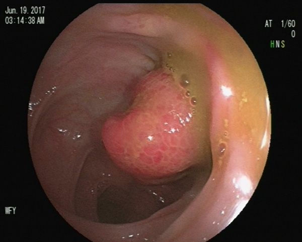
Figure 1. Colonoscopy reveals a great errabund Figure 3. The expelled mass has a smooth surface,
mass removed from the colonic wall; hemorrhagic the color is light brown with a kidney-like shape.
mucosa seen with the mucosal erosion.
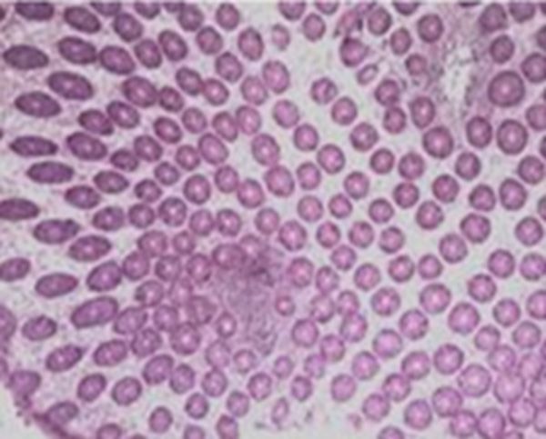
Figure 4. Microscopic image of the expelled mass
Figure 2. Microscopic image of the biopsy from colo-
reveals mature and natural adipocytes (H&E stain,
noscopy shows colonic hyperplastic polyp with ero-
×100).
sion (H&E stain, ×100).
large endo-luminal lesion around 3.6 cm in color, had a smooth surface, and measured
size, left of descending colon level with colonic 50×40 mm. The appearance was similar to
intussusception. The lesion was morphologi- lipoma. The mass was sent for histopathologi-
cally smooth, which was highly suggestive of a cal examinations and the confirmed histopath-
lipoma. Colonoscopy showed a transferable ological diagnosis was submucosal lipoma
mass of 5 cm in diameter about 45 cm from the (Figure 4). A new colonoscopy revealed a mass
anus, within the mucosal erosion, and covered of diameter of 2.5 cm, about 45 cm from the
with hemorrhagic mucosa (Figure 1). The biop- anus and covered with hemorrhagic mucosa
sy obtained from colonoscopy demonstrated a (Figure 5). Biopsy from the new colonoscopy
colonic hyperplastic polyp with erosion (Figure confirmed colonic hyperplastic polyp with ero-
2). sion (Figure 6).
For a definitive diagnosis, laparoscopic surgery Following tumor expulsion, the symptoms even-
was scheduled for June 13. However, during tually disappeared, the intussusception resol-
preoperative preparation, administration of a ved spontaneously and the patient remained
strong oral laxative resulted in expulsion of a asymptomatic. No laparoscopic surgery was
mass from the rectum on defecation. The performed and the patient was discharged
expelled mass (Figure 3) was light brown in from hospital after 48 hours. The patient
6250 Int J Clin Exp Med 2018;11(6):6249-6254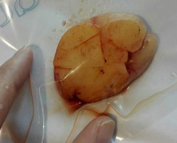
Spontaneous expulsion of colonic lipoma

                                                           Colonic lipomas grow out of the submucosa in
                                                           about 90% of cases with occasional extensions
                                                           into the muscular propria [6]. Chronic irritation,
                                                           inflammation, and fatty tissue accumulating in
                                                           particular areas are the probable causes for
                                                           formation of colonic lipomas [7, 8]. Occasionally,
                                                           the colonic lipoma could separate from its root
                                                           and eventually be expelled from the rectum.
                                                           Spontaneous expulsion of colonic lipomas from
                                                           the rectum have been rarely reported [9-15]. In
                                                           addition, the spontaneous lipoma expulsion
                                                           reported by Kang et al. [3] and Kouritas et al.
                                                           [11] also occurred in the small intestine.

                                                           The mechanism behind this rare spontaneous
Figure 5. The new colonoscopy reveals a mass nar-
rower than earlier with a diameter of 2.5 cm; it is cov-   expulsion of colonic lipomas is still unclear but
ered by hemorrhagic mucosa.                                several factors have been considered relevant.
                                                           First, spontaneous expulsion mainly occurs in
                                                           cases such as giant lipomas stalked with a nar-
                                                           row pedicel [11]. Besides, tension or torsion
                                                           may lead to twisted lipomas, resulting in isch-
                                                           emic necrosis and amputation [3]. Pedunculat-
                                                           ed lipomas, in particular, can be distorted eas-
                                                           ily and are vulnerable to mechanical stress [9].
                                                           Second, when the mucosa ulcer above the lipo-
                                                           ma reaches its maximum diameter, the lower
                                                           covered mass bulges and ruptures into the cav-
                                                           ity, which may be an explanation for this rare
                                                           phenomenon [11]. Another possible reason for
                                                           the lipoma resection could be former endo-
                                                           scopic removal [14, 16, 17] or lipoma intussus-
                                                           ception [3, 18]. In this case, the patient was
                                                           diagnosed with intussusception and the lipoma
Figure 6. Microscopic image of the biopsy from a new       may have developed torsion during bowel peri-
colonoscopy shows colonic hyperplastic polyp with          stalsis. Also, presence of severe diarrhea dur-
erosion (H&E stain, ×100).                                 ing preoperative bowel preparation could have
                                                           contributed to this process. Colonoscopy re-
agreed to receive an endoscope polypectomy                 vealed a diagnosed ploy tissue of 2.5 cm in
after three months.                                        diameter covered with hemorrhagic mucosa.
                                                           Therefore, we speculate that the huge pedun-
Discussion                                                 culated lipoma could have undergone ischemic
                                                           necrosis due to possible peristalsis torsion fol-
Lipomas are soft tissue tumors evolving from               lowed by autoamputation of this lipoma.
the hyperplasia of mature fat cells. Although
the known morbidity of colonic lipoma is                   Most colonic lipomas are symptomless and are
between 0.035% and 4.4%, it is the second                  diagnosed accidentally during surgery or colo-
most frequent benign tumor of the large intes-             noscopy for other conditions [4]. The symptoms
tine [4]. Colonic lipomas are most commonly                are usually correlated with lipoma size, espe-
located within the ascending colon followed by             cially when they increase to more than 2 cm.
the descending colon, the transverse colon,                Pain in the abdominal region, alterations in def-
and rarely the rectum [2, 5]. We, here, described          ecation manner, and gastrointestinal hemor-
a case of spontaneous expulsion of a huge                  rhage are typically reported [4, 9]. The major
colonic lipoma along with a review of 13 global            symptom in the majority of the spontaneous
cases of spontaneously expelled colonic lipo-              lipoma expulsion cases is acute abdominal
ma (Table 1).                                              pain, usually flatulent in nature, and subse-

6251                                                            Int J Clin Exp Med 2018;11(6):6249-6254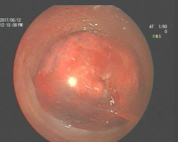
Spontaneous expulsion of colonic lipoma

Table 1. Characteristics of reported cases of spontaneous expulsion of colonic lipoma
                                                                           Diagnostic         Tumor
Authors                   Age   Sex Presentation                                                             Size (cm)      Treatment         Follow-up
                                                                            modality         location
Manheim et al. [23]       47    F   Abdominal pain hematochezia diarrhea Barium enema     Sigmoideum           6×4             No               Well
Robertson et al. [19]     43    M   Abdominal pain hematochezia diarrhea Colonoscopy      Sigmoideum           7×5             No               Well
Misra et al. [18]         32    M   Abdominal pain                       Barium enema   Ascending colon        8×5             No             Unknown
Stebbings et al. [24]     55    F   Abdominal pain hematochezia          Barium enema   Descending colon       5×7             No               Well
Zhou et al. [15]          28    M   Abdominal pain hematochezia           Colonoscopy   Ascending colon      7.2×5.5           No             Unknown
Tzias et al. [13]         53    M   Abdominal pain hematochezia           Colonoscopy     Sigmoideum            6    Endoscopic polypectomy   Unknown
Sidani et al. [17]        45    M   Abdominal pain constipation           Colonoscopy   Right-side colonic    9×10      Alligator forceps       Well
Lazaraki et al. [14]      78    M   Abdominal pain                        Colonoscopy     Sigmoideum           4×3   Endoscopic polypectomy     Well
Jeong et al. [16]         48    M   Abdominal pain                        Colonoscopy     Sigmoideum            8      Snare polypectomy      Unknown
Kouritas et al. [11]      77    F   Abdominal pain melena                 Colonoscopy       Unknown          Unknown           No             Unknown
Ishiyama et al. [12]      38    M   Abdominal pain melena                 Colonoscopy   Transverse colon     7.5×4.5     Electric snare         Well
                                                                         Barium enema
Chahri et al. [10]        45    M   Abdominal pain diarrhea                    CT       Descending colon       7×5             No             Unknown
Rocha et al. [9]          62    M   Abdominal pain constipation                CT       Transverse colon      7.5×5            No             Unknown
Present case              46    M   Abdominal pain hematochezia           Colonoscopy   Descending colon      5×4.5            No               Well
                                                                               CT
CT-Computed tomography.

6252                                                                                                          Int J Clin Exp Med 2018;11(6):6249-6254
Spontaneous expulsion of colonic lipoma
quent few hematochezia [9, 11, 13, 16] that recommendation in doubtful cases is still
lessens after mass defecation. However, few removal of the tumor [1, 22]. Therefore, the
patients experience heavy bleeding and perfo- resection of symptomatic or greater than 2 cm
rations on spontaneous expulsion of lipoma in diameter colonic lipomas is mandatory but if
[19]. Besides, patients with giant lipoma (> 4 the diameter is lower than 2 cm tumor removal
cm) may develop impeding intestinal obstruc- should be reserved only in cases with doubtful
tion because the separated lipoma could block diagnosis [7].
the colorectal tract and thus impede the stool
channel [1, 20]. The patient in our case com- According to the literature [9, 10, 13, 16], most
plained of abdominal distention, acute abdo- symptoms in these cases subside after sponta-
men pain, and melena. However, those symp- neous lipoma expulsion and additional treat-
toms resolved spontaneously after self-ampu- ments are avoidable. Few self-mutilating lipo-
tation of the lipoma. mas are so huge that spontaneous expulsion
becomes impossible. However, the huge mov-
Currently, preoperative diagnosis of colonic able masses can be removed in fragments with
lipoma can be challenging. Colonoscopy is an extensive application of polypectomy snare [16,
important means for diagnosis of colonic lipo- 17]. Robertson et al. [19] reported a case of a
ma [11]. Raising of the mucous membrane 43 year old male patient developing perforation
against the mass with a surgical clamp (“tent of the transverse colon wall subsequent to the
sign”), breach of lipoma with a surgical clamp patient passing a huge tumor, confirmed to be
(the so-called “cushion sign”), or fat expulsion a lipoma. The patient received an extended
after biopsy (“naked fat sign”) [12, 13] are right hemicolectomy and ileocolic anastomosis
methods used due to which lipoma surface and made an uneventful recovery. Recurrence
inflammation, erosion, and ulcer are typical has not been documented, so far [11].
signs of lipoma often lost on colonoscopy. It is
difficult to comprehend the characteristics of In our case, the patient received positive hemo-
adipose tissue because of repeated erosion static treatment after the lipoma discharged
repair and thickening of the surface of the spontaneously. The second colonoscopy re-
mucosa. In addition, Daniele et al. [7] have vealed a narrower mass of a diameter of 2.5
cm, confirmed to be a tissue of polyps, and the
shown that the positive rate of diagnosis using
patient agreed to receive an endoscopic polyp-
diagnostic colonoscopy was 44%. In our case,
ectomy after three months.
the first-time biopsy on colonoscopy shows
colonic hyperplastic polyp with erosion. The fol- Conclusion
lowing biopsy obtained from the spontaneous
tissue expulsion showed normal mature adipo- In spite of the fact that colonic lipomas have
cytes. Therefore, application of barium exami- unusual symptoms, they should be taken into
nation, CT, MRI, and ultrasound examination of consideration for differential diagnosis of
colonic lipoma are also important for early and tumors of the large intestine. Surgical resection
accurate diagnosis [1, 7]. may be a feasible and safe therapy for most
symptomatic lipomas. Occasionally, the colonic
The treatment of colonic lipomas is related to lipoma may separate from its root and be
the size and location of the lipoma, its possible expelled from the rectum; most of these have a
neopathy, possible malignant grade, as well as favorable prognosis. However, we recommend
the surgeon’s decision on surgical or endoscop- that surgeons perform re-colonoscopy inspec-
ic intervention [21]. Endoscopic with snare tion after expulsion and strengthen their aware-
electrocautery removal of colonic lipomas is ness of hemorrhagic focal ulcerations and
recommended for lipomas less than 2 centime- presence of residual tissue. These steps would
ters [7] while surgical removal appears to be help in the timely detection and intervention of
the ideal remedy for large lipomas, particularly hemorrhages and perforations and aid in regu-
when tumors could not be preoperatively lar follow up.
removed [4, 7, 22]. Several research studies
have suggested that it is difficult to differenti- Disclosure of conflict of interest
ate colon lipoma from colon carcinoma. The
risk of misdiagnosis exists and, therefore, the None.
6253 Int J Clin Exp Med 2018;11(6):6249-6254Spontaneous expulsion of colonic lipoma
Address correspondence to: Dr. Xiao-Dong Zhang, [12] Ishiyama S, Tashiro Y, Nagayasu K, Niwa K,
Department of Gastrointestinal Surgery, The First Ono S, Sugimoto K, Hata M, Kamiyama H,
Affiliated Hospital of Wenzhou Medical University, Komiyama H, Takahashi M, Yaginuma Y,
Wenzhou 325000, Zhejiang, China. Tel: 86+1377- Kojima Y, Goto M, Tanaka M, Sengoku H,
7763007, E-mail: amostory@126.com Okuzawa A, Tomiki Y and Sakamoto K.
Spontaneous disappearance of a giant colonic
References lipoma after endoscopic biopsy. Endoscopy
2011; 43 Suppl 2 UCTN: E16.
[1] Nallamothu G and Adler DG. Large colonic [13] Tzias V, Koutsoumpas A and Kamberoglou D.
lipomas. Gastroenterol Hepatol (N Y) 2011; 7: Endoscopic resection of giant colonic lipomas.
490-492. Annals of Gastroenterology 2009; 21: 205.
[2] Kose E, Cipe G, Demirgan S and Oguz S. Giant [14] Lazaraki G, Tzilves D, Tragiannidis D, Pata-
colonic lipoma with prolapse through the rec- kiouta F, Pilpilidis I, Gatopoulou A, Soufleris K
tum treated by external local excision: a case and Katsos I. “Giant” lipoma of the sigmoid
report. Oncol Lett 2014; 8: 1377-1379. colon: spontaneous expulsion 12 days after
[3] Kang B, Zhang Q, Shang D, Ni Q, Muhammad failure of endoscopic resection. Report of a
F, Hou L and Cui W. Resolution of intussuscep- case and review of the literature. Annals of
tion after spontaneous expulsion of an ileal li- Gastroenterology 2008.
poma per rectum: a case report and literature [15] Zhou BF, Lu ZG and Li L. Spontaneous expul-
review. World J Surg Oncol 2014; 12: 143. sion of a colonic lipoma. Chin J Dig 2001; 21:
[4] Ghanem OM, Slater J, Singh P, Heitmiller RF 598.
and DiRocco JD. Pedunculated colonic lipoma [16] Jeong HK, Cho SB, Seo TJ, Lee KR, Lee WS,
prolapsing through the anus. World J Clin Kim HS and Joo YE. Autoamputation of a giant
Cases 2015; 3: 457-461. colonic lipoma. Gut Liver 2011; 5: 380-382.
[5] Andrei LS, Andrei AC, Usurelu DL, Puscasu LI, [17] Sidani SM, Tawil AN and Sidani MS. Extraction
Dima C, Preda E, Lupescu I, Herlea V and of a large self-amputated colonic lipoma: a
Popescu I. Rare cause of intestinal obstruc- case report. Int J Surg 2008; 6: 409-411.
tion-submucous lipoma of the sigmoid colon. [18] Misra SP, Singh SK, Thorat VK, Gulati P,
Chirurgia (Bucur) 2014; 109: 142-147. Malhotra V and Anand BS. Spontaneous expul-
[6] Boler DE, Baca B and Uras C. Laparoscopic re- sion per rectum of an ileal lipoma. Postgrad
section of colonic lipomas: when and why? Am Med J 1988; 64: 718-719.
J Case Rep 2013; 14: 270-275. [19] Robertson DA, Saweirs W and Low-Beer TS.
[7] Crocetti D, Sapienza P, Sterpetti AV, Paliotta A, Spontaneous expulsion of a large colonic tu-
A DEG, Pedulla G and de Toma G. Surgery for mor. Br Med J (Clin Res Ed) 1982; 285: 1084.
symptomatic colon lipoma: a systematic re- [20] Bahadursingh AM, Robbins PL and Longo WE.
view of the literature. Anticancer Res 2014; Giant submucosal sigmoid colon lipoma. Am J
34: 6271-6276. Surg 2003; 186: 81-82.
[8] Tsiaousidou A, Chatzitheoklitos E, Hatzis I, [21] Mouaqit O, Hasnai H, Chbani L, Oussaden A,
Alatsakis M and Katsourakis A. Giant transmu- Maazaz K, Amarti A and Taleb KA. Pedunculated
ral lipoma of the sigmoid colon. Hippokratia lipoma causing colo-colonic intussusception: a
2012; 16: 278-279. rare case report. BMC Surg 2013; 13: 51.
[9] Rocha M, Pereira S and Salgado M. Spon- [22] Goyal V and Ungsunan P. Lipoma of the colon:
taneous wxpulsion per rectum of colonic lipo- should asymptomatic tumors be treated? Am
ma. Clin Gastroenterol Hepatol 2017; 15: A19. Surg 2013; 79: E45-46.
[10] Chahri N, Querol V, Ballesta E, Marti M and [23] Manheim SD and Peskin H. Spontaneous elim-
Garrigo J. Spontaneous expulsion of a large ination of a lipoma from the sigmoid flexure.
left-colon lipoma. Rev Esp Enferm Dig 2013; Journal of the American Medical Association
105: 501-502. 1942; 118: 1214-1215.
[11] Kouritas VK, Baloyiannis I, Koukoulis G, [24] Stebbings WS and Staunton MD. Spontaneous
Mamaloudis I, Zacharoulis D and Efthimiou M. expulsion of a large submucosal colonic lipo-
Spontaneous expulsion from rectum: a rare ma. J R Soc Med 1989; 82: 624-625.
presentation of intestinal lipomas. World J
Emerg Surg 2011; 6: 19.
6254 Int J Clin Exp Med 2018;11(6):6249-6254You can also read