Case Studies in Orthopedic Trauma Emergency Dept to Discharge - Dorothy Christian, NP April 14, 2016 - 30th ANNUAL Medical ...
←
→
Page content transcription
If your browser does not render page correctly, please read the page content below
Case Studies in
    Orthopedic Trauma
Emergency Dept to Discharge

      Dorothy Christian, NP
         April 14, 2016
      26th Annual Medical-Surgical
           Nursing Conference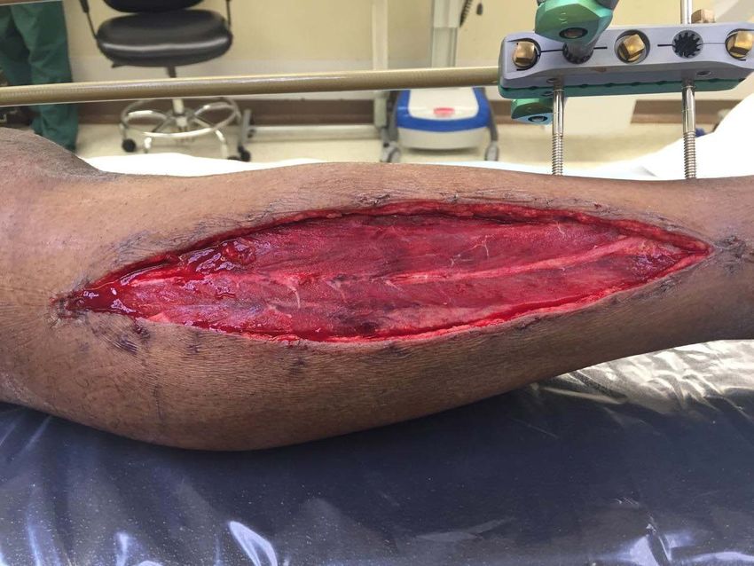
Objectives
• Review AO principles of fracture management

• Identify factors that affect the timing &
  methods of fracture fixation

• Review diagnosis & treatment of compartment
  syndrome, an orthopedic complication found
  most commonly in tibial fractures

• Nurses will be better able to provide bedside
  teaching & anticipatory guidance to their
  orthopedic trauma patients
AO Philosophy & Evolution
Arbeitgemeinschaft fur
  Osteosynthesefragen/Association for the Study of
  Internal Fixation

AO founded in 1958 by a small group of Swiss colleagues
 and is now a worldwide surgical and scientific
 foundation/community

“Focus is on patients with musculoskeletal
  injuries and related disorders. The aim is to
  provide care that allows an early return to
  function and mobility.”
AO Principles
• fracture reduction & fixation to restore anatomical
  relationships

• fracture fixation providing stability as the
  “personality” of the fracture, the patient, & the
  injury requires

• Preservation of the blood supply to soft tissues and
  bone by gentle reduction techniques and careful
  handling;

• Early & safe mobilization and rehabilitation of the
  injured part and the patient as a whole.
Reduction
• Reduction is a procedure to restore a
  fracture or dislocation to the correct
  alignment

• When a bone fractures, the fragments
  displace or angulate

• For the fractured bone to heal without
  deformity the bony fragments must be re-
  aligned to their normal anatomical position
Reduction by Closed or Open methods

Closed reduction refers to manipulation of the bone
  fragments without surgical exposure
  (ie hang fingers in finger-traps to reduce/realign
  wrist fractures)

Open reduction is when fracture fragments are
  exposed surgically by dissecting the tissues.
  (ie internal plating of humerus fracture)

Once the fragments are reduced, the reduction is
  maintained by application of casts, traction or held by
  implants which may in turn be external or internal.
en.wikipedia.org/wiki/Reduction_(orthopedic_surgery)
Goals of External Fixation
• Align bones/fractures

• Get limb/bones out to length
  – muscles pull on bones to deform & shorten them
  – sharpe bony ends can put pressure on skin from
   inside

• Protect soft tissues
  – position external pins as far away from fracture
    site as possible (proximal tibia and into heel bone)
  – Avoid large incisions until soft tissues cool down
Case #1 42 yo M BIBA w Right knee pain s/p falling off of back of work truck (6ft). Unable to bear weight on R leg No LOC, GCS 15 BP158/92, P70, RR16, Pain 8/10 Mechanism of Injury: Fall from 6ft = (high energy)
Past Medical Hx: seizures > on carbamazepine

Past Surg Hx: none

Social Hx: housed, married with 2 children;
           works as cement mason

Substances: tobacco 3cigs/d; occas. marijuana

Last meal: dinner last evening & coffee @ 0700
           this a.m.
PHYSICAL EXAM
• Acute Distress; A&Ox3
• MSK: deformity Right proximal tibia ++ EDEMA,
SKIN INTACT (NO LACERATIONS)
Unable to range knee or test joint stability due to
tenderness/pain
• Motor: wiggles toes, exam limited by pain
• Sensory: intact extremities
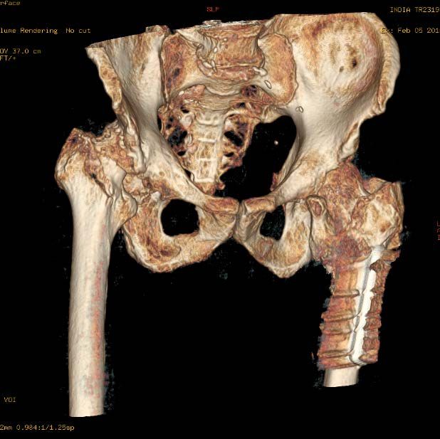
• Pulses: 2+DP/PT Cap Refill: briskHosp Day #1: AP of tibia & knee
Tibial
plateau
fx
Proximal
tibial shaft
comminuted
fxLateral of Tibia
Lateral view of knee shows extension of fx lines
into knee joint = “intra-articular fx”
CT scan is a 3D image used for pre-op planning
this coronal cut shows lat tibial plateau depressed fx
ASSESSMENT

43 yo M s/p 6ft fall off truck with
R proximal tibia fracture & lateral tibial
plateau fracture, with significant early
swelling

These fractures will require surgical fixation

Question is when & how?
PLAN
• NPO
• Pre-Op Labs; CBC, BMP, INR, seizure med
 level, tox screen, EKG, CXR
• Non-WeightBearing RLE in Long Leg splint
• Consent for Surgery
• External-Fixation today in OR
• MONITOR RLE COMPARTMENTS Q4H
Hospital Course
• Routine compartment checks due to tibial
 fx with early swelling
• Developed worsening pain with tense, non-
 compressible “soft tissues” @ around 6hrs
 after admission
• Pt was rushed to OR for two-incision
 (four-compartment) fasciotomies R lower
 leg
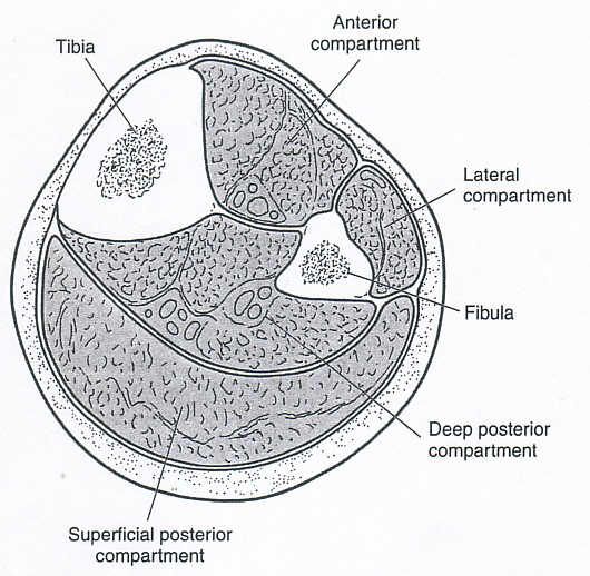
What is compartment syndrome?
How many of you are familiar with the term
compartment syndrome?

How many of you have treated patients with
compartment syndrome?

In a nutshell,
“compartment syndrome is too much stuff in a
confined space & with a risk of tissue damage &
death”
*the muscles surrounded by fascial tissues in the
lower extremity
Compartment Syndrome

“Acute compartment syndrome (ACS) is
defined by a critical pressure increase within
a confined compartmental space, causing a
decline in the perfusion pressure to the
compartment tissue, which without timely
diagnosis & treatment will lead to ischemia,
necrosis & ultimately permanent disability to
the affected region.”

                          (Duckworth & McQueen, JBJS, 2015)
4 Compartments of the Lower Leg • Anterior • Lateral • Superficial posterior • Deep Posterior *arrows show where 2 incisions are made to release soft-tissues
Quick-Facts: Acute Compartment Syndrome

• Fractures comprise ~69% of cases
  – other common causes in trauma cases are

     crush injuries, contusions, gunshot wounds

• Incidence of ACS is ~ 1 to 9% in tibial shaft
  fractures (most common)

• Found in both open & closed tibial shaft
  fractures                          (Duckworth & McQueen, 2011)
ACS is an Orthopedic Emergency
ALL patients with tibial shaft fractures
must be assessed for the development of
the signs & symptoms of compartment
syndrome both at PRESENTATION
AND
AFTER surgical treatment
(i.e. after fracture fixation)Ischemia Time
• Ischemia 8 hrs: IRREVERSIBLE
DAMAGE to muscles/nerves
(ie foot drop)Signs/Symptoms of Compartment Syndrome
• Severe PAIN out of proportion to associated
injury & refractory to analgesia; PAIN ON
PASSIVE STRETCH (passively move the
toes/ankle)
• SWELLING: with elevated compartment
pressures
• PARASTHESIAS: can be due to nerve ischemia as
it runs thru compartment or related to damage to
nerve @ time of injury
• Paralysis: late sign
(Duckworth & McQueen, 2011)Challenges of Diagnosis • Must maintain a high index of suspicion for tibia fx, specific crush injuries, etc. • Must perform serial examinations Ortho providers and RN • Must document changes over time • Be Extra Cautious – in unconscious/obtunded patients – regional anesthesia (nerve block, epidural) – high doses of pain medications • An open fracture does not mean that compartments are decompressed
Role of Nursing Staff

• Recognize the risk of ACS

• Prompt communication w Provider re:
 significant worsening of pain, changes in CSM
 measurements, tight splints/dressings

• Stand-by when provider performing
 compartment checks (trend changes)

• Ask for assistance from Charge, experienced
 staff, if concerned for this complication
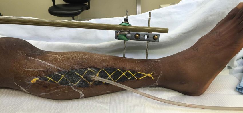
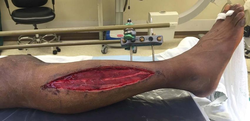
Hosp Day #1: PostOp > fractures stabilized w/
  external fixator & fasciotomies performed

External
fixator
Back to Bedside
• Wound vac on fasciotomy incisions
• Plan for routine return to OR Q2-3 days to
washout wounds & attempt closure of incisions
• DVT ppx: SQ enox
• IV Abx until fasciotomy incisions closed
• RN carry out routine CSM checks; begin pin-
site care POD#2
• Activity:
Elevation in bed, NWB RLE
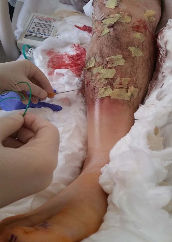
(BRP, OK to start Rehab)HD#3: Medial fasciotomy wound underwent delayed primary closure (DPC)
HD#3: Lateral fasciotomy after routine washout
Lateral fasciotomy wound (peroneal nerve center)
HD#3: Lateral fasciotomy wound with wound-vac as
unable to close this OR visit
Back to Bedside
• Wound vac remains on lateral unclosed incision
• Transition to PO pain meds
• Teach pt pin-site care/enox injection
• Preparation for final wound closure &
  discharge to Home vs SNF
• Patient will return as Outpt for definitive
  fixation (once soft tissues recover) 1-2wks
• NWB RLE x 3mo after final fixation
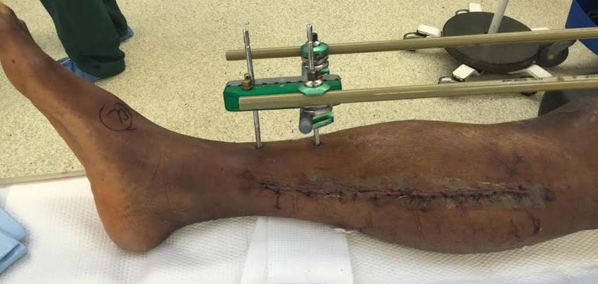
34 Days after Injury Final fixation: ORIF Tibial
plateau & Tibial shaft fx & removal of ex-fix
Factors which affect timing of surgery
Medical Stability: this pt was medically stable

Soft tissues: ++ EDEMA; NOT amenable to
immediate surgical fixation (required ex-fix for
temporary stability)

Complication: compartment syndrome (wait 2-3
wks until fasciotomy incisions closed/skin healed
for Open reduction internal fixation (ORIF)

• Three trips to OR over 8days then DC to home
• Four total trips to OR
• Total Duration 34d fracture to definitive fix
Case #2

49 yr old M BIBA @ 2130 s/p Pedestrian vs Auto;
struck on R side by car travelling ~20mph;
launched 5ft , No LOC, GCS 15
Unable to bear weight on R leg.

BP133/85, P104, RR18, O2Sat 98%RA, Pain 10/10
& located RLE

Mechanism of Injury: Blunt Trauma by vehicle
when walking across street (high-energy injury)
PMHx: +HIV, HL, hx anxiety, depression

Past Surg Hx: appendectomy

Social Hx: housed, lives alone; works as
administrative assistant

Substances: no tobacco; remote hx PSA

Last meal: dinner 3.5h ago
AP & Lateral OPEN midshaft Tibia/Fibula fracture
PE
• Well appearing, NAD, A&Ox3

• MSK: deformity Right tibia/fibula
        anterior LACERATION mid-tibia,
        bone exposed = OPEN FX;
         MILD EDEMA,
        *unable to range or test joint stability due to
        tenderness/pain
• Motor: full, exam limited by pain
• Sensory: intact extremities
• Pulses: 2+DP/PT         Cap Refill: brisk
PLAN
• NPO
• PreOp Labs; CBC, BMP, INR, tox screen
• EKG/CXR
• IV ABX for OPEN FX; tetanus ppx
• Long Leg splint
• STAT CT Scan (pre-op planning)
• Consent for Surgery
• URGENT OR for washout of OPEN fx (goal within 6-8hrs of
injury)
• MONITOR COMPARTMENTS Q4h
Orthobullets, 2016AP & Lat s/p IMN tibia & ORIF R tibial plateau fx
AP of Knee
Back to Bedside

• NWB RLE x 3months for complex tibia fx
• PO splint to protect incisions, offer mild
  compression
• ABx x 24-48h for Open Fracture
• Monitor Compartments Q2-4h x 48h PO
• Elevation in bed (BRP, begin rehab)
• Patient NOT STABLE for transfer or DC until
  Abx & Comp Checks are completed
12 Hrs Post-Op Compartments Measured

*Note shiny swollen
Skin

Splint had been
Progressively
Loosened

• Pain worsening
• Numbness
• Compartments RLE
  very firm
• Chief examined pt
• Compartment pressures
  checked due to high
  suspicion in clinical
  exam
Hospital Course

• Compartment pressures equivocal

• Not taken to OR as exam improved over next
  few hrs

• Patient remained hospitalized to complete
  48h of PO compartment checks & Abx

• Pain controlled w PO meds, worked w Rehab

• Patient stable for Transfer to SNF HD#4
Factors which affect timing of surgery

• Medical Stability: this pt was medically stable;
  Urgent OR on DOA for washout of OPEN fx
• Soft Tissues pre-op: min edema enabled early fx
  fixation
• Soft tissues post-op : ++ edema monitored
  closely x 48hrs for compartment syndrome
• Complications: none
• Total Duration: Admit to Fix to Discharge : 4Days
Case #3
72 yo M found down near outside stairs on
hospital grounds & transported to ED

Injuries: TBI/concussion; R frontal
laceration; rib fxs/R hemothorax
R intertrochanteric hip fracture
R humerus fracture (subacute)

Mechanism of Injury: unknown
Admitted to Trauma service/ICU
Past Medical Hx: HTN, HLD, bilateral inguinal
hernias, BPH, HepC, personality d/o, remote
IVDU & MJ, Vit-D def, genital warts; legally
blind

Past Surg Hx: s/p R cataract extraction 12/15;
s/p Left hip ORIF 2007

Social Hx: lives in SRO; has case mgr

Substances: +tobacco

Last meal: unknown
HD#1 R hip severe degenerative joint disease
& min displaced femoral neck fracture
Barely visible fracture line

                               Severe degenerative joint
                               disease R hip joint:
                               • Asymmetrical (worn
                                 down) femoral head
                               • Asymmetrical
                                 acetabulum
                               • No joint space
Minimally displaced hip fracture with severe
osteoarthritis (degeneration) of the R hip
CT Scan CT scan 3D reformattedRight humerus fx (old): fxs rounded edges w sclerosis (white edges)
Assessment
72 yo blind M, found down with R hip fx & R
humerus fx; previously independent ambulator

PLAN
• NWB RLE will need hip fixed as soon as
  possible
• WBAT RUE (chronic fracture) non-op
  treatment
• OR when Medically Cleared
HD#5      OR for R Total hip replacement
(poor bone quality required extra fixation of
fractured bone intra-op)
R humerus fx (non-op treatment) in fx brace

                         Given the degeneration
                         and chronicity of
                         this humerus fx

                         Minimal pain per patient

                         No surgical intervention
Hospital Course • HD#5 to OR (delayed due to medical issues) • OR was complicated by arrhythmia • Pt. returned to ICU for stabilization • Treated for new onset Afib, GI bleed, new dx hypothyroidism, & delerium • HD#7 transferred to Med Surg Floor • Patient suffered from continued delerium which slowed progress with REHAB
Ortho PLAN:

• WBAT RLE, No hip precautions

• WBAT RUE in Fracture Brace

• DVT PPx enox 40mg SQ daily x14d

• PT/OT ASAP Post-Op

• HD#20 patient transferred to SNF
Role of Nursing
• In this case, delerium played significant
  role in delaying progress to get the patient
  up & out of bed in a timely fashion.
• He was started on new meds for Afib &
  for hypothyroidism & needed meds for pain
• Nursing staff had full time job of
  redirecting the patient until he cleared &
  was able to respond to direction & teaching
  and was “safe” for transfer to SNF at
  Hospital Day #20.
After Discharge:

• Routine outpt clinic f/u

• started on Vitamin D supplement

• Will most likely never get R humerus
  reconstructed as this is a huge
  reconstructive surgery in a patient who
  may not be safe with poor vision (pain/limit
  in function) would be guide for any future
  surgery
Summary
These ortho cases represented different
timelines, yet demonstrate how following the
AO principles of

•   anatomical reduction,
•   fracture stabilization,
•   careful handling of soft-tissues, &
•   early mobilization

will guide most any fracture/trauma case from
hospital admission to discharge.
My hope is that Nursing staff will recognize
these basic orthopedic principles and be able to
apply them to beside nursing from case to case.

Thank you!
References

A.D. Duckworth, M. McQueen; Diagnosis of acute compartment syndrome;
Journal of Bone & Joint Surgery, 2011; 18.

https://www2.aofoundation.org

Babak Shadgan, MD, MSc,* Matthew Menon, MD,† David Sanders, MD,‡
Gregg Berry, MD,§ Claude Martin, Jr., MD, MBA,¶ Paul Duffy, MD,** David
Stephen, MD,†† and Peter J. O’Brien, MD;
Current thinking about acute compartment syndrome of the lower
extremity
Can J Surg. 2010 Oct; 53(5): 329-334.

http://Orthobullets.com/ Orthopedic teaching site for Residents
UptoDate
You can also read