Caudal lumbar spinal cysts in two French Bulldogs - Acta Veterinaria ...
←
→
Page content transcription
If your browser does not render page correctly, please read the page content below
de Nies et al. Acta Vet Scand (2018) 60:14
https://doi.org/10.1186/s13028-018-0368-6                                                          Acta Veterinaria Scandinavica

 CASE REPORT                                                                                                                                    Open Access

Caudal lumbar spinal cysts in two French
Bulldogs
Kiona Sharon de Nies1*, Ralph Alexander Edwards1, Niklas Bergknut2, Martijn Beukers1 and Björn Petrus Meij1

  Abstract
  Background: Spinal cysts are rare findings in veterinary medicine, but they are increasingly recognized due to the
  availability of advanced imaging techniques. Extradural meningeal cysts in French Bulldogs have not been reported
  previously and arachnoid cysts (diverticula) have not been reported at the caudal lumbar (L6–L7) region in dogs.
  Case presentation: Two French Bulldogs, aged 5 and 8 years, were referred for evaluation of lower back pain and
  bilateral hind limb neurological deficits. Neurologic examination revealed ataxia and postural deficits in both dogs.
  Magnetic resonance imaging (MRI) showed cauda equina compression due to a cyst-like lesion at the level of L6–L7
  in both cases. The dogs underwent dorsal laminectomy and the meningeal cyst was completely removed in one dog
  and in the other dog the spinal arachnoid diverticula was marsupialized. In Case 1, histopathology of the cysts was
  performed and MRI was repeated. Both dogs were pain free during follow-up evaluations.
  Conclusions: Based on radiological, intra-operative and histopathological findings, the first case was diagnosed as a
  meningocele connected by a pedicle to the caudal tip of the dural sac forming a dural diverticulum categorized as an
  extradural spinal cyst type Ib, and Case 2 as a type III intradural arachnoid diverticula. It is concluded that spinal cysts
  should be included in the differential diagnosis of cauda equina syndrome and lower back pain in French Bulldogs.
  Results of these cases may be useful for diagnostic and treatment management.
  Keywords: Dorsal laminectomy, French Bulldog, Lumbar spine, Magnetic resonance imaging, Spinal cysts

Background                                                                             classified as extradural or intradural cysts [4]. Extradural
A cyst is defined as ‘a closed epithelium-lined sac or cap-                            cysts (of non-meningeal origin) are recognized in three
sule containing liquid, air or a semi-solid substance’ [1].                            types: (1) synovial cysts containing a lining of synovial-
Cystic-like lesions that lack an epithelial lining are not                             like epithelial cells, (2) ganglion cysts, originating from
true cysts and therefore the term diverticula is a more                                vertebral ligaments, consisting of a collagenous capsule
appropriate term [1, 2]. Spinal cysts and diverticula asso-                            surrounding myxoid material and (3) discoid cysts origi-
ciated with spinal cord dysfunction are rare conditions in                             nating from the annulus fibrosus containing degenerative
veterinary medicine, but they are increasingly recognized                              fibrous material without a synovial lining [3, 4]. Intra-
due to the availability of advanced imaging techniques                                 dural cysts arise from the meninges and contain cerebro-
like magnetic resonance imaging (MRI) and computed                                     spinal fluid (CSF). These include arachnoid cysts that lack
tomography (CT) [3, 4]. The aetiology of spinal cysts                                  epithelial lining. For this reason they are not true cysts
remains unclear but most are considered congenital                                     and the term spinal arachnoid diverticula (SAD) is more
lesions with a genetic predisposition, although acquired/                              appropriate [1, 2, 6]. Meningeal cysts/diverticula can also
traumatic cysts have been reported [5]. Spinal cysts are                               be presented with an extradural position.
                                                                                          Lowrie et al. [1] introduced a classification scheme
                                                                                       used for humans into the veterinary literature. Based
*Correspondence: k.s.denies@uu.nl                                                      on pathology, meningeal cysts were classified as type I:
1
  Department of Clinical Sciences of Companion Animals, Faculty                        extradural cyst without spinal nerve root involvement,
of Veterinary Medicine, Utrecht University, Yalelaan 108, 3584 CM Utrecht,
The Netherlands                                                                        whereof type Ia is thought to arise from herniation of the
Full list of author information is available at the end of the article

                                          © The Author(s) 2018. This article is distributed under the terms of the Creative Commons Attribution 4.0 International License
                                          (http://creativecommons.org/licenses/by/4.0/), which permits unrestricted use, distribution, and reproduction in any medium,
                                          provided you give appropriate credit to the original author(s) and the source, provide a link to the Creative Commons license,
                                          and indicate if changes were made. The Creative Commons Public Domain Dedication waiver (http://creativecommons.org/
                                          publicdomain/zero/1.0/) applies to the data made available in this article, unless otherwise stated.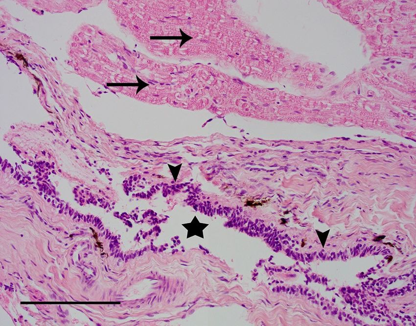
de Nies et al. Acta Vet Scand (2018) 60:14                                                                      Page 2 of 8

arachnoid through a dural defect and type Ib is defined as   recovery (STIR) sequences (Fig. 1). At the level of L6–L7
meningocele connected by a pedicle to the caudal tip of      an oval cyst-like structure was seen with dorsal compres-
the dural sac forming a dural diverticula. Type II extra-    sion of the cauda equina. The content of this structure
dural arachnoid diverticula with nerve root involvement      was isointense with CSF. An ill-defined T2W hyperin-
are also known as Tarlov cysts and are presented as CSF      tensity was present within the spinal cord at the level of
filled dilatations between the perineum and endoneu-         T12, most likely consistent with oedema and less likely
rium. Type III are intradural arachnoid diverticula with-    fibrocartilaginous embolism due to the insidious onset
out epithelial cell lining and they communicate freely       and progressive ataxia. Kyphosis and multiple thoracic
with the subarachnoid space [1, 4, 7].                       hemivertebrae were noted, consistent with breed related
   Spinal cysts and diverticula in humans have been          vertebral column deformities, without evidence of spi-
reported to occur primarily in the lumbar spine [8, 9],      nal cord compression. The radiological diagnosis was a
whereas in veterinary literature both non-meningeal          cyst-like structure with dorsal compression of the cauda
cysts, meningeal type II cysts, and SAD were reported to     equina. In addition, all visible intervertebral discs showed
occur at two main locations; the cervical spine and thora-   decreased hypointense signal compatible with interver-
columbar junction [1–4, 10, 11]. Synovial cysts have also    tebral disc degeneration. Mild disc protrusion was found
been reported in the lumbosacral junction [4, 12, 13]. In    at multiple levels, but there was no disc-associated com-
French Bulldogs, SAD have only been reported in the          pression of the spinal cord or cauda equina.
thoracolumbar region [10]. To our knowledge, types Ia           Since the dog was not responsive to medication, the
and Ib have not been reported in dogs.                       owners opted for surgery. The dog received premedica-
   In this case report we describe two French Bulldogs,      tion with dexmedetomidine (4 µg/kg, IV) and butorpha-
one presenting with a meningeal cyst type Ib and one         nol (0.1 mg/kg, IV). Propofol (2 mg/kg, IV) was used to
with a type III (intradural arachnoid diverticula) in the    induce anaesthesia. General anaesthesia was maintained
caudal lumbar area. The clinical and MRI findings, surgi-    with isoflurane, ketamine (10 µg/kg/min, IV) and dex-
cal treatment, histopathology, and follow up are reported.   medetomidine (1 µg/kg/h, IV). Medication included
                                                             cefazolin (20 mg/kg, IV), buprenorphine (10 µg/kg, IV),
Case presentation                                            and dexamethasone (0.2 mg/kg, IV). A standard dorsal
Case 1                                                       laminectomy from the midpoint of L6 extending over L7
An 8-year-old female French Bulldog weighing 12.4 kg         to S1 was performed as described previously [14], using
was referred to the Department of Clinical Sciences of       a high speed burr and Kerrison rongeurs. After removal
Companion Animals at Utrecht University with a his-          of the ligamentum flavum with a beaver scalpel knife, the
tory of intermittent faecal incontinence and progres-        cystic structure was exposed (Fig. 2a). The cyst was care-
sive abnormal gait in the hind limbs since 3 months.         fully freed with a ball-tipped probe, the cranial side of the
Conservative treatment with corticosteroids initially        cyst was attached to the dural sac and the caudal side of
showed improvement but after tapering the dosage,            the cyst was attached to one of the smaller caudal nerves
clinical signs recurred. The owners felt that the dog was    of the cauda equina (Fig. 2b). The appearance of this cau-
in pain because they found the dog progressively more        dal nerve was similar to the other nerves. The cyst was
depressed/lethargic. On general examination, the dog         sharply dissected from the caudal nerve and the dural sac
showed signs related to brachycephalic obstructive air-      (Fig. 2c). The lumen of the cyst was not continuous with
way syndrome. Orthopaedic examination elicited pain on       the dural sac and therefore the cyst could be removed
palpation and extension of the L6 to S3 region. Neurolog-    completely without collapse (Fig. 2d). Before routine clo-
ical examination identified hind limb ataxia and ambula-     sure, a morphine splash-block was deposited in the surgi-
tory paraparesis and postural deficits in the hind limbs,    cal field and an autologous free fat graft was transplanted
more severe on the left side. Spinal reflexes were nor-      over the neural tissue to prevent dural adhesions [15].
mal in all limbs. The neuroanatomic localization based          The entire cyst was fixed in 10% neutral buffered for-
on the neurological deficits included T3–L3 and based        malin, processed by routine methods in toto, embed-
on the lumbosacral pain on palpation and faecal incon-       ded in paraffin, sliced and stained with haematoxylin
tinence it was extended until S3. Magnetic resonance         and eosin (HE) and periodic acid Schiff (PAS). Macro-
imaging (MRI, type Ingenia 1.5T, Philips, Eindhoven, The     scopically, the cyst was stalked, thin-walled, measured
Netherlands) was performed under general anaesthe-           1.3 × 0.5 cm and filled with clear fluid. Histologically,
sia of the thoracolumbar, lumbar and lumbosacral spine       several optically empty slit-like spaces were lined with
extending from T6 to Cd3 with sagittal and/or transverse     cuboidal epithelium and embedded in connective tissue
T1-weigted, T2-weighted, T2*-weighted, fluid attenua-        adjacent to a peripheral nerve that showed minor degen-
tion inversion recovery (FLAIR) and short tau inversion      erative changes (Fig. 3). There was sporadic perineural
de Nies et al. Acta Vet Scand (2018) 60:14 Page 3 of 8 Fig. 1 Magnetic resonance images of the caudal lumbar vertebral column of Case 1, including T2-weighted sagittal (a), T1-weighted sagittal (b), T2-weighted transverse (c), and FLAIR transverse (d) images. The meningeal cyst is marked with an arrow on all images and L7 is labelled with an asterisk on images (a) and (b) and perivascular lymphoplasmacellular infiltrations. ataxia was still noticeable, but less severe and the same After PAS staining, cilia were not detected in the epithe- was true for the faecal incontinence. The dog no longer lial lining. Thus, the histological findings were compatible showed signs of depression/lethargy, ambulatory para- with a meningeal cyst type Ib of the classification scheme paresis was no longer present, and no pain could be proposed by Lowrie et al. [1]. elicited during palpation and/or extension of the lower The dog was discharged 1 day after surgery with the fol- back. MRI of the thoracic, lumbar and lumbosacral lowing oral medication: tramadol (2–5 mg/kg, q6–8 h), spine was performed, including sagittal and transverse gabapentin (10 mg/kg, q12 h) and prednisolone was T1W and T2W sequences, transverse T2 * W and sag- tapered down and stopped over a period of a few weeks. ittal STIR sequences (Fig. 4). The rounded cystic struc- The owners were advised to keep the dog quiet and on ture, as described in the preoperative MRI examination, leash walks for at least 6 weeks. Physical therapy was was no longer visible. Dorsal to the cauda equina there started at 2 weeks after surgery to support rehabilitation. was hyperintensity visible at the level of L6–L7 and the At 3 months follow-up examination, the dog was LS-junction over a length of 19.6 mm (at both T1W without medication and the owners had noted progres- and T2W sequences) consistent with the fat graft which sive improvement in the clinical signs. The hind limb slight displaced the cauda equina ventrally [15]. The soft
de Nies et al. Acta Vet Scand (2018) 60:14 Page 4 of 8
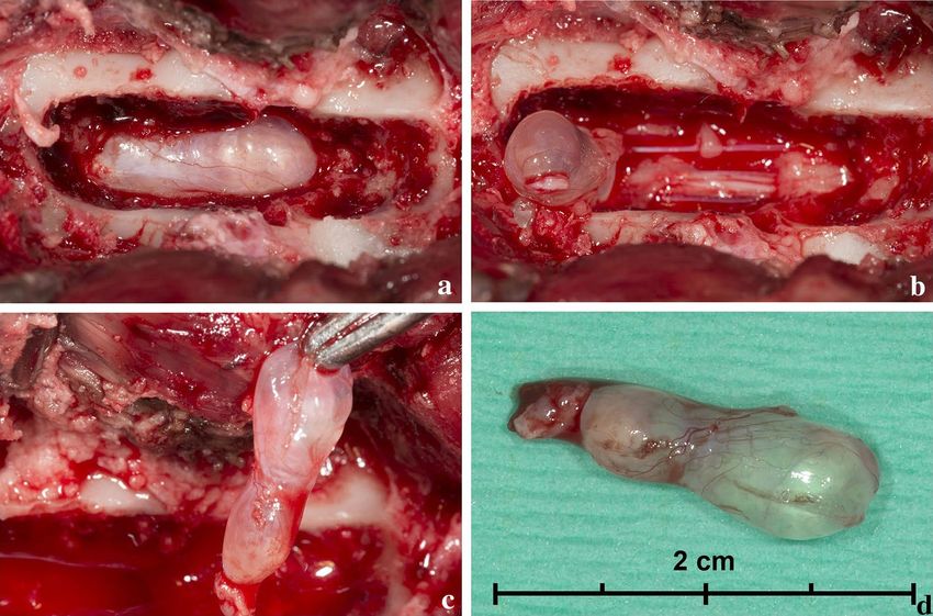
Fig. 2 Intraoperative photographs of the surgical removal of the meningeal cyst in Case 1. Dorsal laminectomy at L6–L7 revealed a spinal cyst (a).
The cyst was released from the cauda equina caudally (b) and retracted cranially (c) before being removed completely without collapse (d)
Case 2
A 5-year-old male French Bulldog weighing 10.3 kg was
referred to the Department of Clinical Sciences of Com-
panion Animals at Utrecht University with a 3-year his-
tory of hind limb ataxia and ambulatory paraparesis
without clinical signs of pain. MRI of the cervical and
thoracolumbar spine performed 1 year previously in
another institution showed intervertebral disc degenera-
tion at multiple levels and mild disc protrusion at T12–
T13 but without compression of neural structures. The
dog was treated conservatively with corticosteroids with-
out clinical improvement. Ambulatory paraparesis per-
sisted and in addition to the motor deficits, passive urine
incontinence developed not related to overflow. Results
of initial physical examination were unremarkable; how-
Fig. 3 Histopathology images of Case 1. Collapsed optic empty cyst ever, a complete neurological examination revealed
(asterisk) lined by cuboidal to cylindrical epithelium (arrowheads), ataxia with dysmetria and hypermetria of both hind
surrounded by fibrous tissue. Peripheral nerve tissue (arrow) is shown
at the top of the image. HE staining, bar = 100 µm
limbs, as well as muscle atrophy and hypotony. The dog
had proprioceptive deficits in both hind limbs, but spinal
reflexes were normal in all limbs. Based on the neurologi-
cal deficits, the urinary incontinence, and the results of
tissue on the dorsal side of the L6–L7–S1 spine showed neurological examination, the neuroanatomic localiza-
irregularity and the muscles were heterogeneously hyper- tion was determined as T3–S3.
intense, consistent with changes secondary to surgery Magnetic resonance imaging was repeated from C1
(Fig. 4). to S1, including sagittal and transverse T1-weigted
and T2-weighted sequences, and a transverse FLAIRde Nies et al. Acta Vet Scand (2018) 60:14 Page 5 of 8 Fig. 4 Magnetic resonance images of the caudal lumbar vertebral column of Case 1 post surgery, including T2-weighted sagittal (a) and T1-weighted sagittal (b) images. L7 is labelled with an asterisk. The meningeal cyst is absent and post-surgical changes are seen in the soft tissues dorsal to the region of interest. N.B. The oval structure at approximately the previous position of the meningeal cyst (see Fig. 1a, b) is a fat graft (T1-weighted and T2-weighted hyperintense) sequence of the lumbar spine. Multiple intervertebral was covered with an autologous fat graft to prevent discs showed decreased hypointense signal compatible adhesions. with intervertebral disc degeneration. At the level of C2– The dog was discharged 1 day after surgery with the C3, T12–T13, T13–L1 and L1–L2 mild disc protrusion following oral medication: amoxicillin/clavulanic acid was found with visible compression of the ventral suba- (12.5 mg/kg, q12 h), tramadol (2–5 mg/kg, q6–8 h), and rachnoid space, but without compression or dislocation carprofen (2 mg/kg q12 h). The owners were advised to of the spinal cord. A fusiform cystic lesion dorsal to the keep the dog quiet and on leash walks for 6 weeks. cauda equina at the level of L6–L7 was found with an ill- The dog was re-examined at 2 weeks after surgery. The defined cranial and caudal border. The cystic structure dog showed moderate ataxia and postural reaction defi- was isointense to CSF and compressed the cauda equina cits in the hind limbs. The dog was both urinary and fae- (Fig. 5). Based on these imaging characteristics a SAD cal continent. All spinal reflexes including perineal reflex was suspected. There was mild dilation of the central were normal. Physical therapy and hydrotherapy was canal from T12 to L5. started to support rehabilitation. At 6 weeks postopera- Anaesthesia and medication for the surgical pro- tively, the motor function of the dog had recovered sig- cedure were identical to Case 1 except that this dog nificantly. Although, the dog was still ataxic, the dog was received carprofen (4 mg/kg, IV) instead of corticoster- evidently less paretic in the hind limbs. Postural deficits oids in the postoperative period. The surgical technique were improved in both hind limbs and all spinal reflexes was the same as for Case 1. After dorsal laminectomy were normal. Also, the muscle mass over the hind limbs from halfway L6 over L7 to S1, a dilated dural sac was had progressively increased. exposed which was macroscopically identified as SAD. A durotomy was performed over a length of approximately Discussion and conclusions 3 cm, which resulted in free flow of CSF into the spinal In this study, a meningeal cyst and a SAD are reported canal and collapse of the diverticula. No tissue was avail- in the lumbar region of French Bulldogs. Although spinal able for histological examination. Marsupialization of the cysts have been described more frequently in veterinary SAD was performed by placing interrupted sutures with literature in recent years, prevalence of canine arachnoid polydioxanone 5-0 attaching the dural sac to the lateral diverticula in the caudal lumbar region is very rare [1, wall of the laminectomy. Before closure the laminectomy 10]. Schmöckel and Rapp [13] described a case series of
de Nies et al. Acta Vet Scand (2018) 60:14                                                                                                 Page 6 of 8

 Fig. 5 Magnetic resonance images of the caudal lumbar vertebral column of Case 2, including T2-weighted sagittal (a), T1-weighted sagittal (b),
 T2-weighted transverse (c), and FLAIR transverse (d) images. The subarachnoid diverticula is marked with an arrow on all images and L7 is labelled
 with an asterisk on images (a) and (b)

synovial cyst at the lumbosacral junction in three Ger-                      aetiology of spinal cysts still remains unclear, theories are
man Shepherd dogs. One case, reported by Webb et al.                         different between the various type of cysts/diverticula.
[12] and two dogs of a case series reported by Sale and                      Extradural cysts (of non-meningeal origin) were linked to
Smith [16], presented a synovial/ganglion cyst associ-                       increased mechanical stress and instability [2, 4, 12, 13,
ated with the L6–L7 articulations. Here we describe a                        16], whereas meningeal cysts are generally considered
case of a meningeal spinal cyst type Ib and a case of a                      congenital lesions with a genetic predisposition [1–4, 10,
type III spinal arachnoid diverticula at the L6–L7 region,                   11]. However, SAD may also be acquired or traumatic,
which both have not been reported before. Although the                       and seems to be related to concurrent spinal disorders
de Nies et al. Acta Vet Scand (2018) 60:14                                                                       Page 7 of 8

like vertebral malformations and intervertebral disc dis-      study extended from C1 to S1. Multiple mild protrusions
ease [1, 10].                                                  of intervertebral discs at the cervical, thoracic and the
   Congenital vertebral column deformities like hemiver-       lumbar regions were seen, but without evident compres-
tebrae and degenerative intervertebral disc disease            sion of the spinal cord. Therefore, we cannot completely
have been reported previously in French Bulldogs and           exclude a possible relation of some of the neurological
other chondrodystrophic breeds [17, 18]. In agreement          signs to the intervertebral disc protrusions, but it seems
with previous studies, instability is suspected to play        less likely since there was no significant compression of
an important role as possible underlying cause of the          the spinal cord or cauda equina. Also, the occurrence of
evolution of lumbar spinal cyst [3, 4, 10]. In the French      a spinal cyst is usually a gradual and chronic problem;
Bulldogs in our case series, congenital vertebral abnor-       therefore the cyst may have affected the dural sac and
malities, i.e. kyphosis and hemivertebrae and interverte-      the cauda equina nerves already for a prolonged time,
bral disc disease, were also evident on MRI and it is likely   leading to permanent changes in the neural tissue. It is
that the aetiology and evolution of the lumbar spinal          not possible to state whether the spinal cyst/diverticula,
cysts is in some way genetically linked to spinal deformity    the concurrent spinal disorder, or a combination of both
and abnormal biomechanics of the spine in French Bull-         attributed to the neurologic signs. Although, it remains
dogs. Mauler et al. [10] found high rates (61.5%) of con-      unexplained why the dogs did not recover completely,
current diseases in French Bulldogs with SAD [10] and a        both dogs showed an acceptable clinical outcome and the
predisposition to acquired SAD was presumed [1, 4, 10]         owners were satisfied with the result.
resembling our Case 2.                                           In accordance to radiological, intra-operative and his-
   Progressive postoperative clinical improvement sug-         topathological findings our first case was categorized as
gested the causality between the cyst and some of the          an extradural meningeal type Ib cyst [1, 4]. Type Ib spinal
clinical signs, although this is difficult to confirm due to   cysts are closely related to myelomeningocele for which
the absence of a control group. Both cases showed clini-       Bulldogs are predisposed [1]. Only few case reports [17,
cal signs consistent with a neuroanatomical localization       18] have reported myelomeningocele. Ployart et al. [17]
T3–L3 (hind limb ataxia and paraparesis, postural defi-        described a case of myelomeningocele and a dermoid
cits of the hind limbs, and normal spinal reflexes repre-      sinus-like lesion at the L7 site in the French Bulldogs,
sent upper motor neuron signs related to spinal cord           which is different from our clinical case. Whether the
segments), and could be further extended until S3 (pain        cyst in Case 1 was a type Ib or a type II (perineural or
reaction on palpation and extension of the L6 to S3 seg-       Tarlov cyst) [1] remains under discussion. Histopatho-
ment, paraparesis, intermittent faecal incontinence in         logical findings showed some infiltrations of perineu-
Case 1, and passive urinary incontinence in Case 2, repre-     ral tissue, but there was no clear evidence of nerve root
sent lower motor neuron signs related to cauda equina).        involvement. Therefore a type Ib defined as meningoceles
Persistent neurological deficits, i.e. moderate hind limb      connected by a pedicle to the caudal tip of the dural sac
ataxia and, although less frequent, persistent intermittent    forming a dural diverticulum [1] is the most likely histo-
faecal incontinence (Case 1) may be explained by two           logical diagnosis. In Case 2 there was no tissue available
possible reasons. First, clinical signs of hind limb ataxia    for histology, but radiologic and intra-operative findings
may be explained by other detected spinal deformi-             demonstrated a type III intradural arachnoid diverticula
ties, i.e. the intramedullary hyperintensity at the level of   [1]. A type Ib spinal cyst (case 1) has not been reported
T12 representing spinal cord oedema, and abnormali-            previously in dogs [1] and SAD (case 2) has not been
ties common to the brachycephalic breed, i.e. hemiver-         reported in the caudal lumbar (L6–L7) region in dogs
tebrae, kyphosis, and degenerative disc disease [19]. In       [10, 21].
a recent publication, Ryan et al. [20] studied in retro-         In conclusion, this is the first report of caudal lumbar
spect neurologically normal French Bulldogs and found          spinal cysts/diverticula in French Bulldogs and gives us
a high prevalence of vertebral malformations. In our           more knowledge and insights into (congenital) spinal
cases, we could not exclude any clinical relation to this      deformities and abnormalities related to the French Bull-
finding (our patients both showed neurological deficits);      dogs breed. Spinal cysts/diverticula should be included
however, since vertebral column deformities in French          in the differential diagnosis of cauda equina syndrome
Bulldogs are common incidental findings, it appears to         and lower back pain in French Bulldogs. However, fur-
be less likely. In the first case, the MRI study extended      ther studies are indicated to evaluate the prevalence and
from T6 to Cd3, abnormalities cranial to the T6 seg-           possible genetic relation of spinal cysts/diverticula to the
ment which could be related to the neurological signs like     French Bulldog breed.
ataxia could not be excluded. In the second case, the MRI
de Nies et al. Acta Vet Scand (2018) 60:14                                                                                                               Page 8 of 8

Abbreviations                                                                      2.    Sharp NW, Wheeler S. Miscellaneous conditions. Small animal spinal
CSF: cerebrospinal fluid; CT: computed tomography; FLAIR: fluid attenuation              disorders. 2nd ed. New York: Elsevier Ltd; 2005. p. 323–6.
inversion recovery; HE: haematoxylin and eosin; IV: intravenous; MRI: magnetic     3.    Bismuth C, Ferrand FX, Millet M, Buttin P, Fau D, Cachon T, et al. Original
resonance imaging; PAS: periodic acid Schiff; SAD: spinal arachnoid diverticula;         surgical treatment of thoracolumbar subarachnoid cysts in six chondro-
STIR: short tau inversion recovery.                                                      dystrophic dogs. Acta Vet Scand. 2014;56:32.
                                                                                   4.    da Costa RC, Cook LB. Cystic abnormalities of the spinal cord and verte-
Authors’ contributions                                                                   bral column. Vet Clin N Am Small Anim Pract. 2016;46:277–93.
KSdN, BPM and NB performed clinical examination, surgery and clinical care of      5.    Rylander H, Lipsitz D, Berry WL, Sturges BK, Vernau KM, Dickinson PJ, et al.
the two dogs. RAE and MB performed the diagnostic imaging. KSdN and RAE                  Retrospective analysis of spinal arachnoid cysts in 14 dogs. J Vet Intern
drafted the manuscript. All authors gave substantial input to the manuscript.            Med. 2002;16:690–6.
All authors read and approved the final manuscript.                                6.    Rohdin C, Nyman HT, Wohlsein P, Hultin Jäderlund K. Cervical spinal
                                                                                         intradural arachnoid cysts in related, young pugs. J Small Anim Pract.
Author details                                                                           2014;55:229–34.
1
  Department of Clinical Sciences of Companion Animals, Faculty of Veterinary      7.    Nabors MW, Pait TG, Byrd EB, Karim NO, Davis DO, Kobrine AI, et al.
Medicine, Utrecht University, Yalelaan 108, 3584 CM Utrecht, The Netherlands.            Updated assessment and current classification of spinal meningeal cysts.
2
  Department Neurology, North Downs Specialist Referrals, Bletchingley, UK.              J Neurosurg. 1988;68:366–77.
                                                                                   8.    Kadono Y, Yuguchi T, Ohnishi YI, Iwatsuki K, Yoshimine T. A symptomatic
Acknowledgements                                                                         spinal extradural arachnoid cyst with lumbar disc herniation. Case Rep
Presented in abstract form at the 50th Edition of the European Veterinary                Orthop. 2015;2015:1–5.
Conference Voorjaarsdagen, The Hague, April 2017.                                  9.    Zhenbo Z, Huanting L, Jin W, Haifeng G, Yuan F, Ming L. Hemilamino-
    We are very grateful to Jooske IJzer, DVM, Ph.D. for performing the histopa-         plasty for the treatment of lumbar intraspinal synovial cysts (LISCs) and
thology and the interesting discussions.                                                 literature review. Eur Spine J. 2016;25:3393–402.
                                                                                   10.   Mauler DA, De Decker S, De Risio L, Volk HA, Dennis R, Gielen I, et al. Sig-
                                                                                         nalment, clinical presentation, and diagnostic findings in 122 dogs with
Competing interests                                                                      spinal arachnoid diverticula. J Vet Intern Med. 2014;28:175–81.
The authors declare that they have no competing interests.                         11.   Gnirs K, Ruel Y, Blot S, Begon D, Rault D, Delisle F, et al. Spinal subarach-
                                                                                         noid cysts in 13 dogs. Vet Radiol Ultrasound. 2003;44:402–8.
Availability of data and materials                                                 12.   Webb AA, Pharr JW, Lew LJ, Tryon KA. MR imaging findings in a dog with
All data generated or analysed during this study are included in this article.           lumbar ganglion cysts. Vet Radiol Ultrasound. 2001;42:9–13.
                                                                                   13.   Schmökel H, Rapp M. Lameness caused by an extradural lumbosacral
Consent for publication                                                                  foraminal synovial cyst in three German shepherd dogs. Vet Comp
The Utrecht University Small Animal Clinic signs an agreement (‘Akkoord                  Orthop Traumatol. 2016;29:83–8.
verklaring’) with every client with an animal when entering the clinic. In this    14.   Meij BP, Bergknut N. Degenerative lumbosacral stenosis in dogs. Vet Clin
agreement, the owner consents to a range of items but also to the use of                 N Am Small Anim Pract. 2010;40:983–1009.
medical information for case report documentation as long as the welfare           15.   Quist JJ, Dhert WJA, Meij P, Visser WJ, Oner FC, Hazewinkel HAW, et al. The
of the animal is not compromised and the identity of client and animal is                prevention of peridural adhesions. J Bone Jt Surg. 1998;80(3):520–6.
protected.                                                                         16.   Sale CSH, Smith KC. Extradural spinal juxtafacet (synovial) cysts in three
                                                                                         dogs: case report. J Small Anim Pract. 2007;48:116–9.
Ethics approval and consent to participate                                         17.   Ployart S, Doran I, Bomassi E, Bille C, Libermann S. Myelomeningo-
Since this study concerned the treatment of patients with clinical signs (and            coele and a dermoid sinus-like lesion in a French bulldog. Can Vet J.
not experimental animals) it did not require official or institutional ethical           2013;54:1133–6.
approval. The animals were handled and treated according to the high ethical       18.   Song RB, Glass EN, Kent M, Sánchez MD, Smith DM, de Lahunta A. Surgi-
standards of the Utrecht University Small Animal Clinic and its’ veterinarians           cal correction of a sacral meningomyelocele in a dog. J Am Anim Hosp
and the Dutch legislation.                                                               Assoc. 2014;50:436–43.
                                                                                   19.   Schlensker E, Distl O. Heritability of hemivertebrae in the French bulldog
Funding                                                                                  using an animal threshold model. Vet J. 2016;207:188–9.
The authors declare that there were no funding and support.                        20.   Ryan R, Gutierrez-Quintana R, ter Haar G, De Decker S. Prevalence of
                                                                                         thoracic vertebral malformations in French bulldogs, pugs and English
                                                                                         bulldogs with and without associated neurological deficits. Vet J.
Publisher’s Note                                                                         2017;221:25–9.
Springer Nature remains neutral with regard to jurisdictional claims in pub-       21.   Moissonnier P. Facet joints and cysts. Proc. 24th ECVS Annu. Sci. Meet.
lished maps and institutional affiliations.                                              Cerv. Spine Dis. Forum. 2015.
Received: 28 October 2017 Accepted: 23 February 2018

                                                                                         Submit your next manuscript to BioMed Central
                                                                                         and we will help you at every step:
References                                                                                • We accept pre-submission inquiries
1. Lowrie ML, Platt SR, Garosi LS. Extramedullary spinal cysts in dogs. Vet               • Our selector tool helps you to find the most relevant journal
    Surg. 2014;43:650–62.
                                                                                          • We provide round the clock customer support
                                                                                          • Convenient online submission
                                                                                          • Thorough peer review
                                                                                          • Inclusion in PubMed and all major indexing services
                                                                                          • Maximum visibility for your research

                                                                                          Submit your manuscript at
                                                                                          www.biomedcentral.com/submit
You can also read