Chapparvovirus DNA Found in 4% of Dogs with Diarrhea - MDPI
←
→
Page content transcription
If your browser does not render page correctly, please read the page content below
viruses
Article
Chapparvovirus DNA Found in 4% of Dogs
with Diarrhea
Elizabeth Fahsbender 1,2 , Eda Altan 1,2 , M. Alexis Seguin 3 , Pauline Young 3 , Marko Estrada 3 ,
Christian Leutenegger 3 and Eric Delwart 1,2, *
 1    Vitalant Research Institute, San Francisco, CA 94118, USA; efahsbender@vitalant.org (E.F.);
      EAltan@vitalant.org (E.A.)
 2    Dept. of Laboratory Medicine, University of California, San Francisco, CA 94118, USA
 3    IDEXX Reference Laboratories, -Inc., West Sacramento, CA 95605, USA; alexis-seguin@idexx.com (M.A.S.);
      Pauline-Young@idexx.com (P.Y.); Marko-Estrada@idexx.com (M.E.);
      Christian-Leutenegger@idexx.com (C.L.)
 *    Correspondence: Eric.Delwart@ucsf.edu

 Received: 22 March 2019; Accepted: 24 April 2019; Published: 27 April 2019

 Abstract: Feces from dogs in an unexplained outbreak of diarrhea were analyzed by viral
 metagenomics revealing the genome of a novel parvovirus. The parvovirus was named cachavirus
 and was classified within the proposed Chapparvovirus genus. Using PCR, cachavirus DNA was
 detected in two of nine tested dogs from that outbreak. In order to begin to elucidate the clinical
 impact of this virus, 2,053 canine fecal samples were screened using real-time PCR. Stool samples
 from 203 healthy dogs were positive for cachavirus DNA at a rate of 1.47%, while 802 diarrhea
 samples collected in 2017 and 964 samples collected in 2018 were positive at rates of 4.0% and 4.66%
 frequencies, respectively (healthy versus 2017-2018 combined diarrhea p-value of 0.05). None of
 83 bloody diarrhea samples tested positive. Viral loads were generally low with average real-time
 PCR Ct values of 36 in all three positive groups. The species tropism and pathogenicity of cachavirus,
 the first chapparvovirus reported in feces of a placental carnivore, remains to be fully determined.

 Keywords: parvovirus; viral metagenomics; canine chapparvovirus

1. Introduction
      Canine diarrhea is one of the most common illnesses treated by veterinarians with many
possible causes of canine diarrhea, including bacteria, parasites, and viruses [1]. One of the
most important dog enteric viruses is canine parvovirus 2 (CPV-2) in the Carnivore protoparvovirus
species 1 [2]. Parvoviruses are small, icosahedral, nonenveloped, single-stranded DNA viruses that are
pathogenic to a variety of mammals [3–5]. The vertebrate-infecting parvoviruses are classified in the
subfamily Parvovirinae in the Parvoviridae family (which also includes the insect infecting subfamily
Densovirinae). The Parvovirinae subfamily is currently subdivided into eight officially recognized genera
(Dependoparvovirus, Copiparvovirus, Bocaparvovirus, Amdoparvovirus, Aveparvovirus, Protoparvovirus,
Tetraparvovirus, and Erythroparvovirus [6]). The recently proposed genus Chapparvovirus is currently
comprised of a rat parvovirus 2 (KX272741) [7], Eidolon helvum fruit bat parvovirus 1 (MG693107.1) [8],
and E. helvum bat parvovirus 2 (JX885610) [9], Desmodus rotundus bat parvovirus (NC032097.1) [10],
simian parvo-like virus 3 (KT961660.1) [11], Turkey parvovirus TP1-2012/Hun (KF925531) [12], porcine
parvovirus 7 (KU563733) [13], murine chapparvovirus (MF175078) [14], Tasmanian devil-associated
chapparvovirus strains 1–6 (MK513528-MK53533) [15], red-crowned crane-associated parvovirus
(KY312548, KY312549, KY312550, KY312551) [16], and chicken chapparvovirus 1 and 2 (MG846441
and MG846642) [17]. A close relative of murine chapparvovirus, initially reported in the feces of

Viruses 2019, 11, 398; doi:10.3390/v11050398                                    www.mdpi.com/journal/viruses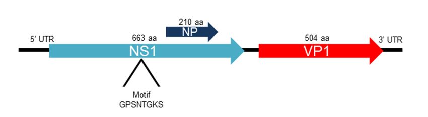
Viruses 2019, 11, 398                                                                                2 of 8

a wild Mus musculus from New York City [14], called murine kidney parvovirus (MH670588) was
recently shown to cause nephropathy in immunocompromised laboratory mice [18]. A recent survey of
eukaryotic genomes for chapparvovirus sequences has also shown the presence of a likely exogeneous
chapparvovirus genome in a fish (Gulf pipefish or Syngnathus scovelli) and of mostly defective germline
sequences in another fish (Tiger tail seahorse or Hippocampus comes) as well as in multiple invertebrates,
indicating an ancient origin for chapparvoviruses [19]. A phylogenetic analysis of NS1 also indicated
chapparvoviruses fall outside the traditional vertebrate-infecting Parvovirinae subfamily clade and
closer to that of a subset of members of the subfamily Densovirinae [19].
     Here an unexplained diarrhea outbreak among dogs was analyzed using viral metagenomics
after diagnostic tests were negative for common canine enteric pathogens. The genome of a novel
chapparvovirus was characterized and used to perform an epidemiological study to measure its
prevalence and possible clinical significance.

2. Materials and Methods

2.1. Sample Collection and Pathogen Screening
      Nine stool samples from dogs suffering from an infectious diarrhea outbreak in Colorado in
October 2017 were submitted to IDEXX Reference Laboratories, Inc. (Sacramento, CA, USA) for
pathogen testing. Fourteen dogs were involved in the initial outbreak which were identified by clinical
signs that started with steatorrhea, progressed to hemorrhagic diarrhea with additional symptoms
of lethargy, fever, and low lymphocyte counts pointing to a possible viral infection. At the time of
feces collection, the nine sampled dogs were at various stages of the disease, with two of the dogs
relapsing a month after initially experiencing parvo-like clinical signs. These stool samples were all
negative for Giardia spp., Cryptosporidium spp., Salmonella spp., Clostridium perfringens enterotoxin
gene (quantitative), Clostridium perfringens Alpha-toxin gene (quantitative), Canine enteric coronavirus
(alphacoronavirus), Canine Parvovirus 2 and Canine Distemper virus using the IDEXX canine diarrhea
profile real-time PCR tests.

2.2. Metagenomic Analysis
     Stool samples were grouped into three pools of three and vortexed in phosphate buffer saline (PBS)
with zirconia beads followed by microfuge centrifugation at 14,000 rpm for 10 min. The supernatants
were passed through a 0.45 µm filter (Millipore, Burlington, MA, USA) and digested with a mixture
of nuclease enzymes to enrich for viral particles [20,21]. RNA was extracted using the MagMAX kit
(ThermoFisher, Waltham, MA, USA) which was transcribed into cDNA using a random RT-PCR step.
The library was generated using the transposon-based Nextera™ XT Sample Preparation Kit (Illumina,
San Diego, CA, USA) which was deep sequenced with the MiSeq platform (250 bases, paired-end reads)
with dual barcoding. After demultiplexing the reads, they were trimmed and de novo assembled to
produce contigs [22]. Both singlets and contigs were compared to all eukaryotic viral protein sequences
in GenBank’s non-redundant database using BLASTx [23].

2.3. Genome Assembly and Diagnostic PCR
     Pairwise identity matrices using the amino acid sequence of the NS1 wasgenerated using
Geneious R11 (Newark, NJ, USA). Amino acid sequences from the NS1 region of all available
chapparvoviruses were aligned using MUSCLE and a Maximum likelihood tree was created using the
Jones–Taylor–Thorton matrix-based model with 1,000 bootstrap replicates in MEGA6.0 [24–26].
     A set of nested PCR primers were designed to screen for cachavirus in the nine stool samples
from the diarrheal outbreak. DNA was extracted from each individual stool sample using the QIAamp
MinElute Virus Spin kit (Qiagen, Hilden, Germany) and nested PCR assay primers were used to screen
for cachavirus DNA. The first round of primers CPV_625F (5’-CAA CTA GCC GAA TGC AGG GA-3’)
and CPV_948R (5’-CGA TAA CAT CCC CGG ACT GG-3’) were designed to target 323 nt of the NS1
Viruses 2019, 11, 398 3 of 8
region. The second round of primers CPV18_687FN (5’-AGC TCA GTT TGG CCC AGA TC-3’) and
CPV_911RN (5’-AGAGGGATCGCTGGATCTGT-3’) targeted a 224 nt region within the amplicon of
the first round of primers. The PCR (containing a final concentration of 0.2 µm of each primer, 0.2 mM
of dNTPs, 0.625 U of Amplitaq Gold® DNA polymerase (Applied Biosystems, Waltham, MA, USA),
1× PCR Gold buffer II, 1.5 mM of MgCl2 and 1 µL of DNA template in a 25 µl reaction) proceeded as
follows: 95 ◦ C for 5 min, 40 cycles of (95 ◦ C for 30 s, (52 ◦ C for the first round and 54 ◦ C for the second
round of primers) for 30 s, and 72 ◦ C for 30 s), followed by a final extension at 72 ◦ C for 7 min. PCR
products of the correct size were verified by gel electrophoresis and Sanger sequencing.
2.4. Prevalence
A proprietary real-time PCR assay with an amplification efficiency of 95% and an r2 value of
0.99 was developed by IDEXX. One gram of feces was added to 3 mL of lysis buffer and 600 µL
extracted into 200 µL nucleic acid eluate. Five µL of the eluate was tested in a PCR reaction with a
limit of detection of 10 copies DNA for a sensitivity of 1,600 copies per gram of feces. A chi-square test
comparing the proportion of healthy dogs that tested positive for cachavirus and those testing positive
with diarrhea in 2017 and 2018 was performed in order to determine if the difference in frequency was
statistically significant.
3. Results
Nine canine diarrheal samples from an unexplained outbreak of diarrhea were analyzed by viral
metagenomics using three pools of three diarrhea samples each. Based on the BLASTx results, one of the
three pools showed the presence of viral sequences most closely related to different chapparvoviruses
reported from different vertebrates (0.05% of all reads). Other eukaryotic viral sequences observed
were from Gyrovirus 4 (0.0003% of all reads), which has been reported in both chicken meat and
human stool [27], indicating that it likely represents a dietary contaminant, and Torque teno canis virus
(0.002% of reads), a common commensal canine blood virus [28].
Using de novo assembly and PCR paired with Sanger sequencing, a near complete genome of
4,123 bases containing the two main open reading frames of chapparvoviruses was generated (Figure 1,
panel A). The available genome consisted of a 516 bases partial 5’UTR followed by an ORF encoding a
663 aa non-structural protein (NS) possessing the ATP binding Walker loop motif GPSNTGKS followed
by a second ORF encoding a 505 aa viral capsid (VP) finishing with a 108 bases partial 3’UTR (Figure 1,
panel A). When NS1 and VP1 proteins were compared to all available parvovirus sequences, the
closest relative was from a Cameroonian fruit bat chapparvovirus (MG693107.1) [8] with an amino
acid identity of 61 and 63% respectively (Table S1). A 210 amino acid ORF that is missing a start
codon and is overlapping the NS1 ORF was also detected showing 57% identity to its homologue
protein in mouse kidney parvovirus (AXX39021) [18] (Figure 1, panel A). This NP ORF is widely
conserved among chapparvoviruses [19]. The 5’ UTR DNA sequence was 68% identical to that of the
bat parvovirus sequence (MG693107.1)). The virus was named cachavirus (canine chapparvovirus)
strain 1A (CachaV-1A).
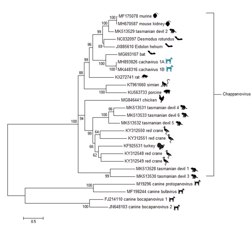
Distance matrices of the NS1 showed that the cachavirus is sufficiently divergent based on
ICTV criteria [6] (members of same species showing >85% NS1 identity) to qualify as a member
of a tentative new species Carnivore chapparvovirus species 1 in the proposed Chapparvovirus genus
(Table S1). A phylogenetic analysis of the NS1 ORF confirms its closest currently known relative is
from a Cameroonian fruit bat (Figure 1, panel B).
Using a nested PCR, the other 8 samples were tested for the presence of this virus which was
detected in a second diarrheic sample from that outbreak.Viruses 2019, 11, x FOR PEER REVIEW 4 of 8
Viruses 2019, 11, 398 4 of 8
A.
B.
Figure 1. (A) ORF map and location of nucleotide-binding Walker loop motif. (B) Maximum likelihood
Figure
tree of NS11.aa(A) ORF map
sequences and location of nucleotide-binding
of chapparvoviruses. Bar, 0.5 amino acidWalker loop motif.
substitutions (B)Bootstrap
per site. Maximum
likelihood
values below tree of NS1
60 were aa sequences of chapparvoviruses. Bar, 0.5 amino acid substitutions per site.
removed.
Bootstrap values below 60 were removed.
A larger set of canine fecal samples were then tested using a real-time PCR assay. Of 2,053 fecal
samples A tested,
larger set of canine
a total of 80 fecal
were samples
positive were
(Tablethen
1). tested using a submissions
Fecal sample real-time PCR assay.
from theOf 2,053
same fecal
time
samples
frame as thetested, a total
outbreak of 80 were
(Sept-Oct 2017)positive (Tablein1).
were tested Fecal
order to sample
determine submissions from the
the prevalence same time
of CachaV-1
framethat
during as the outbreak
time. Healthy (Sept-Oct
samples 2017) were flotation
from fecal tested insamples
order tosubmitted
determineinthe prevalence
2018 of CachaV-1
for preventive care
during that
screening weretime. Healthy
available. A samples
second setfrom fecal flotation
of diarrhea samplessamples submitted
that were in 2018
collected duringforthe
preventive
same time care
screening
frame as thewere available.
healthy samplesA second
was alsosetanalyzed
of diarrhea samples
to check forthat were collected
differences during the
in prevalence same
across time
time,
asframe
was a as
setthe healthy
of 83 bloody samples
diarrheawas also analyzed to check for differences in prevalence across time, as
samples.
was a setstool
Three of 83samples
bloody out diarrhea
of 203samples.
healthy animals tested positive, 32 were positive out of 802 diarrhea
Threefrom
submissions stoolSeptember
samples out of 203 of
to October healthy animals
2017, and tested
45 were positive,
positive out of32965were positive
diarrhea out of 802
submissions
diarrhea
from submissions
September to OctoberfromofSeptember
2018. None to of
October
the 83ofbloody
2017, and 45 were
diarrhea positive
samples out were
tested of 965positive
diarrhea
submissions
(Table 1). Whenfrom September
the fraction to October
of PCR positiveoffecal
2018. None of
samples thecompared
was 83 bloodybetween
diarrheathesamples
healthytested were
animals
positive
(1.47% (Table
positive) and1).those
When withthe fractiona of
diarrhea, PCR positive
statistically fecal difference
significant samples was(p < 0.05)
compared
could bebetween
detectedthe
healthy
with the 965animals
diarrhea(1.47%
casespositive)
from 2018 and thosepositive;
(4.66% p = 0.037),
with diarrhea, a statistically
but not with significant difference
the 803 diarrhea cases(p <
0.05) could be detected with the 965 diarrhea cases from 2018 (4.66% positive; p = 0.037), but not with
the 803 diarrhea cases collected in 2017 (4.0% positive; p = 0.08). When 2017 and 2018 diarrhea samplesViruses 2019, 11, 398 5 of 8
collected in 2017 (4.0% positive; p = 0.08). When 2017 and 2018 diarrhea samples were combined (4.35%
positive) and compared to the healthy group (1.47% positive), we measured a p-value of 0.05.
Cachavirus viral load as reflected by the Ct value of the real-time PCR were low across all four
cohorts, with Ct values ranging from 29 to 39 with an average value of 36 for all positive groups.
The five dog samples with the lowest Ct values (highest viral load) were then analyzed by viral
metagenomics. All five samples yielded cachavirus reads, but one yielded a near complete genome
(cachavirus [1B]). This sample also yielded 0.001% reads that were related to anelloviruses. None of
the other four animals showed the presence of other known mammalian viruses. The cachavirus-1B
genome showed 98% overall nucleotide identity with the index IDEXX-1A strain. The NS1 and VP
encoded protein showed 99 % identity.
Table 1. Real-time PCR results of cachavirus from four cohorts. A total of 2,053 samples were tested.
Diarrhea Diarrhea Total Diarrhea
Healthy Bloody
Submissions Submissions submissions
Stool Diarrhea
Sept-Oct 2017 Sept-Oct 2018 (2017 + 2018)
number 203 83 802 965 1767
# Tested positive 3 0 32 45 77
Frequency 1.47% 0% 3.99% 4.66% 4.35%
Average Ct 36.49 - 36.48 36.38 36.15
p-value of frequency
when compared to - - 0.08 0.037 0.05
healthy cohort
4. Discussion
There are currently five other known canine parvovirus species belonging to two genera of the
Parvoviridae family. Canine parvovirus 2 (CPV2) in the Carnivore protoparvovirus 1 species is a highly
pathogenic virus that is closely related to feline parvovirus (FPV), the cause of feline panleukopenia, and
can infect other carnivores such as coyotes, wolfs, raccoons and pumas [29]. Canine bufavirus, a second
protoparvovirus (in the species Carnivore protoparvovirus 2) was reported in 2018 in fecal and respiratory
samples from both healthy and dogs with signs of respiratory illness [30]. That same protoparvovirus
was recently reported as a frequent component of juvenile cats fecal and respiratory samples [31].
The canine minute virus (CnMV) in the Carnivore bocaparvovirus 1 species is less pathogenic than CPV2
but can cause diarrhea in young pups and is frequently found in the context of co-infections [32].
Distantly related to CnMV, a second canine bocavirus in the Carnivore bocaparvovirus 2 species was
sequenced in dogs with respiratory diseases [33]. A third bocavirus was then characterized from the
liver of a dog with severe hemorrhagic gastroenteritis [34].
Here, we describe the near complete genomes of two closely related cachaviruses, members of a
new tentative species (Carnivore chapparvovirus 1) in a proposed genus Chapparvovirus, the third genera
of viruses from the Parvoviridae family now reported in canine samples. The chapparvovirus was
found in only two animals of the initial nine sampled. Many of the dogs in the outbreak analyzed were
sampled more than 10 days after onset of clinical signs, increasing the possibility that they were no
longer shedding viruses. Additionally, diarrhea is one of the top reasons for veterinary visits and some
patients may have coincidentally presented with diarrhea from some other cause.
The two samples positive for CachaV-1 presented in the same week and were in the group
of patients with the most severe clinical signs, requiring plasma transfusion and more aggressive
supportive care. One of the two dogs, sampled at nine days after onset, died two days later. Because
of the variable and often delayed feces sampling, it was therefore not possible to determine a clear
disease association in this small group of diarrheic dogs (i.e., not all affected animals were shedding
cachavirus).
A possible role for the cachavirus infection in canine diarrhea was further tested by comparing
cachavirus DNA PCR detection in larger groups of healthy and diarrheic animals including a group ofViruses 2019, 11, 398 6 of 8
animals with bloody diarrhea. A statistically significant difference (p = 0.037) was seen when diarrhea
samples from 2018 were compared to the feces from healthy animals collected the same year. When
2017 diarrheic samples were compared to e 2018 healthy samples, the p-value was 0.08. When 2017
and 2018 diarrhea samples were combined and compared to the healthy samples, the p-value was
0.05. The association of cachavirus with diarrhea is therefore borderline and the detection of viral
DNA remains limited to ~4% of cases of diarrhea. The limited number of healthy samples available for
PCR limited the statistical power of this analysis and a larger sample size will be required for further
testing of disease association. The absence of detectable cachavirus DNA in 83 other cases of bloody
diarrhea was unexpected given the similar signs that developed in the initial outbreak. Detection of
viral DNA in feces may be related to timing of sample collection as shedding of the intestinal lining
during hemorrhagic diarrhea may preclude viral replication and fecal shedding.
The detection of this virus in multiple fecal samples, the absence of prior cachavirus reports from
tissues or fecal samples from other animals, and the confirmed vertebrate (murine) tropism of another
chapparvovirus (mouse kidney parvovirus) [18], support the tentative conclusion that cachavirus
infects dogs. Given its relatively low viral load and only borderline association with diarrhea, this virus’
possible role in canine diarrhea or other diseases will require further epidemiological studies. Because
viral nucleic acids in fecal samples may also originate from ingestion of contaminated food (rather
than replication in gut tissues), the tropism of cachavirus for dogs will require further confirmation
such as specific antibody detection, viral culture in canine cells, and/or evidence of replication in vivo
such as RNA expression in enteric tissues of dogs shedding cachavirus DNA.
Supplementary Materials: The following are available online at http://www.mdpi.com/1999-4915/11/5/398/s1,
Table S1: Percent identity between NS1 proteins of chapparvoviruses
Author Contributions: Conceptualization, E.F., M.A.S., P.Y., M.E. and C.L. and E.D.; Data curation, E.F., E.A.,
M.A.S., P.Y., M.E. and C.L.; Formal analysis, E.F., M.A.S., P.Y., and C.L.; Methodology, E.F.; Supervision, E.D.;
Validation, M.E.; Writing—original draft, E.F.; Writing—review and editing, E.F. and E.D.
Acknowledgments: The authors would like to acknowledge IDEXX and the Vitalant Research Institute for funding.
Conflicts of Interest: At the time of this research, M.A.S., P.Y., C.L. and M.E. were employees of IDEXX Reference
Laboratories, a division of IDEXX Laboratories, Inc., a company that provides veterinary diagnostics. IDEXX
funded a portion of the work described in the article. The other authors declare no conflicts of interest.
References
1. Cave, N.J.; Marks, S.L.; Kass, P.H.; Melli, A.C.; Brophy, M.A. Evaluation of a routine diagnostic fecal panel
for dogs with diarrhea. J. Am. Vet. Med. Assoc. 2002, 221, 52–59. [CrossRef]
2. Nandi, S.; Kumar, M. Canine parvovirus: Current perspective. Indian J. Virol. 2010, 21, 31–44. [CrossRef]
3. Conteville, L.C.; Zanella, L.; Marín, M.A.; Filippis, A.M.B.d.; Nogueira, R.M.R.; Vicente, A.C.P.;
Mendonça, M.C.L.d. Parvovirus B19 1A complete genome from a fatal case in Brazil. Mem. Inst. Oswaldo
Cruz 2015, 110, 820–821. [CrossRef]
4. Pérez, R.; Calleros, L.; Marandino, A.; Sarute, N.; Iraola, G.; Grecco, S.; Blanc, H.; Vignuzzi, M.; Isakov, O.;
Shomron, N. Phylogenetic and genome-wide deep-sequencing analyses of canine parvovirus reveal
co-infection with field variants and emergence of a recent recombinant strain. PLOS ONE 2014, 9, e111779.
[CrossRef] [PubMed]
5. Miranda, C.; Parrish, C.R.; Thompson, G. Canine parvovirus 2c infection in a cat with severe clinical disease.
J. Vet. Diagn. Invest. 2014, 26, 462–464. [CrossRef]
6. Cotmore, S.F.; Agbandje-McKenna, M.; Canuti, M.; Chiorini, J.A.; Eis-Hubinger, A.M.; Hughes, J.; Mietzsch, M.;
Modha, S.; Ogliastro, M.; Penzes, J.J.; et al. ICTV Virus Taxonomy Profile: Parvoviridae. J. Gen. Virol. 2019,
23, 367–368. [CrossRef]
7. Yang, S.; Liu, Z.; Wang, Y.; Li, W.; Fu, X.; Lin, Y.; Shen, Q.; Wang, X.; Wang, H.; Zhang, W. A novel rodent
Chapparvovirus in feces of wild rats. Virol. J. 2016, 13, 133. [CrossRef] [PubMed]
8. Yinda, C.K.; Ghogomu, S.M.; Conceicao-Neto, N.; Beller, L.; Deboutte, W.; Vanhulle, E.; Maes, P.; Van Ranst, M.;
Matthijnssens, J. Cameroonian fruit bats harbor divergent viruses, including rotavirus H, bastroviruses, and
picobirnaviruses using an alternative genetic code. Virus Evol. 2018, 4, vey008. [CrossRef] [PubMed]Viruses 2019, 11, 398 7 of 8
9. Baker, K.S.; Leggett, R.M.; Bexfield, N.H.; Alston, M.; Daly, G.; Todd, S.; Tachedjian, M.; Holmes, C.E.;
Crameri, S.; Wang, L.F.; et al. Metagenomic study of the viruses of African straw-coloured fruit bats: Detection
of a chiropteran poxvirus and isolation of a novel adenovirus. Virology 2013, 441, 95–106. [CrossRef] [PubMed]
10. Souza, W.M.; Romeiro, M.F.; Fumagalli, M.J.; Modha, S.; de Araujo, J.; Queiroz, L.H.; Durigon, E.L.;
Figueiredo, L.T.; Murcia, P.R.; Gifford, R.J. Chapparvoviruses occur in at least three vertebrate classes and
have a broad biogeographic distribution. J. Gen. Virol. 2017, 98, 225–229. [CrossRef]
11. Kapusinszky, B.; Ardeshir, A.; Mulvaney, U.; Deng, X.; Delwart, E. Case-Control Comparison of Enteric
Viromes in Captive Rhesus Macaques with Acute or Idiopathic Chronic Diarrhea. J. Virol. 2017, 91. [CrossRef]
[PubMed]
12. Reuter, G.; Boros, A.; Delwart, E.; Pankovics, P. Novel circular single-stranded DNA virus from turkey faeces.
Arch. Virol. 2014, 159, 2161–2164. [CrossRef] [PubMed]
13. Palinski, R.M.; Mitra, N.; Hause, B.M. Discovery of a novel Parvovirinae virus, porcine parvovirus 7,
by metagenomic sequencing of porcine rectal swabs. Virus Genes 2016, 52, 564–567. [CrossRef]
14. Williams, S.H.; Che, X.; Garcia, J.A.; Klena, J.D.; Lee, B.; Muller, D.; Ulrich, W.; Corrigan, R.M.; Nichol, S.;
Jain, K.; et al. Viral Diversity of House Mice in New York City. MBio 2018, 9, 01354–01317. [CrossRef]
[PubMed]
15. Chong, R.; Shi, M.; Grueber, C.E.; Holmes, E.C.; Hogg, C.; Belov, K.; Barrs, V.R. Fecal viral diversity of captive
and wild Tasmanian devils characterized using virion-enriched metagenomics and meta-transcriptomics.
J. Virol. 2019. [CrossRef] [PubMed]
16. Wang, Y.; Yang, S.; Liu, D.; Zhou, C.; Li, W.; Lin, Y.; Wang, X.; Shen, Q.; Wang, H.; Li, C.; et al. The fecal
virome of red-crowned cranes. Arch. Virol. 2019, 164, 3–16. [CrossRef] [PubMed]
17. Lima, D.A.; Cibulski, S.P.; Tochetto, C.; Varela, A.P.M.; Finkler, F.; Teixeira, T.F.; Loiko, M.R.; Cerva, C.;
Junqueira, D.M.; Mayer, F.Q.; et al. The intestinal virome of malabsorption syndrome-affected and unaffected
broilers through shotgun metagenomics. Virus Res. 2019, 261, 9–20. [CrossRef]
18. Roediger, B.; Lee, Q.; Tikoo, S.; Cobbin, J.C.A.; Henderson, J.M.; Jormakka, M.; O’Rourke, M.B.; Padula, M.P.;
Pinello, N.; Henry, M.; et al. An Atypical Parvovirus Drives Chronic Tubulointerstitial Nephropathy and
Kidney Fibrosis. Cell 2018, 175, 530–543.e524. [CrossRef] [PubMed]
19. Pénzes, J.J.; de Souza, W.M.; Agbandje-McKenna, M.; Gifford, R.J. An ancient lineage of highly divergent
parvoviruses infects both vertebrate and invertebrate hosts. bioRxiv 2019. [CrossRef]
20. Allander, T.; Emerson, S.U.; Engle, R.E.; Purcell, R.H.; Bukh, J. A virus discovery method incorporating
DNase treatment and its application to the identification of two bovine parvovirus species. Proc. Nat. Acad.
Sci. USA 2001, 98, 11609–11614. [CrossRef] [PubMed]
21. Victoria, J.G.; Kapoor, A.; Li, L.; Blinkova, O.; Slikas, B.; Wang, C.; Naeem, A.; Zaidi, S.; Delwart, E.
Metagenomic Analyses of Viruses in Stool Samples from Children with Acute Flaccid Paralysis. J. Virol. 2009,
83, 4642–4651. [CrossRef]
22. Deng, X.; Naccache, S.N.; Ng, T.; Federman, S.; Li, L.; Chiu, C.Y.; Delwart, E.L. An ensemble strategy
that significantly improves de novo assembly of microbial genomes from metagenomic next-generation
sequencing data. Nucleic Acids Res. 2015, 43. [CrossRef] [PubMed]
23. Zaretskaya, I.; Johnson, M.; McGinnis, S.; Raytselis, Y.; Merezhuk, Y.; Madden, T.L. NCBI BLAST: a better
web interface. Nucleic acids research 2008, 36, W5–W9. [CrossRef]
24. Tamura, K.; Stecher, G.; Peterson, D.; Filipski, A.; Kumar, S. MEGA6: Molecular Evolutionary Genetics
Analysis version 6.0. Mol. Boil. Evol. 2013, 30, 2725–2729. [CrossRef]
25. Edgar, R.C. MUSCLE: Multiple sequence alignment with high accuracy and high throughput. Nucleic Acids
Res. 2004, 32, 1792–1797. [CrossRef]
26. Jones, D.T.; Taylor, W.R.; Thornton, J.M. The rapid generation of mutation data matrices from protein
sequences. Comput. Appl. Biosci. 1992, 8, 275–282. [CrossRef]
27. Chu, D.K.; Poon, L.L.; Chiu, S.S.; Chan, K.H.; Ng, E.M.; Bauer, I.; Cheung, T.K.; Ng, I.H.; Guan, Y.; Wang, D.;
et al. Characterization of a novel gyrovirus in human stool and chicken meat. J. Clin. Virol. 2012, 55, 209–213.
[CrossRef] [PubMed]
28. Okamoto, H.; Takahashi, M.; Nishizawa, T.; Tawara, A.; Fukai, K.; Muramatsu, U.; Naito, Y.; Yoshikawa, A.
Genomic characterization of TT viruses (TTVs) in pigs, cats and dogs and their relatedness with species-specific
TTVs in primates and tupaias. J. Gen. Virol. 2002, 83, 1291–1297. [CrossRef] [PubMed]Viruses 2019, 11, 398 8 of 8
29. Allison, A.B.; Kohler, D.J.; Fox, K.A.; Brown, J.D.; Gerhold, R.W.; Shearn-Bochsler, V.I.; Dubovi, E.J.;
Parrish, C.R.; Holmes, E.C. Frequent cross-species transmission of parvoviruses among diverse carnivore
hosts. J. Virol. 2013, 87, 2342–2347. [CrossRef]
30. Vito, M.; Gianvito, L.; Eszter, M.K.; Szilvia, M.; Renáta, V.-K.; Eszter, K.; Barbara Di, M.; Michele, C.; Nicola, D.;
Canio, B.; et al. Novel Parvovirus Related to Primate Bufaviruses in Dogs. Emerg. Infect. Dis. J. 2018, 24,
1061. [CrossRef]
31. Diakoudi, G.; Lanave, G.; Capozza, P.; Di Profio, F.; Melegari, I.; Di Martino, B.; Pennisi, M.G.; Elia, G.;
Cavalli, A.; Tempesta, M.; et al. Identification of a novel parvovirus in domestic cats. Vet. Microbiol. 2019,
228, 246–251. [CrossRef] [PubMed]
32. Carmichael, L.E.; Schlafer, D.H.; Hashimoto, A. Pathogenicity of minute virus of canines (MVC) for the
canine fetus. Cornell Vet. 1991, 81, 151–171. [PubMed]
33. Kapoor, A.; Mehta, N.; Dubovi, E.J.; Simmonds, P.; Govindasamy, L.; Medina, J.L.; Street, C.; Shields, S.;
Lipkin, W.I. Characterization of novel canine bocaviruses and their association with respiratory disease.
J. Gen. Virol. 2012, 93, 341–346. [CrossRef] [PubMed]
34. Li, L.; Pesavento, P.A.; Leutenegger, C.M.; Estrada, M.; Coffey, L.L.; Naccache, S.N.; Samayoa, E.; Chiu, C.;
Qiu, J.; Wang, C.; et al. A novel bocavirus in canine liver. Virol. J. 2013, 10, 10–54. [CrossRef] [PubMed]
© 2019 by the authors. Licensee MDPI, Basel, Switzerland. This article is an open access
article distributed under the terms and conditions of the Creative Commons Attribution
(CC BY) license (http://creativecommons.org/licenses/by/4.0/).You can also read