Honey Bee as Alternative Medicine to Treat Eleven Multidrug-Resistant Bacteria Causing Urinary Tract Infection during Pregnancy - MDPI
←
→
Page content transcription
If your browser does not render page correctly, please read the page content below
Scientia
           Pharmaceutica

Article
Honey Bee as Alternative Medicine to Treat Eleven
Multidrug-Resistant Bacteria Causing Urinary Tract
Infection during Pregnancy
Mabrouka Bouacha 1, *        ID
                                  , Hayette Ayed 2 and Nedjoud Grara 2
 1   Laboratory of Biochemistry and Microbiology, Department of Biochemistry, Faculty of Sciences,
     University of Badji Mokhtar, 23000 Annaba, Algeria
 2   Department of Biology, Faculty of Biology, University 8 May 1945, 24000 Guelma, Algeria;
     rymadafri@yahoo.fr (H.A.); grara120@yahoo.fr (N.G.)
 *   Correspondence: bouacha.mabrouka@yahoo.fr; Tel.: +213-664-42-94-86

 Received: 3 March 2018; Accepted: 10 April 2018; Published: 13 April 2018

 Abstract: Medicinal benefits of honey bee have been recognized in the medical community since
 ancient times as a remedy for many diseases and infections. This study aimed to investigate the
 in vitro susceptibility of 11 multidrug-resistant bacterial strains, isolated from urinary tract infections
 of pregnant women, to six honey samples collected from different localities in the east of Algeria.
 The evaluation of the antibacterial activity was performed by the well method followed by the
 broth dilution method using two-fold dilutions of each honey sample ranging from 2.5 to 80% (w/v).
 The results obtained in this study revealed that all tested honeys exhibited potent antibacterial
 activity against the tested strains. The diameters of inhibition ranged from 19.67 to 53.33 mm,
 with minimum inhibitory concentrations (MICs) ranging from 2.5 to 40% (w/v) and minimum
 bactericidal concentration (MBCs) varied between 2.5 and 80% (w/v). Gram-positive bacteria were
 found to be more susceptible than Gram-negative bacteria with diameters ranging from 43.33 to
 53.33 mm; MIC and MBC values ranged from 2.5 to 5% (w/v). The P. aeruginosa strain was found
 to be less susceptible than other strains with inhibitory diameters ranging from 19.67 to 27.33 mm;
 MICs ranged from 20 to 40% and MBCs ranged from 20 to 80% (w/v). This contribution has provided
 a broad overview of the antibacterial activity of Algerian honey and shown that honey bee has great
 potential for therapeutic use as an alternative therapy for urinary tract infection treatment which is
 safe and efficient during pregnancy.

 Keywords: honey bee; urinary tract infections; pregnant women; antibacterial activity;
 alternative medicine

1. Introduction
      Urinary tract infections (UTIs) are among the most common bacterial infections,
affecting approximately 150 million people worldwide each year [1]. UTIs are most frequently caused
by uropathogens from fecal flora (i.e., Escherichia coli) that ascend the urethra to infect the bladder [2].
      UTIs are considered as a serious public health burden and significantly disturb the quality of life
of affected persons. UTIs are accompanied by complications such as pyelonephritis and bacteraemia
which, if not treated carefully, may lead to development of permanent renal damage and significant
mortality [3].
      These infections can affect both men and women of all ages; however, women are more likely to
experience this infection than men [4]. It was estimated that around 11% of women report at least one
physician-diagnosed UTI per year and 20–30% report multiple recurrences (rUTI). This is mostly due

Sci. Pharm. 2018, 86, 14; doi:10.3390/scipharm86020014                       www.mdpi.com/journal/scipharm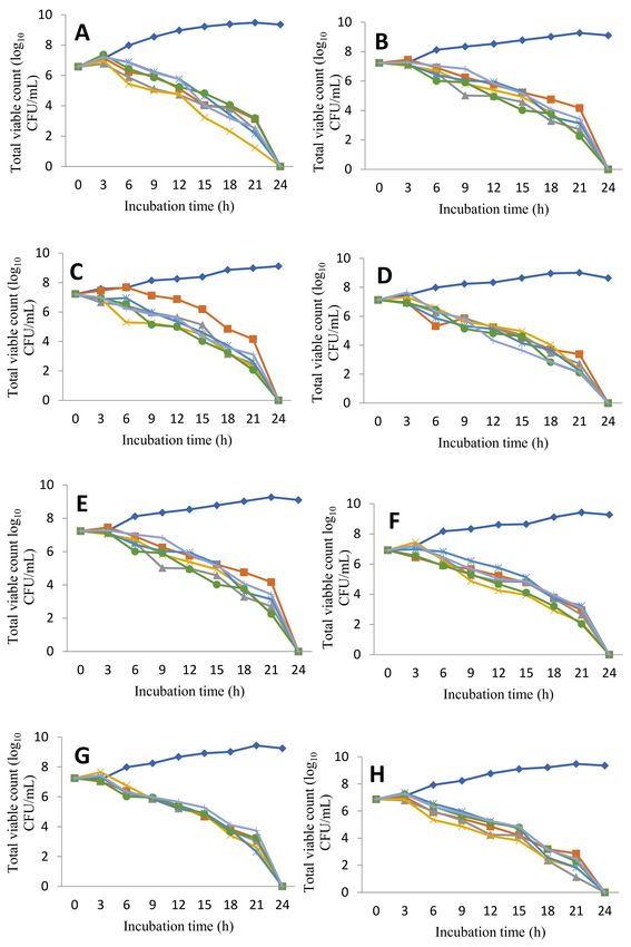
Sci. Pharm. 2018, 86, 14                                                                             2 of 11

to their shorter female urethral length compared with male urethra as a result of an easier point of
access for bacterial pathogens.
       On the other hand, during pregnancy, the urinary tract of women undergoes anatomical and
physiological changes that can result in symptoms and conditions affecting both the mother and the
fetus [5]. The hormonal and anatomo-physiological changes facilitate the growth and dissemination of
bacteria in the maternal urinary tract [4,5]. They are mostly limited to the lower urinary tract and often
arise from a single type of bacteria: Escherichia coli, which is a floral member of the colon. Other common
urinary tract infection pathogens include enterobacterial strains (Klebsiella spp., Enterobacter spp. and
Proteus mirabilis), and staphylococci and Pseudomonas aeruginosa can be isolated [6].
       All bacteriuria in pregnancy should be treated and antimicrobial choices should reflect safety
for both the mother and the fetus [7]. In simple urinary infection, nitrofurans and trimethoprim are
antibiotics of first choice, and fosfomycin and β-lactamins are a second-line of treatment. In outpatient
practice, fluoroquinolones are normally reserved for the treatment of complicated urinary tract
infections. These medicines are considered safe and effective for the mother and the fetus during
pregnancy [8,9]; however, recent research studies have demonstrated increasing resistance to these
drugs [10,11]. Currently, the evolution and spread of antibiotic resistance bacteria is of great concern
to the global health community [12]. To overcome this issue, scientists are searching for alternative
efficient antimicrobial agents. The use of traditional medicine to treat a number of health problems
(i.e., infections) in a natural way has been applied since ancient times. Honey bee (Apis mellifera) is one
of the oldest traditional medicines, considered to be a traditional remedy to treat burns, infected and
non-healing wounds and ulcers, boils, pilonidal sinus, venous and diabetic foot ulcers, and it has been
shown that honey bee is capable of clearing infection from the wound and improving tissue healing.
In addition, honey bee may possess anti-inflammatory activity and stimulate immune responses within
a wound. The overall effect is to reduce infection and to enhance wound healing in burns, ulcers and
other cutaneous wounds [13,14].
       Honey bee contains about 200 substances, including amino acids, vitamins, minerals and enzymes,
but it primarily contains sugar and water. The main carbohydrate constituents of honey are fructose
and glucose [15].
       The potential antibacterial agent in honey bee exhibits inhibitory effects towards approximately
60 bacterial species causing infections in the human body; including aerobes and anaerobes,
Gram-positive and Gram-negative bacteria [16]. Different factors are responsible for the antimicrobial
activity of honey, which include its sugar content, which is high enough to hinder microbial growth.
This is believed to be a result of its osmotic effect, which prevents the growth of bacteria and therefore
promotes healing. Honey bee is hygroscopic, meaning that it draws moisture out of the environment
and dehydrates the bacteria with the aid of its hyperosmolar properties [17]. The low pH level of
honey (mean 4.4) is unsuitable for bacterial growth and it can reduce wound colonization or infection.
In addition, the antibacterial activity of honey bee may be due to hydrogen peroxide activity, which
is continuously produced by the action of glucose oxidase; it is able to interact with bacterial cell
proliferative signals, and thus affects bacterial growth even when honey is diluted [16–18]. Some floral
sources such as flavonoids and aromatic acids provide additional antibacterial components in honey
bee [19].
       The Algerian populations have used honey bee frequently as a cure for several diseases. It is
worth mentioning here that the spread of the use of honey in traditional and modern medicine has
origins linked to the religious beliefs of Muslim people, where many Quranic and Islamic texts reveal
that honey is a proven remedy [20].
       The antibacterial activity of honeys from different countries has been extensively studied [21].
However, to the best of our knowledge, this is the first study that reported the clear effect of honey
against many pathogens isolated from urinary tract infection during pregnancy. It aimed to evaluate
the antibacterial activity of honey bee against the antibiotic-resistant bacteria isolated from the urine of
pregnant women suffering from urinary tract infections.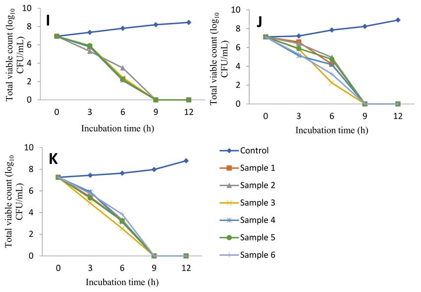
Sci. Pharm. 2018, 86, 14 3 of 11
2. Materials and Methods
2.1. Honey Bee Samples
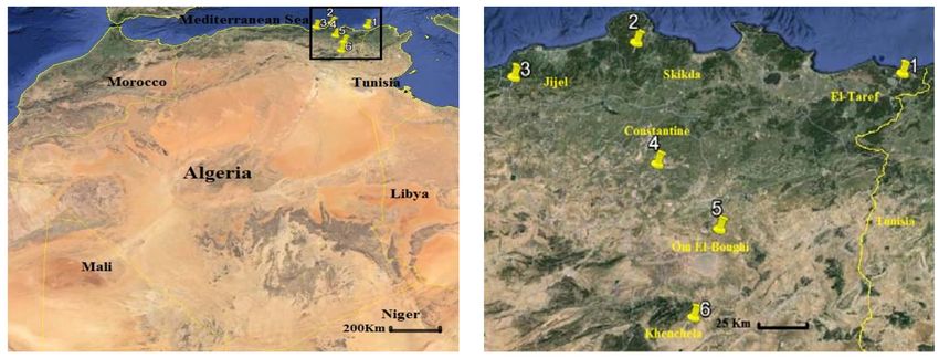
Honey samples of the current study were collected by farmers from six localities in the east
of Algeria during 2017. A map indicating the locations of the honey samples is shown in Figure 1.
Six freshly harvested, natural, untreated and unpasteurized honey samples were used. Each sample
was collected in a sterile universal container and kept at 4 ◦ C in the dark until usage.
The following honey concentrations were prepared in sterile saline solution: 2.5%, 5%, 10%,
20%, 40%, 80% (w/v) and undiluted honey. Each honey sample was filtered through a 0.22 µm filter
(Millipore, Nunc, Paramus, NJ, USA).
Figure 1. Geographic locations of the study area including honey harvesting sites.
2.2. Strains
Honey samples were screened for their antibacterial activity against 11 bacterial strains
isolated from the urine of pregnant women suffering from urinary infection at Ibn Rochd
Hospital, Annaba, situated in east of Algeria. These strains are as follows: Escherichia coli,
Enterobacter aerogenes, Klebsiellaoxytoca, Klebsiella pneumoniae, Proteus mirabilis, Proteus vulgaris,
Citrobacter koseri, Pseudomonas aeruginosa, Enterococcus faecalis, Staphylococcus aureus and Staphylococcus
saprophyticus. All tested strains were identified by conventional methods of microbiology (Gram staining,
oxidase and catalase test, analytical profile index (API) 20E, API 20NE, API STAPH and API 20 STREP)
(Biomerieux, Paris, France).
An inoculum of each strain was prepared, and the turbidity of the suspension was adjusted to
achieve 0.5 McFarland (equivalent to that of 1.5 × 10⁸ colony-forming units (CFU)/mL) with the
absorbance range of 0.08 to 0.1 by UV-Vis spectrophotometer at wave length of 620 nm.
All bacterial strains were subjected to antibiotic sensitivity tests by the Kirby Bauer’s disc diffusion
method according to the Clinical and Laboratory Standards Institute [22] using Mueller Hinton agar
medium (Biomerieux, Paris, France). The tested strains were selected because they are resistant to
antibiotics used in the treatment of urinary infection during pregnancy.
2.3. Honey Samples Analysis
2.3.1. Sensory Analysis
Sensory analysis is a tool to evaluate color and flavors of honey in order to understand if
there is a relationship between the color, flavor and antibacterial activity of different honey samples.
Sensory analysis is achieved according to the methodology described previously [23]. The samples
were tested by a panel of 20 assessors that evaluate the taste and the color of honey samples.Sci. Pharm. 2018, 86, 14 4 of 11
2.3.2. pH Measurement
The pH measurement was carried out by a pH meter (HI 98127, Hanna instruments) of a 50% (w/v)
solution of each honey sample.
2.3.3. Color Intensity
The absorbance of the honey samples was determined by the method of Beretta et al. [24].
The honey samples were diluted to 50% (w/v) with warm water (45–50 ◦ C) and the solution was
filtered using a 0.45 µm filter to eliminate large particles. The absorbance was measured using a UV-Vis
spectrophotometer (T80 UV/VIS, PG instrument, England), at 450 and 720 nm and the difference in
absorbance was expressed as mAU.
2.4. Antibacterial Activity
2.4.1. Wells Assay
Wells (6 mm of diameter) were prepared in Mueller Hinton agar plates: these plates were
inoculated by bacterial suspension and 150 µL of tested honey was added to each well. A hole filled
with sterile water served as control. Plates were incubated at 37◦ C for 24h. The antibacterial activity
of the samples was compared on the basis of the radius of a clear inhibition zone around the wells.
The results are shown as mean values from triplicate measurements.
2.4.2. Spectrophotometric Assay for MIC Determination
Minimum inhibitory concentrations (MICs) were determined using test described previously by
Patton et al. [25]. A volume of 0.5 mL of standardized culture was added to 4.5 mL of tested honey,
at each of the concentrations stated above. Control tubes containing broth only (negative or sterility
control) or bacteria and broth (positive control) were used. Tubes were incubated in the dark at 37◦ C
with shaking at 150 rpm for 24 h. The optical density was determined just prior to incubation (T0 )
and again after 24h incubation (T24 ) at 620 nm. The percent inhibition of growth was thus determined
using the formula: percent inhibition (%) = 1− (OD test/OD of corresponding control) × 100. The MIC
is reported as the lowest concentration of honey which results in 100% inhibition of growth of the
test bacteria.
2.4.3. Minimum Bactericidal Concentration Determination
The minimum bactericidal concentration (MBC) values were read as the lowest concentration of
honey required for a 99.9% reduction in the viable strains. For determining MBC values, an aliquot
(0.1 mL) of MIC mixtures that showed no growth was inoculated onto Mueller Hinton plates and
incubated at 37 ◦ C for 24 h.
2.4.4. Time-Kill Assay
The time-kill assays were performed according to the method described previously [26]. 2 mL
of honey was taken in sterile test tubes, one tube was inoculated with 20 µL of broth culture of the
test bacterium in an initial concentration of approximately 107 CFU/mL and the other tube was not
inoculated (control). The tubes were incubated at 37◦ C with constant stirring (200rpm). Broth aliquots
were collected at different time points, serially diluted in saline solution, plated on nutrient agar media
and grown for 24h at 37 ◦ C to determine the total CFUs in each tube.
2.5. Statistical Analysis
The results are reported as mean ± standard deviation (m ± SD). Multiple comparisons of
means (Tukey HSD tests) were performed after each analysis of variance (ANOVA) to distinguishSci. Pharm. 2018, 86, 14 5 of 11
homogeneous groups among six origins using the software GraphPad Prism version 5 (Trial),
Mars 12(2007).
3. Results
3.1. Antibiotic Sensitivity
The results of the susceptibility of urinary infection strains to prescribed antibiotics are
summarised in Table 1. The results showed that all tested bacteria exhibited varying degrees of
multidrug resistance of standard antibiotics used in urinary infections. The uropathogenic strains
revealed the presence of high levels of multiple antimicrobial resistances.
Table 1. Susceptibility of urinary infection strains to prescribed antibiotics.
Susceptibility to Prescribed Antibiotics
Strain β-Lactams Cephalosporin Fluoroquinolone Aminglycosides Others
OX AM AMC CN CF OF NA CIP GM TM NIT FOS SXT
E. coli ND R R R R S S S S R R S R
E. aerogenes ND R R R R S S S R R R R R
K. oxytoca ND R R R R S S S S R R R R
K. pneumoniae ND R R R R R R R R R R R R
P. mirabilis ND R R R R R R R R R R R R
P. vulgaris ND R R R R R R R R R R R R
C. koseri ND R R R R S S S S R R R R
P. aeruginosa ND R R R R R R R R R R R R
S. aureus R ND ND R R R R R S R R R R
S. saprophyticus S ND ND R R S S S S R R S R
E. faecalis R ND ND R R R R R R R R R R
Susceptibility
33.3 0 0 0 0 45.4 45.4 45.4 45.4 0 0 18.2 0
Percentage (%)
OX: oxacilline, AM: amoxicillin, AMC: amoxicillin–clavulanic-acid, CN: cefazolin, CF: cefexim, OF: ofloxacin, NA:
nalidixicacid, CIP: ciplofloxacin, GM: gentamicine, TM: tobramicine, NIT: nitrofuranto ne, FOS: fosfomicine, SXT:
sulfamethoxazol-trimethoprim, R: resistant, S:susceptible, ND: not determined.
3.2. Honey Analysis
The taste, pH values and color intensity of honey samples are reported in Table 2. Sample 2 has
a bitter taste, whereas other samples have a sweet taste. As shown, all tested honey samples were
acidic; the pH values ranged from 3.19 to 4.54. The color intensity of honey samples varied from 352 to
982 mAU for the light honey samples and from 1654 to 1965 mAU for the dark honey samples.
Table 2. Taste, pH and color intensity of honey samples.
Honey Sample Taste pH Color Color Intensity (mAU)
Sample 1 Sweet 3.44 Light brown 742
Sample 2 Bitter 4.54 Dark brown 1965
Sample 3 Sweet 3.86 Brown 829
Sample 4 Sweet 3.19 Dark brown 1654
Sample 5 Sweet 3.54 Brown 982
Sample 6 Sweet 4.02 Light brown 352
3.3. Inhibitory Diameters Determination
The results of inhibitory diameters of all honey bee samples are shown in Table 3. All tested honeys
had a measurable antibacterial activity against all tested bacteria; the diameters of inhibition ranged
from 19.67 to 53.33 mm. The results demonstrated that all honey samples had similar antibacterial
activities (p > 0.05). However, there were highly significant differences for the susceptibility of
different strains (p < 0.001): the inhibitory diameters of P. aeruginosa strain were less than other strains,Sci. Pharm. 2018, 86, 14 6 of 11
which ranged from 19.67 to 27.33 mm. However, Gram-positive bacteria had the most important
inhibitory diameters, ranging from 43.33 to 53.33 mm.
Table 3. Mean diameter (mm) of inhibition by honey samples against uropathogenic strains.
Diameter of Inhibition (mm±SD)
Strain
Sample 1 Sample 2 Sample 3 Sample 4 Sample 5 Sample 6
E. coli 30.33 ± 1.56 32 ± 0.67 36.67 ± 1.11 33.67 ± 0.44 30.00 ± 0.67 30.00 ± 0.67
E. aerogenes 30.33 ± 1.11 31.33 ± 0.89 37.00 ± 0.67 33.67 ± 0.89 31.33 ± 1.11 31.00 ± 0.67
K. oxytoca 28.33 ± 1.11 31.33 ± 0.44 36.00 ± 1.33 33.33 ± 0.44 29.00 ± 1.33 30.67 ± 1.56
K. pneumoniae 29.67 ± 1.11 32.33 ± 0.44 37.33 ± 0.89 35.00 ± 0.67 29.33 ± 0.44 29.33 ± 0.44
P. mirabilis 30.00 ± 0.67 31.33 ± 0.89 36.33 ± 1.11 33.33 ± 1.11 30.33 ± 0.44 28.67 ± 0.89
P. vulgaris 30.33 ± 1.11 32.33 ± 0.44 35.67 ± 1.56 31.00 ± 0.67 29.67 ± 0.44 29.67 ± 0.89
C. koseri 29.00 ± 1.33 30.33 ± 0.44 34.67 ± 0.89 31.67 ± 1.56 29.00 ± 0.67 29.00 ± 0.67
P. aeruginosa 19.67 ± 1.56 24.33 ± 0.89 27.33 ± 0.89 26.00 ± 0.67 20.33 ± 0.89 20.33 ± 0.44
S. aureus 47.667 ± 1.11 48.67 ± 0.89 50.67 ± 1.11 49.67 ± 0.44 47.67 ± 0.44 47.33 ± 1.56
S. saprophyticus 48.33 ± 0.89 50.00 ± 0.67 53.33 ± 0.44 50.33 ± 0.89 47.67 ± 0.44 47.67 ± 0.89
E. faecalis 43.67 ± 0.89 44.00 ± 0.67 46.67 ± 0.44 44.00 ± 0.67 43.33 ± 0.89 43.67 ± 0.44
3.4. Determination of MICs and MBCs
From Table 4, it can be seen that the growth of all tested bacteria was inhibited by different
samples of honey at concentrations of 5 to 20% (w/v), except P. aeruginosa, which has an MIC ranging
from 20 to 40% (w/v) and an MBC ranging from 20 to 80% (w/v). However, Gram-positive bacteria
were inhibited by all tested honey bee at concentration ranged from 2.5% to 5% (w/v).
Table 4. Minimum inhibitory concentration (MIC) and minimum bactericidal concentration (MBC)
values of honey samples against uropathogen strains (% w/v).
Sample 1 Sample 2 Sample 3 Sample 4 Sample 5 Sample 6
Strain
MIC MBC MIC MBC MIC MBC MIC MBC MIC MBC MIC MBC
E. coli 10 10 10 10 5 10 10 10 10 10 10 10
E. aerogenes 10 10 10 10 5 10 10 10 10 10 10 10
K. oxytoca 20 20 10 20 5 20 10 20 20 20 10 20
K. pneumoniae 20 20 10 20 5 20 5 20 20 20 20 20
P. mirabilis 10 10 10 10 5 10 10 10 10 10 20 10
P. vulgaris 10 10 10 10 5 10 10 10 20 10 20 10
C. koseri 20 40 10 40 5 40 10 40 20 40 20 40
P. aeruginosa 40 80 20 40 20 40 20 20 40 80 40 80
S. aureus 2.5 2.5 2.5 2.5 2.5 2.5 2.5 2.5 2.5 2.5 2.5 2.5
S. saprophyticus 2.5 2.5 2.5 2.5 2.5 2.5 2.5 2.5 2.5 2.5 2.5 2.5
E. faecalis 5 5 5 5 2.5 5 5 5 5 5 5 5
3.5. Time Kill Curve
The results of the time-kill curve are indicated in Figure 2. The bactericidal action of honey bee
seems to be different according to the bacterial strain; some strains were destroyed after nine hours
of incubation (Gram-positive bacteria), but others were destroyed by the same sample of honey bee
after 24 h.Sci. Pharm. 2018, 86, 14 7 of 11
Figure 2. Cont.Sci. Pharm. 2018, 86, 14 8 of 11
Figure 2. Time-kill curve showing in vitro bactericidal effect of six Algerian honey bee samples on
multidrug resistant bacteria causing urinary tract infection during pregnancy. (A) E. coli, (B) E.aerogens,
(C) K. oxytoca, (D) K. pneumoniae, (E) P. mirabilis, (F) P. vulgaris, (G) C. koseri, (H) P. aeruginosa, (I) S. aureus,
(J) S. saprophyticus, (K) E.faecalis.
4. Discussion
Antibacterial susceptibility testing, as illustrated in Table 1, revealed that all tested strains
exhibited a high level of resistance to standard antibiotics used in urinary tract infections; similar results
were previously reported [9]. However, some strains had poor sensitivity to gentamycin (45.4%) and
to flouroquinolones (45.5%). Nevertheless, widespread usage may lead to resistance against these
antibiotics. There have been no large-scale studies of the safety or risk of antibiotic use during
pregnancy. However, the use of sulfonamide, trimethoprim and nitrofuran is known to increase
the risk of neural-tube defects, cardiovascular defects, oral defects and urinary tract defects of
birth [27,28]. Indeed, the use of aminoglycoside is known to be nephrotoxic to the fetus and should
therefore be avoided during pregnancy. In addition, the use of macrolides (excluding erythromycin),
quinolones, tetracyclines, during early pregnancy was associated with an increased risk of spontaneous
abortion [29,30]. Thus, there is an urgent requirement for the development of new drugs constituting
an effective and safe treatment which are not dangerous for both mother and fetus.
From Table 2, the analysis of honey samples showed that all of the tested Algerian honey samples
were acidic in nature, with pH values that varied between 3.19 and 4.54. It was reported previously
that the pH values of Algerian honey ranged from 3.70 to 4.00 [31] and 3.96 to 4.34 [32]. The acidity
of any honey is directly related to the floral sources that created it. Honey bee contains a number of
different acids, including about 18 amino acids, many different organic acids, as well as aliphatic and
aromatic acids. The aromatic acids greatly contribute to the flavor of honey bee.
The absorbance of 50% (w/v) honey solutions was varied from 352 to 982 mAU for the light
honeys and from 1654 to 1965 mAU for the dark and brown honeys. Honey samples from other
countries were reported to have absorbance values between 25 and 3413 mAU in Italian honey [24],
between 254 and 2034 mAU in Bangladeshi honey [33] and between 524–1678 mAU in IndianSci. Pharm. 2018, 86, 14 9 of 11
honeys [34]. This marked difference of color intensity might be a reliable index of the presence
of pigments with antioxidant activities, such as carotenoids and some flavonoids, which are known to
have antioxidant properties [35].
All honey samples have a sweet taste except Sample 2, which exhibited the highest color intensity,
and has a bitter taste. The differing taste and color of honey bee directly depend on the flower foraged
by the bee. There are a huge variety of flavors and colors in honey bee, depending on its origin.
The analysis of the diameter of inhibition values (in Table 3) showed that all tested honeys had
potent antibacterial activity against all examined bacteria, and there highly significant differences
for the susceptibility of different strains (pSci. Pharm. 2018, 86, 14 10 of 11
is related to the complex composition of honey bee, which contains a combination of components that
may act in a synergistic manner to compromise the resistance; this makes honey bee a very promising
topical antimicrobial agent against the infection of antibiotic-resistant bacteria.
Author Contributions: (M.B) carried out the experimental part and wrote the manuscript. (M.B) and (H.A.)
conceived of the presented idea. (H.A.) and (N.G.) developed the theory and performed the computations.
(M.B) and (N.G.) verified the analytical methods and supervised the findings of this work. All authors discussed
the results and contributed to the final manuscript.
Conflicts of Interest: The authors declare no conflict of interest.
References
1. Curtiss, N.; Meththananda, I.; Duckett, J. Urinary tract infection in obstetrics and gynaecology.
Obstet. Gynaecol. Reprod. Med. 2017, 27, 261–265. [CrossRef]
2. Flores-Mireles, A.L.; Walker, J.N.; Caparon, M.; Hultgren, S.J. Urinary tract infections: Epidemiology,
mechanisms of infection and treatment options. Nat. Rev. Microbiol. 2015, 13, 269–284. [CrossRef] [PubMed]
3. Turpen, H.C. Frequent Urinary Tract Infection. Physician Assist. Clin. 2018, 3, 55–67. [CrossRef]
4. Michelim, L.; Bosi, G.R.; Comparsi, E. Urinary Tract Infection in Pregnancy: Review of Clinical Management.
J. Clin. Nephrol. Res. 2016, 3, 1030–1037.
5. Souza, R.B.; Trevisol, D.J.; Schuelter-Trevisol, F. Bacterial sensitivity to fosfomycin in pregnant women with
urinary infection. Braz. J. Infect. Dis. 2015, 19, 319–323. [CrossRef] [PubMed]
6. Usta, T.A.; Dogan, O.; Ates, U.; Yucel, B.; Onar, Z.; Kaya, E. Comparison of single-dose and multiple-dose
antibiotics for lower urinary tract infection in pregnancy. Int. J. Gynaecol. Obstet. 2011, 114, 229–233.
[CrossRef] [PubMed]
7. Glaser, A.P.; Schaeffer, A.J. Urinary tract infection and bacteriuria in pregnancy. Urol. Clin. N. Am. 2015,
42, 547–560. [CrossRef] [PubMed]
8. Ronald, A.; Nicolle, L.E.; Harding, G. Single dose treatment failure in women with acute cystitis.
Infection 1992, 20, S276–S279. [CrossRef] [PubMed]
9. Sibi, G.; Pinki, K.; Kabungulundabungi, N. Antibiotic sensitivity pattern from pregnant women with urinary
tract infection in Bangalore, India. Asian Pac. J. Trop. Med. 2014, 7, S116–S120. [CrossRef]
10. Hecini-Hannachi, A.; Bentchouala, C.; Lezzar, A.; Laouar, H.; Benlabed, K.; Smati, F. Multidrug-resistant
bacteria isolated from patients hospitalized in intensive care unit in university hospital of Constantine,
Algeria (2011–2015). Afr. J. Microbiol. Res. 2016, 10, 1328–1336. [CrossRef]
11. Laxminarayan, R.; Matsoso, P.; Pant, S.; Brower, C.; Rottingen, J.A.; Klugman, K.; Davies, S. Access to
effective antimicrobials: A worldwide challenge. Lancet 2016, 387, 168–175. [CrossRef]
12. Taylor, P.K.; Amy, T.Y.Y.; Robert, E.W.H. Antibiotic resistance in Pseudomonas aeruginosa biofilms: Towards the
development of novel anti-biofilm therapies. J. Biotech. 2014, 12, 1–10. [CrossRef] [PubMed]
13. Manndal, M.D.; Mandal, S. Honey: Its medicinal property and antibacterial activity. Asian Pac. J. Trop. Biomed.
2011, 154–160. [CrossRef]
14. Lusby, P.E.; Coombes, A.B.; Wilkinson, J.M. Honey: A Potent Agent for Wound Healing? J. Wound Ostomy
Cont. Nurs. 2002, 29, 295–300. [CrossRef]
15. Eteraf-Oskouei, T.; Najafi, M. Traditional and Modern Uses of Natural Honey in Human Diseases: A Review.
Iran. J. Basic Med. Sci. 2013, 16, 731–742. [PubMed]
16. Kateel, R.; Gopalakrishna, B.K.; Baliga, S.; Augustine, A.J.; Ullal, S.; Adhikari, P. Antibacterial action of
Tropical honey on various bacteria obtained from diabetic foot ulcer. Complement. Ther. Clin. Pract. 2017,
30, 29–32. [CrossRef] [PubMed]
17. Yaghoobi, R.; Kazerouni, A.; Kazerouni, O. Evidence for Clinical Use of Honey in Wound Healing as
an Antibacterial, Anti-inflammatory Anti-oxidant and Anti-viral Agent: A Review. Jundishapur J. Nat.
Pharm. Prod. 2013, 8, 100–104. [CrossRef] [PubMed]
18. Pasias, I.N.; Kiriakou, I.K.; Kaitatzis, A.; Koutelidakis, A.E.; Proestos, C. Effect of late harvest and floral
origin on honey antibacterial properties and quality parameters. Food Chem. 2018, 242, 513–518. [CrossRef]
[PubMed]
19. Molan, P.C. The role of honey in the management of wounds. J. Wound Care 1999, 8, 414–418. [CrossRef]
[PubMed]Sci. Pharm. 2018, 86, 14 11 of 11
20. Khan, I.U.; Dubey, W.; Gupta, V. Medicinal Properties of Honey: A Review. Int. J. Pure Appl. Biosci. 2014,
2, 149–156.
21. Deng, J.; Liua, R.; Lua, Q.; Haoa, P.; Xua, A.; Zhanga, J.; Tan, J. Biochemical properties, antibacterial and
cellular antioxidant activities of buckwheat honey in comparison to manuka honey. Food Chem. 2018,
252, 243–249. [CrossRef] [PubMed]
22. Clinical and Laboratory Standards Institute (CLSI). Performance Standards for Antimicrobial Susceptibility
Testing, M100, 27th ed.; Replaces M100-S26; CLSI: Wayne, PA, USA, 2017; Available online: http://www.
facm.ucl.ac.be/intranet/CLSI/CLSI-2017-M100-S27.pdf (accessed on 3 February 2018).
23. Piana, M.L.; Persano, O.L.; Bentabol, A.; Bruneau, E.; Bogdanov, S.; Guyot, D.C. Sensory analysis applied to
honey: State of the art1. Apidologie 2004, 35, S26–S37. [CrossRef]
24. Beretta, G.; Granata, P.; Ferrero, M.; Orioli, M.; Facino, R.M. Standardization of antioxidant properties of
honey by a combination of spectrophotometric/fluorimetric assays and chemometrics. Anal. Chim. Acta
2005, 533, 185–191. [CrossRef]
25. Patton, T.; Barett, J.; Brennan, J.; Moran, N. Use of a spectrophotometric bioassay for determination of
microbial sensitivity to manuka honey. J. Microbiol. Methods 2005, 64, 84–95. [CrossRef] [PubMed]
26. Kim, S.; Hong, I.; Woo, S.; Jang, H.; Pak, S.; Han, S. Isolation of abscisic acid from Korean acacia honey with
anti-Helicobacter pylori activity. Pharmacogn. Mag. 2017, 13 (Suppl. S2), 170–173. [CrossRef]
27. Crider, K.S.; Cleves, M.A.; Reefhuis, J.; Berry, R.J.; Hobbs, C.A.; Hu, D.J. Antibacterial medication use during
pregnancy and risk of birth defects. Arch. Pediatr. Adolesc. Med. 2009, 163, 978–985. [CrossRef] [PubMed]
28. Hernandez-Diaz, S.; Werler, M.M.; Walker, A.M.; Mitchell, A.A. Neural tube defects in relation to use of folic
acid antagonists during pregnancy. Am. J. Pidemiol. 2001, 153, 961–968. [CrossRef]
29. Muanda, F.; Sheehy, O.; Bérard, A. Use of antibiotics during pregnancy and risk of spontaneous abortion.
CMAG 2017, E625–E633. [CrossRef] [PubMed]
30. Buchholtz, K.; Carsten, T.L.; Hassager, C.; Bruun, N.E. Severity of gentamicin’s nephrotoxic effect on patients
with infective endocarditis: A prospective observational cohort study of 373 Patients. Clin. Infect. Dis. 2009,
48, 65–71. [CrossRef] [PubMed]
31. Khalil, M.I.; Moniruzzaman, M.; Boukraâ, L.; Benhanifia, M.; Islam, M.A.; Islam, M.N.; Sulaiman, S.A.;
Gan, S.H. Physicochemical and antioxidant properties of Algerian honey. Molecules 2012, 17, 11199–11215.
[CrossRef] [PubMed]
32. Mehdi, Y.; Mebrek, S.; Djebara, S.; Aissaoui, Y.; Benhamed, K.; Benali, A.I.; Benali, M.; Belbraout, S.
Characterization of Algerian honey from Tiaret region and immunoassay study of its immunomodulatory
effect in BALB/c Mice. J. Food Res. 2016, 5, 1–8. [CrossRef]
33. Asiful, I.; Khalil, I.; Nazmul, I.; Moniruzzaman, M.; Mottali, A.; Sulaiman, S.A.; Gan, S.H. Physicochemical
and antioxidant properties of Bangladeshi honeys stored for more than one year. BMC Complement.
Altern. Med. 2012, 12, 177–187. [CrossRef]
34. Saxena, S.; Gautam, S.; Sharma, A. Physical, biochemical and antioxidant properties of some Indian honeys.
Food Chem. 2010, 118, 391–397. [CrossRef]
35. Islem, M.R.; Pervin, T.; Hossain, H.; Saha, B.; Hossain, S.J. Physicochemical and antioxidant properties
of honeys from the Sundarbans mangrove forests of Bangladesh. Prev. Nutr. Food Sci. 2017, 22, 335–344.
[CrossRef] [PubMed]
36. Al-Nahari, A.M.; Almasaudi, S.B.; El Sayed, A.M.; Barbour, E.; Al Jaouni, S.K.; Harakeh, S. Antimicrobial activities
of Saudi honey against Pseudomonas aeruginosa. Saudi J. Biol. Sci. 2015, 22, 521–525. [CrossRef] [PubMed]
37. Al-Namma, R.T. Evaluation of in vitro inhibitory effect of honey on some microbial isolate. J. Bacteriol. Res.
2009, 1, 64–67.
38. Nazzaro, F.; Fratianni, F.; De Laura, M.; Coppola, R.; De Feo, V. Effect of essential oils on pathogenic bacteria.
Pharmaceuticals 2013, 6, 1451–1474. [CrossRef] [PubMed]
39. Nikaido, H. Prevention of drug access to bacterial targets: Permeability barriers and active efflux. Science 1994,
264, 382–388. [CrossRef] [PubMed]
© 2018 by the authors. Licensee MDPI, Basel, Switzerland. This article is an open access
article distributed under the terms and conditions of the Creative Commons Attribution
(CC BY) license (http://creativecommons.org/licenses/by/4.0/).You can also read