The Blood Pressure Pendulum following Spinal Cord Injury: Implications for Vascular Cognitive Impairment - MDPI
←
→
Page content transcription
If your browser does not render page correctly, please read the page content below
International Journal of
            Molecular Sciences

Review
The Blood Pressure Pendulum following Spinal
Cord Injury: Implications for Vascular
Cognitive Impairment
Rahul Sachdeva 1,2 , Tom E. Nightingale 1,2 and Andrei V. Krassioukov 1,2,3, *
 1    International Collaboration on Repair Discoveries (ICORD), University of British Columbia, Vancouver,
      BC V5Z 1M9, Canada; sachdeva@icord.org (R.S.); tnightingale@icord.org (T.E.N.)
 2    Department of Medicine, Division of Physical Medicine and Rehabilitation, University of British Columbia,
      Vancouver, BC V5Z 1M9, Canada
 3    GF Strong Rehabilitation Center, Vancouver Coastal Health, Vancouver, BC V5Z 2G9, Canada
 *    Correspondence: andrei.krassioukov@vch.ca; Tel.: +1-604-714-4113; Fax: +1-604-737-6359

 Received: 26 March 2019; Accepted: 16 May 2019; Published: 18 May 2019

 Abstract: Cognitive impairment following spinal cord injury (SCI) has received considerable attention
 in recent years. Among the various systemic effects of SCI that contribute towards cognitive decline
 in this population, cardiovascular dysfunction is arguably one of the most significant. The majority of
 individuals with a cervical or upper-thoracic SCI commonly experience conditions called orthostatic
 hypotension and autonomic dysreflexia, which are characterized by dangerous fluctuations in
 systemic blood pressure (BP). Herein, we review the potential impact of extreme BP lability on
 vascular cognitive impairment (VCI) in individuals with SCI. Albeit preliminary in the SCI population,
 there is convincing evidence that chronic hypotension and hypertension in able-bodied individuals
 results in devastating impairments in cerebrovascular health, leading to VCI. We discuss the pertinent
 literature, and while drawing mechanistic comparisons between able-bodied cohorts and individuals
 with SCI, we emphasize the need for additional research to elucidate the mechanisms of cognitive
 impairment specific to the SCI population. Lastly, we highlight the current and potential future
 therapies to manage and treat BP instability, thereby possibly mitigating VCI in the SCI population.

 Keywords: spinal cord injury; vascular cognitive impairment; orthostatic hypotension; autonomic
 dysreflexia; cerebrovascular health

1. Introduction
      It is now widely recognized that cognitive impairment is a serious consequence of SCI. The majority
of spinal injuries occur in early adulthood and individuals can survive for decades with a potentially
permanent impairment [1]. Cognitive functioning is a major concern not just in the rehabilitation phase,
but also during re-employment and reintegration into society [2], having implications for those aging
with SCI. A recent study shows that individuals with SCI are at an alarming 13-fold higher risk of
cognitive impairment compared to able-bodied controls [3]. Furthermore, our recent systematic review
suggests that despite substantial variability in the reported incidence, up to 60% individuals with SCI
suffer from at least some degree of impairment in one or more cognitive domains, such as memory,
attention, concentration, or executive function [4]. A number of studies over past four decades have
unraveled various comorbid factors that are responsible for impaired cognition after SCI (reviewed
in [4]). For the purpose of this minireview, we focus on major cardiovascular dysfunctions following
SCI that are key contributors to vascular cognitive impairment (VCI).
      Considering the segmentally differentiated autonomic innervation of the heart and vasculature,
neurological level (and severity) of SCI determines the extent of subsequent cardiovascular dysfunction

Int. J. Mol. Sci. 2019, 20, 2464; doi:10.3390/ijms20102464                           www.mdpi.com/journal/ijms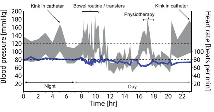
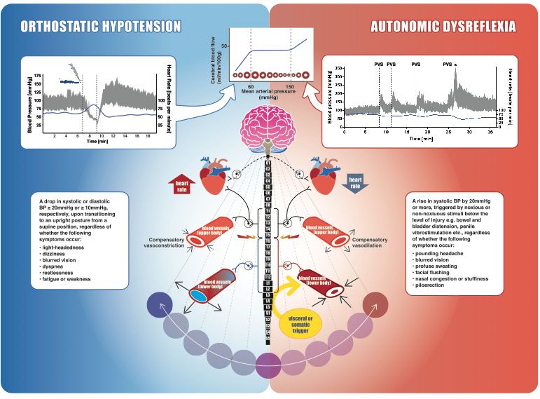
Int. J. Mol. Sci. 2019, 20, 2464                                                                      2 of 8

(Figure 1). With disruption of supraspinal sympatho-excitatory drive to spinal sympathetic preganglionic
neurons (located between first thoracic and second lumbar spinal segments), the vast majority of
individuals with tetraplegia or high paraplegia (SCI above T6) experience debilitating cardiovascular
impairments as a result of sympathetic decentralization. In addition to a consistently low resting
blood pressure (BP), decentralization of sympathetic cardiovascular control also predisposes these
individuals to drastic BP fluctuations, where systolic BP can drop down to 50 mmHg or lower during
an orthostatic challenge (e.g., assuming an upright posture) or can rapidly spike up to 300 mmHg as
a reflex response to noxious or non-noxious stimuli originating below the spinal lesion (e.g., distended
bladder). These hypotensive and hypertensive crises, called orthostatic hypotension and autonomic
dysreflexia (Figure 1), are essentially the result of either insufficient or excessive vasoconstriction,
respectively, and are generally present within the same individual. While a single episode of extreme BP
can have dangerous consequences for the cerebrovasculature, these hypertensive and hypotensive events
occur as frequently as 41 and 28 times per day, respectively [5], and thus are a significant chronic burden.
originating below the spinal lesion (e.g., distended bladder). These hypotensive and hypertensive
  crises, called orthostatic hypotension and autonomic dysreflexia (Figure 1), are essentially the result
  of either insufficient or excessive vasoconstriction, respectively, and are generally present within the
  same individual. While a single episode of extreme BP can have dangerous consequences for the
  cerebrovasculature,
Int.                          these hypertensive and hypotensive events occur as frequently as 41 and 28 times
     J. Mol. Sci. 2019, 20, 2464                                                                           3 of 8
  per day, respectively [5], and thus are a significant chronic burden.

       Figure1. 1.AnAn
     Figure             overview
                           overviewof spinal   cordcord
                                        of spinal    injuryinjury
                                                            (SCI), (SCI),
                                                                    autonomic    cardiovascular
                                                                           autonomic              innervation,
                                                                                         cardiovascular            blood pressure
                                                                                                            innervation,     blood
     (BP)   instability,    and   cerebral    autoregulation.     The   schematic    diagram    in
       pressure (BP) instability, and cerebral autoregulation. The schematic diagram in the middle  the  middle     demonstrates
     autonomic
       demonstrates  control   of the cardiovascular
                           autonomic                       system. Parasympathetic
                                         control of the cardiovascular                    control of the control
                                                                            system. Parasympathetic         heart (dashed      line),
                                                                                                                      of the heart
     mediated       by  the  vagus   nerve,    usually   remains   intact  following    SCI.  Neurons    within
       (dashed line), mediated by the vagus nerve, usually remains intact following SCI. Neurons within the        the  brainstem
     provide
       brainstemsympathetic      tonic controltonic
                      provide sympathetic         to spinal  sympathetic
                                                       control              preganglionic
                                                                to spinal sympathetic        neurons. Heart
                                                                                          preganglionic         and upper-body
                                                                                                            neurons.   Heart and
     blood    vesselsblood
       upper-body         are innervated
                                vessels arevia     spinal segments
                                                innervated   via spinalT1–T5,
                                                                          segmentswhereas
                                                                                      T1–T5,the  trunk and
                                                                                               whereas         lowerand
                                                                                                          the trunk      extremity
                                                                                                                             lower
     vasculature      receive  innervation     from  T6–L2.   The  splanchnic   bed  (liver, spleen,
       extremity vasculature receive innervation from T6–L2. The splanchnic bed (liver, spleen, and   and  intestines)   is densely
     innervated,
       intestines) is  highly  compliant,
                          densely   innervated,and highly
                                                    contains   approximately
                                                            compliant,            one-quarter
                                                                         and contains            of the total
                                                                                          approximately        blood volume
                                                                                                             one-quarter          at
                                                                                                                             of the
     rest,  making     it the primary    capacitance    bed.  An  SCI  disrupting   the  sympathetic    control
       total blood volume at rest, making it the primary capacitance bed. An SCI disrupting the sympathetic       of these  vessels
     (i.e., at orofabove
       control       theseT6)   makes
                            vessels      them
                                     (i.e., at orhighly
                                                  above vulnerable    to vasodilation
                                                         T6) makes them                 and extreme
                                                                            highly vulnerable           constriction,
                                                                                                 to vasodilation     andleading
                                                                                                                          extreme to
     BP   instability. leading
       constriction,      Orthostatic
                                   to BPhypotension       (shown on the
                                            instability. Orthostatic        left): cardiovascular
                                                                        hypotension     (shown on changes
                                                                                                      the left): in  a participant
                                                                                                                  cardiovascular
     with a motor-complete cervical SCI (C5, American Spinal Injury Association Impairment Scale (AIS) A)
     during a head-up-tilt assessment. Beat-by-beat BP is shown in grey, and heart rate is shown in blue. BP
     plummeted immediately upon initiation of 60◦ upright tilt from the supine position and the tilt was
     terminated after 2 min. Mean arterial pressure was recorded as 25 mmHg at its lowest, well below the
     lower limit of cerebral autoregulation (top middle inset). Rebound hypertension was also apparent
     when the participant was returned to the supine position, further emphasizing the instability in blood
     pressure regulation. Autonomic dysreflexia (shown on the right): cardiovascular changes in a male
     with motor-incomplete SCI (C6, AIS C) during a sperm retrieval procedure with penile vibrostimulation
     (PVS), which is a visceral/somatic trigger originating below the spinal lesion. The dashed lines indicate
     each time the PVS is applied and is followed by significant and rapid increases in BP. * indicates
     ejaculation. In this case, systolic BP almost triples and mean arterial pressure is ~250 mmHg, well
     above the upper limit of cerebral autoregulation (top Figure). Cerebral autoregulation curve (shown
     on the top): cerebral blood flow (CBF) is shown in relation to cerebral artery lumen diameter and mean
     arterial pressure. The dashed lines represent the lower and upper limits of CBF autoregulation, which
     are exceeded by our clinical orthostatic and autonomic dysreflexia examples. Red circles represent
     the cerebral arteries (either vasodilating or vasoconstricting to counteract changes in systemic blood
     pressure), and the blue solid line represents cerebral blood flow.
Int. J. Mol. Sci. 2019, 20, 2464 4 of 8
2. BP Lability and Cognitive Impairment after SCI: Lessons Learned from Able-Bodied
Individuals
Across a wide range of systemic BPs (i.e., 60–150 mmHg mean arterial pressure), cerebral vasculature
maintains a fairly uniform brain perfusion via a process called autoregulation. Furthermore, increased
local metabolic demands in the brain (such as during a cognitive task) are typically coupled with
an increase in regional cerebral blood flow (CBF). This phenomenon, called neurovascular coupling,
ensures adequate substrate delivery and removal of metabolites within the activated brain region.
Neurovascular coupling and cerebral autoregulation, along with CO2 vasoreactivity, maintains the
spatiotemporal adequacy of cerebral perfusion [6]. Despite cerebral autoregulation, chronic hypotension
in able-bodied individuals has been shown to result in cerebral hypoperfusion [7], as well as impaired
neurovascular coupling [8]. More importantly, certain brain regions, e.g., the basal ganglia, hippocampus
and cortex, are likely to be more susceptible to ischemic damage [7,9], resulting in significant deficits
in cognitive domains such as memory, attention, and reaction time [10,11]. Similar to able-bodied
hypotensive individuals, those with upper-thoracic and cervical SCI also exhibited significantly lower
CBF at rest that correlated with reduced cognitive performance compared to controls. Indeed, raising
systemic BP was shown to increase resting CBF, independent of the mechanism (e.g., either by an alpha-1
agonist, midodrine hydrochloride, or by a nitric oxide synthase inhibitor, nitro-L-arginine methyl
ester) [12]. Interestingly, the experiments from our laboratory showed that although resting CBF was
similar between SCI and age-matched controls, neurovascular coupling was significantly impaired during
a cognitive task, but was improved by raising BP with the administration of midodrine hydrochloride [13],
highlighting the association between low BP and impaired cognitive performance.
Conversely, several studies have substantiated the association between chronic hypertension
and cognitive decline in non-SCI individuals. Linked with impaired vascular tone, enhanced blood
brain barrier permeability, and profound structural remodeling, abnormally high BP for a prolonged
period of time detrimentally alters both structural and functional properties of arteries [14]. Among
these aforementioned aspects, structural maladaptations in response to chronic hypertension are better
understood. With higher intralumenal pressure and increased tangential stress on the artery wall,
cerebral arteries become thicker as an adaptive response to protect downstream vasculature against
increased BP. This, however, results in reduced lumen diameter and increased wall-to-lumen ratio, which
is a major predictor for end-organ damage [15]. Consequently, in able-bodied hypertensive individuals,
this results in reduced CBF at rest in cortical (occipitotemporal and prefrontal) and hippocampal
regions [16]. Furthermore, regional increase in CBF during memory tasks (neurovascular coupling)
is also impaired in hypertensive subjects compared to normotensive controls [17]. In individuals
with SCI above T6, extreme bouts of transient hypertension (i.e., autonomic dysreflexia) are prevalent
and occur numerous times per day (mean: 11 times/day) [5]. Using a rodent model, our laboratory
has shown that predisposing animals to cardiovascular impairment via a high-thoracic SCI leads to
deleterious structural and functional maladaptations in cerebrovasculature. Specifically, in rats with T3
SCI, the middle cerebral artery showed a significant reduction in distensibility, increased stiffness, and
increased wall-to-lumen ratio [18]. This was further associated with a reduction in CBF (at rest as well
as during a hypercapnic challenge) and significantly impaired short-term memory [19]. More recent
work from our laboratory showed that cerebrovascular impairments are more pronounced when the
rats with T3 SCI are exposed to daily repetitive autonomic dysreflexia via colorectal distension [20].
Clinical evidence has also demonstrated that the intensity of autonomic dysreflexia, quantified using
a questionnaire, is inversely associated with performance in executive function tests [21]. The existing
evidence linking repeated bouts of autonomic dysreflexia with cognitive impairment following SCI is
still preliminary. However, given the plethora of studies supporting hypertension-related cognitive
decline in able-bodied individuals, it is tempting to speculate that similar mechanisms would also
underlie cognitive impairment due to transient hypertensive episodes in SCI—an avenue worthy of
future research.Int. J. Mol. Sci. 2019, 20, 2464 5 of 8
It is also noteworthy that unlike the able-bodied individuals with either persistent hypotension
or hypertension, individuals with cardiovascular impairment secondary to high-level SCI generally
experience both extreme ends of the BP spectrum (Figure 2). The pendulum-like swings in BP in
those with a high-level SCI (i.e., during orthostatic hypotension and autonomic dysreflexia) easily
reach the values beyond the autoregulatory limit [5], predisposing an individual to either ischemic or
hemorrhagic stroke. A recent report from our laboratory demonstrated, in an individual with a chronic,
motor-complete SCI at the T4 spinal segment, that excessive hypertension during an episode of
autonomic dysreflexia (likely due to urinary tract infection) exceeded the autoregulatory limit, resulting
in cortical and subcortical vasogenic edema, a condition called posterior reversible encephalopathy
syndrome [22]. While discussing the upper end of Lassen’s autoregulation curve (Figure 1), it is also
important to consider the lower end, beyond which vasodilation becomes ineffective and arteries
tend to collapse due to low intralumenal pressure [23]. Chronic hypertension is known to increase
the lower limit of autoregulation, likely via maladaptive remodeling of cerebrovasculature, and thus
even less severe episodes of hypotension can be problematic [24]. It is reasonable to envision that this
phenomenon may have serious implications for individuals with SCI that suffer both hypertensive and
hypotensive episodes concomitantly, presenting a unique double-edged sword for this population.
Adding further to the cardiovascular disease risk is the fact that owing to the amplification of various
physical, physiological, and environmental risk factors, cardiovascular (and, in turn, cerebrovascular)
disease progression is immensely accelerated after SCI [25]. In fact, based on our analysis of a Canadian
Community Health Survey, it was found that even after controlling for risk factors such as age and
sex, SCI was independently associated with a nearly 3-fold higher risk of cardiovascular disease and
4-fold higher risk of stroke [26]. This is especially concerning in light of the mounting evidence from
the non-SCI population, which suggests that essentially any of the stroke etiologies can result in VCI,
Int. J. Mol. Sci. 2019, 20, x 5 of 8
ranging from mild cognitive impairment to dementia [27].
Figure 2. Ambulatory BP monitoring data collected from a research participant with a motor-complete
Figure 2. Ambulatory
cervical SCI (C5, AIS BP monitoring
B). These data collected
data demonstrate from
transient, a research participant
pendulum-like with
shifts in BP (in a in
grey) motor-
response to various stimuli throughout a normal day. Multiple episodes of autonomic
complete cervical SCI (C5, AIS B). These data demonstrate transient, pendulum-like shifts in BP (in dysreflexia
grey) = 25)
(n in and orthostatic
response hypotension
to various stimuli (n = 33) were aobserved
throughout normal inday.
this Multiple
case, with episodes
systolic BPofranging
autonomic
from 71 to 180 mmHg (mean arterial pressure: 53 to 132 mmHg). Triggers for these
dysreflexia (n = 25) and orthostatic hypotension (n = 33) were observed in this case, with systolic conditions are BP
annotated on the figure. The bowel routine in particular demonstrates aberrant BP changes, in both
ranging from 71 to 180 mmHg (mean arterial pressure: 53 to 132 mmHg). Triggers for these conditions
directions, in response to suppository insertion, digital stimulation, and pressure applied to the
are annotated on the figure. The bowel routine in particular demonstrates aberrant BP changes, in
abdomen (autonomic dysreflexia) and transferring to and from the commode (orthostatic hypotension).
bothHeart
directions, in response to suppository insertion, digital stimulation, and pressure applied to the
rate is represented by the blue solid line.
abdomen (autonomic dysreflexia) and transferring to and from the commode (orthostatic
hypotension). Heart rate is represented by the blue solid line.
3. Therapeutic Perspectives
Preventing and/or controlling volatile BP fluctuations to mitigate VCI following SCI can beInt. J. Mol. Sci. 2019, 20, 2464 6 of 8
3. Therapeutic Perspectives
Preventing and/or controlling volatile BP fluctuations to mitigate VCI following SCI can be
approached in a number of ways. In terms of preclinical validation, this could be achieved by:
(1) restoration of supraspinal control through neural regeneration [28], (2) prevention of secondary spinal
cord damage through early neuroprotection [29], (3) reduction of aberrant sprouting of nociceptive
afferent fibers that trigger autonomic dysreflexia episodes [30], or a logical combination of these
approaches. This topic has been previously reviewed by our group [31]. From a clinical perspective,
a variety of pharmacological and nonpharmacological options are available for management of
autonomic dysreflexia and orthostatic hypotension that could reduce cardiovascular disease burden
and decelerate the VCI trajectory following SCI [32–34]. A major limitation (other than the obvious side
effects) of currently available pharmacotherapies is that most of the drugs are slow-acting (i.e., they take
several minutes to reach effective plasma concentrations and get metabolized) and also lead to sustained,
undesirable cardiovascular effects. The extreme cardiovascular events following SCI are more transient;
hence, it is reasonable to question the efficacy of presently available treatments. One potential solution
to this could be the employment of neuromodulation strategies such as epidural or transcutaneous
spinal cord stimulation, which have demonstrated the capability to almost instantaneously modulate
BP [35–38]. These studies, although promising, need further systematic exploration prior to widespread
clinical implementation.
4. Conclusions
We are only beginning to explore the interplay between cardiovascular and cognitive impairments
following SCI. Given the wealth of research in the non-SCI population, many principles can potentially
be extrapolated in order to expedite our understanding of the precise mechanisms involved. Future
research is necessary to develop effective strategies to prevent or ameliorate cognitive impairment in
persons with SCI. Advances in these areas will significantly impact independence and quality of life in
this population.
Funding: Krassioukov’s laboratory is supported by funds from the Canadian Institute for Health Research, Heart
and Stroke Foundation; Canadian Foundation for Innovation; BC Knowledge Development Fund; Wings for Life
Foundation; Craig H. Neilsen Foundation; and Seed grants from International Collaboration on Repair Discoveries
(ICORD). Sachdeva is supported by Postdoctoral Fellowships from the Craig H. Neilsen Foundation, Canadian
Institutes of Health Research, and University of British Columbia (Bluma Tischler Postdoctoral Fellowship).
Nightingale is supported by a Michael Smith Foundation for Health Research/ICORD Postdoctoral Trainee Award.
Acknowledgments: We sincerely thank Cheryl Niamath and Matthias Walter (ICORD) for their creative assistance
in figure design.
Conflicts of Interest: The authors declare no conflict of interest. The funders had no role in the design of the
study; in the collection, analyses, or interpretation of data; in the writing of the manuscript, or in the decision to
publish the results.
References
1. Savic, G.; DeVivo, M.J.; Frankel, H.L.; Jamous, M.A.; Soni, B.M.; Charlifue, S. Long-term survival after
traumatic spinal cord injury: A 70-year British study. Spinal. Cord. 2017, 55, 651–658. [CrossRef] [PubMed]
2. Craig, A.; Nicholson Perry, K.; Guest, R.; Tran, Y.; Middleton, J. Adjustment following chronic spinal cord
injury: Determining factors that contribute to social participation. Br. J. Health Psychol. 2015, 20, 807–823.
[CrossRef]
3. Craig, A.; Guest, R.; Tran, Y.; Middleton, J. Cognitive Impairment and Mood States after Spinal Cord Injury.
J. Neurotrauma 2017, 34, 1156–1163. [CrossRef]
4. Sachdeva, R.; Gao, F.; Chan, C.C.H.; Krassioukov, A.V. Cognitive function after spinal cord injury: A systematic
review. Neurology 2018, 91, 611–621. [CrossRef]
5. Hubli, M.; Gee, C.M.; Krassioukov, A.V. Refined assessment of blood pressure instability after spinal cord
injury. Am. J. Hypertens. 2015, 28, 173–181. [CrossRef]Int. J. Mol. Sci. 2019, 20, 2464 7 of 8
6. Willie, C.K.; Colino, F.L.; Bailey, D.M.; Tzeng, Y.C.; Binsted, G.; Jones, L.W.; Haykowsky, M.J.; Bellapart, J.;
Ogoh, S.; Smith, K.J.; et al. Utility of transcranial Doppler ultrasound for the integrative assessment of
cerebrovascular function. J. Neurosci. Methods 2011, 196, 221–237. [CrossRef] [PubMed]
7. Duschek, S.; Meinhardt, J.; Schandry, R. Reduced cortical activity due to chronic low blood pressure: An EEG
study. Biol. Psychol. 2006, 72, 241–250. [CrossRef] [PubMed]
8. Duschek, S.; Schandry, R. Cognitive performance and cerebral blood flow in essential hypotension.
Psychophysiology 2004, 41, 905–913. [CrossRef]
9. Roman, G.C. Brain hypoperfusion: A critical factor in vascular dementia. Neurol. Res. 2004, 26, 454–458.
[CrossRef] [PubMed]
10. Duschek, S.; Matthias, E.; Schandry, R. Essential hypotension is accompanied by deficits in attention and
working memory. Behav. Med. 2005, 30, 149–158. [CrossRef]
11. Duschek, S.; Weisz, N.; Schandry, R. Reduced cognitive performance and prolonged reaction time accompany
moderate hypotension. Clin. Auton. Res. 2003, 13, 427–432.
12. Wecht, J.M.; Weir, J.P.; Radulovic, M.; Bauman, W.A. Effects of midodrine and L-NAME on systemic and
cerebral hemodynamics during cognitive activation in spinal cord injury and intact controls. Physiol. Rep.
2016, 4, e12683. [CrossRef]
13. Phillips, A.A.; Warburton, D.E.; Ainslie, P.N.; Krassioukov, A.V. Regional neurovascular coupling and
cognitive performance in those with low blood pressure secondary to high-level spinal cord injury: Improved
by alpha-1 agonist midodrine hydrochloride. J. Cerebral Blood Flow MeTable 2014, 34, 794–801. [CrossRef]
14. Pires, P.W.; Dams Ramos, C.M.; Matin, N.; Dorrance, A.M. The effects of hypertension on the cerebral
circulation. Am. J. Physiol. Heart Circ. Physiol. 2013, 304, H1598–H1614. [CrossRef]
15. Izzard, A.S.; Rizzoni, D.; Agabiti-Rosei, E.; Heagerty, A.M. Small artery structure and hypertension: Adaptive
changes and target organ damage. J. Hypertens. 2005, 23, 247–250. [CrossRef]
16. Beason-Held, L.L.; Moghekar, A.; Zonderman, A.B.; Kraut, M.A.; Resnick, S.M. Longitudinal changes in
cerebral blood flow in the older hypertensive brain. Stroke 2007, 38, 1766–1773. [CrossRef]
17. Jennings, J.R.; Muldoon, M.F.; Ryan, C.; Price, J.C.; Greer, P.; Sutton-Tyrrell, K.; van der Veen, F.M.; Meltzer, C.C.
Reduced cerebral blood flow response and compensation among patients with untreated hypertension.
Neurology 2005, 64, 1358–1365. [CrossRef]
18. Phillips, A.A.; Matin, N.; Frias, B.; Zheng, M.M.; Jia, M.; West, C.; Dorrance, A.M.; Laher, I.; Krassioukov, A.V.
Rigid and remodelled: Cerebrovascular structure and function after experimental high-thoracic spinal cord
transection. J. Physiol. 2016, 594, 1677–1688. [CrossRef]
19. Jia, M.; Phillips, A.A.; Yung, A.; Kozlowski, P.; Krassioukov, A.V. Cerebrovascular Endothelial Function is
Impaired after Experimental Spinal Cord Injury. Faseb. J. 2016, 30, 998.
20. Phillips, A.A.; Matin, N.; Jia, M.; Squair, J.W.; Monga, A.; Zheng, M.M.Z.; Sachdeva, R.; Yung, A.; Hocaloski, S.;
Elliott, S.; et al. Transient Hypertension after Spinal Cord Injury Leads to Cerebrovascular Endothelial
Dysfunction and Fibrosis. J. Neurotrauma 2018, 35, 573–581. [CrossRef]
21. Krassioukov, A.; Gao, F.; Li, J.; Pak, M.; Chan, C. Cognitive function among spinal cord injured individuals
with autonomic dysreflexia: A pilot study. Top Spinal Cord Inj. Rehabil. 2012, 18, 206.
22. Squair, J.W.; Phillips, A.A.; Harmon, M.; Krassioukov, A.V. Emergency management of autonomic dysreflexia
with neurologic complications. CMAJ 2016, 188, 1100–1103. [CrossRef] [PubMed]
23. Lassen, N.A. Control of cerebral circulation in health and disease. Circ. Res. 1974, 34, 749–760. [CrossRef]
[PubMed]
24. Jones, J.V.; Fitch, W.; MacKenzie, E.T.; Strandgaard, S.; Harper, A.M. Lower limit of cerebral blood flow
autoregulation in experimental renovascular hypertension in the baboon. Circ. Res. 1976, 39, 555–557.
[CrossRef] [PubMed]
25. Phillips, A.A.; Krassioukov, A.V. Contemporary Cardiovascular Concerns after Spinal Cord Injury:
Mechanisms, Maladaptations, and Management. J. Neurotrauma 2015, 32, 1927–1942. [CrossRef]
26. Cragg, J.J.; Noonan, V.K.; Krassioukov, A.; Borisoff, J. Cardiovascular disease and spinal cord injury: Results
from a national population health survey. Neurology 2013, 81, 723–728. [CrossRef]
27. Dichgans, M.; Leys, D. Vascular Cognitive Impairment. Circ. Res. 2017, 120, 573–591. [CrossRef]
28. Sachdeva, R.; Gopaul, R.; Jia, M.; Monga, A.; Ramer, M.; Krassioukov, A.V. A Triple Combination Approach
Involving Nerve Transplantation, Glial Scar Digestion and Passive Exercise Promotes Cardiovascular
Recovery after Spinal Cord Injury. Faseb J. 2017, 31, 1077.Int. J. Mol. Sci. 2019, 20, 2464 8 of 8
29. Squair, J.W.; Ruiz, I.; Phillips, A.A.; Zheng, M.M.Z.; Sarafis, Z.K.; Sachdeva, R.; Gopaul, R.; Liu, J.; Tetzlaff, W.;
West, C.R.; et al. Minocycline Reduces the Severity of Autonomic Dysreflexia after Experimental Spinal Cord
Injury. J. Neurotrauma 2018, 35, 2861–2871. [CrossRef]
30. Krenz, N.R.; Meakin, S.O.; Krassioukov, A.V.; Weaver, L.C. Neutralizing intraspinal nerve growth factor
blocks autonomic dysreflexia caused by spinal cord injury. J. Neurosci. 1999, 19, 7405–7414. [CrossRef]
31. Squair, J.W.; West, C.R.; Krassioukov, A.V. Neuroprotection, Plasticity Manipulation, and Regenerative
Strategies to Improve Cardiovascular Function following Spinal Cord Injury. J. Neurotrauma 2015, 32, 609–621.
[CrossRef]
32. Krassioukov, A.; Warburton, D.E.; Teasell, R.; Eng, J.J. A systematic review of the management of autonomic
dysreflexia after spinal cord injury. Arch. Phys. Med. Rehabil. 2009, 90, 682–695. [CrossRef]
33. Krassioukov, A.; Eng, J.J.; Warburton, D.E.; Teasell, R. A Systematic Review of the Management of Orthostatic
Hypotension After Spinal Cord Injury. Arch. Phys. Med. Rehabil. 2009, 90, 876–885. [CrossRef]
34. Mills, P.B.; Fung, C.K.; Travlos, A.; Krassioukov, A. Nonpharmacologic management of orthostatic
hypotension: A systematic review. Arch. Phys. Med. Rehabil. 2015, 96, 366–375.e6. [CrossRef] [PubMed]
35. Phillips, A.A.; Squair, J.W.; Sayenko, D.G.; Edgerton, V.R.; Gerasimenko, Y.; Krassioukov, A.V. An Autonomic
Neuroprosthesis: Noninvasive Electrical Spinal Cord Stimulation Restores Autonomic Cardiovascular
Function in Individuals with Spinal Cord Injury. J. Neurotrauma 2018, 35, 446–451. [CrossRef] [PubMed]
36. West, C.R.; Phillips, A.A.; Squair, J.W.; Williams, A.M.; Walter, M.; Lam, T.; Krassioukov, A.V. Association of
Epidural Stimulation With Cardiovascular Function in an Individual With Spinal Cord Injury. Jama Neurol.
2018, 75, 630–632. [CrossRef] [PubMed]
37. Darrow, D.; Balser, D.; Netoff, T.I.; Krassioukov, A.; Phillips, A.; Parr, A.; Samadani, U. Epidural Spinal Cord
Stimulation Facilitates Immediate Restoration of Dormant Motor and Autonomic Supraspinal Pathways
after Chronic Neurologically Complete Spinal Cord Injury. J. Neurotrauma 2019. [CrossRef]
38. Aslan, S.C.; Legg Ditterline, B.E.; Park, M.C.; Angeli, C.A.; Rejc, E.; Chen, Y.; Ovechkin, A.V.; Krassioukov, A.;
Harkema, S.J. Epidural Spinal Cord Stimulation of Lumbosacral Networks Modulates Arterial Blood Pressure
in Individuals With Spinal Cord Injury-Induced Cardiovascular Deficits. Front. Physiol. 2018, 9, 565.
[CrossRef]
© 2019 by the authors. Licensee MDPI, Basel, Switzerland. This article is an open access
article distributed under the terms and conditions of the Creative Commons Attribution
(CC BY) license (http://creativecommons.org/licenses/by/4.0/).You can also read