Generalized sarcoidosis in two horses - ild care foundation
←
→
Page content transcription
If your browser does not render page correctly, please read the page content below
wetenschap
Generalized sarcoidosis in two horses
E.P.R. Reijerkerk i, E.J.B.Veldhuis Kroeze ii and M.M. Sloet van afwijkende huid de typische veranderingen: uitgebreide
Oldruitenborgh-Oosterbaan i lymfohistiocytaire infiltratie en veel meerkernige reuscellen van
het Langhanstype.
Bij bloedonderzoek werd bij beide patiënten een normaal aantal
Tijdschr Diergeneeskd 2008; 133: 654-661 witte bloedcellen met een rechtsverschuiving gevonden. Casus B
had ook een anemie en een iets hoge totaal-eiwitconcentratie met
Oorspronkelijk artikel een duidelijk verhoogde beta- en gammafractie. Beide paarden
reageerden in het verloop van de aandoening onvoldoende of niet
summary op behandeling met systemische corticosteroïden en beide
Equine sarcoidosis is a rare disorder usually characterized paarden werden op verzoek van de eigenaar geeuthanaseerd.
by exfoliative dermatitis, moderate to severe wasting, and
sarcoidal granulomatous inflammation of multiple organ introduction
systems. It has an unknown aetiopathogenesis. The Equine sarcoidosis, also known as ‘equine idiopathic
condition is not related to equine sarcoid. This case report systemic granulomatous disease’ (14), ‘generalized
describes generalized cutaneous and systemic sarcoidosis granulomatous disease’ (11), ‘systemic granulomatous
in an 11-year-old Trakehner mare (case A) and in a disease’ (17), ‘equine histiocytic disease’ (11), and ‘equine
7-year-old Dutch Warmblood gelding (case B). histiocytic dermatitis’ (3), is a rare disease of unknown
Case A was presented with cutaneous sarcoidosis on aetiopathogenesis (13). Although the name is similar, the
the head and body and was diagnosed on the basis of condition is not related to equine cutaneous sarcoid (9).
histological examination of skin. Case B presented with Sarcoidosis also has been reported in humans and cattle (2,
multiple subcutaneous nodules (2-15 cm in diameter) and 6, 12, 14), based on histopathological findings. The clinical
the diagnosis was established at postmortem examination. manifestations are quite different in the different species.
Both horses showed distinctive histology of the skin with Equine sarcoidosis is suggested to be the result of an
extensive lymphohistiocytic infiltration and Langhans- exaggerated immunological response, with exogenous
type multinucleated giant cells. Haematology and infectious agent(s) or allergens as the antigenic stimulus
biochemistry revealed a normal total white blood cell (8, 16). No (infectious) agent has been identified as yet (13,
count with a right shift in both horses. Case B was anae 16), and no breed, sex, or age predilection has been
mic and had a slightly elevated total protein concentration established (9, 11, 13, 15, 16). The disease is characterized
with hyperglobulinaemia. Both horses were unresponsive by exfoliative dermatitis, severe wasting, and granuloma
to corticosteroids and were euthanized. tous inflammation of one or more internal organs (9, 13,
16). The skin, lungs, lymph nodes, and gastrointestinal
samenvatting tract are most commonly affected (1, 5, 11, 13, 16). Less
Gegeneraliseerde sarcoidose bij twee paarden commonly affected organs or tissue(s) are the liver, spleen,
Equine sarcoidose is een zeldzaam voorkomende aandoening die kidneys, skeletal system, heart, adrenal- and thyroid
wordt gekenmerkt door exfoliatieve dermatitis en granuloma- glands, pancreas, and nervous system (13, 16).
teuse infiltratie van meerdere orgaansystemen en/of organen. Stannard (17) classified equine sarcoidosis in two
Equine sarcoidose en equine sarcoid zijn twee geheel verschil- forms: the cutaneous form and the nodular form. The
lende ziektebeelden. Equine sarcoidose heeft een onbekende most commonly occurring cutaneous form starts clini
etiologie en pathogenese. Deze casusbespreking beschrijft twee cally with skin lesions, with generalized scaling and
casus met gegeneraliseerde cutane en systemische sarcoidose, een crusting combined with variable alopecia of the face and
elf jaar oude Trakehner merrie (casus A) en een zeven jaar oude limbs, often with sparing of the mane and the tail (9, 13).
kwpn-ruin (casus B). The nodular form, with nodules or tumour-like masses, is
Casus A toonde eerst de klinische verschijnselen van cutane less common and is often combined with extensive scaling
sarcoidose op het hoofd en het lichaam en de diagnose werd and crusting (11). Recently, a third form, the ‘localized
gesteld op basis van een huidbiopt. Casus B begon met subcutane form’, has been described (13, 15). In these patients the
nodula (2 tot 15 centimeter diameter) en vertoonde pas later de typical hyperkeratotic, crusted, and alopecic areas remain
typische huidveranderingen. De diagnose bij dit paard werd localized (for many years) and occur most often on the
gesteld bij postmortaal onderzoek. lower limbs, and occasionally elsewhere, in otherwise
Beide paarden toonden bij histologisch onderzoek van de systemically healthy horses that are mostly performing
well (15). Non-cutaneous clinical signs, such as a persis
i Department of Equine Sciences, Faculty of Veterinary Medicine, tent low-grade fever, poor appetite, and decreased perfor
Utrecht University, Yalelaan 114, 3584 cm Utrecht, the Netherlands. mance, are described in horses with systemic sarcoidosis
ii Department of Pathobiology, Faculty of Veterinary Medicine, Utrecht (9, 13, 16). Peripheral lymphadenopathy and variable
University, Yalelaan 1, 3584 cl Utrecht, the Netherlands. organ signs may also be detected (9, 11, 16). The presence
Corresponding author: e.p.r.reijerkerk@uu.nl of mediastinal and pulmonary masses may account for the
654 Tijdschrift voor Diergeneeskunde • Deel 133 • Aflevering 16 • 15 augustus 2008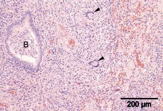
wetenschap
60 39.5
50 39
amount/minute
degrees Celcius
40 38.5
30 38
20 37.5
10 37
0 36.5
D1 D1 D2 D2 D3 D3 D4 D4 D5
mo ev mo ev mo ev mo ev mo
respiratory rate D1 = Day 1
heart rate mo = morning
rectal temperature ev = evening
Figure 1. Respiratory rate, heart rate, and rectal temperature of case A
during 5 days’ hospitalization before euthanasia. 2a
characteristic poor performance and dyspnoea (9),
whereas liver and gastrointestinal involvement may
account for clinical signs such as icterus and diarrhoea (9).
Other organs are rarely involved.
Horses with localized sarcoidosis have no clinical
systemic signs other than the affected skin on mostly the
lower limb(s), which can become crusty, thick and painful,
sometimes resulting in lameness (15). In contrast to most
other lower limb dermatoses, sarcoidosis involves both
pigmented and unpigmented skin. Pruritus has been
reported as a clinical sign in human sarcoidosis and is also
reported as a minor presenting sign in some horses (4, 15).
The diagnosis of sarcoidosis is based on clinical and
histological findings combined with exclusion of other
diseases. Differential diagnoses include dermatophilosis, 2b
dermatophytosis, immune-mediated diseases (e.g., Figure 2. An 11-year-old Trakehner mare with generalized scaling and
pemphigus foliaceus, systemic lupus erythematosus, crusting on head, neck, and body resulting from sarcoidosis.
exfoliative eosinophilic dermatitis, erythema multiforme,
food allergies, drug eruptions, and multisystemic eosino a 4-month history of a skin problem on the head, neck,
philic epitheliotrophic disease), seborrhoea, verrucous abdomen, and around the tail and perineum. The horse
sarcoids, and toxicoses (hairy vetch, arsenic, iodine, also showed a slight localized oedematous reaction on the
aluminium, and silicon) (9, 13, 16). Biopsy of the skin back and abdomen. When the skin problem started, the
lesions is essential for confirmation of the diagnosis. horse had a raised rectal temperature. The horse was
Typical histopathological lesions include perifollicular treated with povidone iodine shampoo a, seborrhoea
and mid- or superficial dermal granulomata, a multifocal shampoo b, and insect repellents and was clothed while at
nodular to diffuse non-caseating granulomatous dermati pasture with an insect blanket. Later the horse became
tis with histiocytes, multinucleated giant cells, and lethargic and lost its appetite. A few days before referral,
limited lymphocytic infiltration (9, 13). The clinical the horse coughed occasionally. The mare had a history of
management of generalized or localized sarcoidosis is recurrent colic and had been operated on for right dorsal
often problematic (11). A few horses show spontaneous displacement 6 months before the onset of the dermato
remission for no apparent reason (9, 12, 16), but horses logical problem. One month before the skin problem
with the localized form in particular may show a good started, the mare had shown signs of ‘bronchitis’. These
response to long courses of systemic corticosteroids (e.g., symptoms disappeared after treatment with clenbuterol c,
dexamethasone or prednisolone) (15, 16). prednisolone d, and acetylcysteine e. Antibiotic treatment
This case report provides the first description of two resolved the horse’s lameness but did not improve the skin
cases of generalized equine sarcoidosis in the Netherlands.
a Betadine Shampoo®, Mundipharma, Basel, Switserland.
History b Sebomild P®, Virbac Nederland BV, Barneveld, The Netherlands.
c Ventipulmin®, Boehringer Ingelheim, Alkmaar, The Netherlands.
An 11-year-old Trakehner mare was referred to the d Prednisolon®, Alfasan, Woerden, The Netherlands.
Department of Equine Sciences at Utrecht University with e Lysox®, Florence, Italy.
Tijdschrift voor Diergeneeskunde • Deel 133 • Aflevering 16 • 15 augustus 2008 655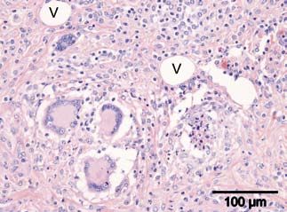
wetenschap

                                                                             Blood variable              Horse A   Horse B      Reference values
                                                                             pcv (l/l)                   0.37      0.26         0.32-0.42
                                                                             wbc (109/l)                 8.2       9.7          7-10
                                                                             lymphocytic (%)             13        14           30-35
                                                                             segmented gran (%)          78        84           35-60
                                                                             band gran (%)               0         0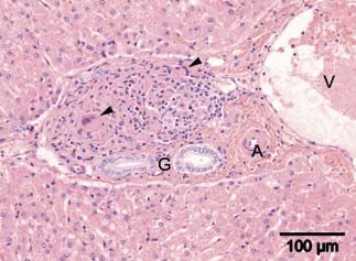
wetenschap
5a
4a
5b
Figure 5. At postmortem examination of case A, except the skin, the only
macroscopic signs of granulomatous disease noticed were severely swol-
len and firm bronchial lymph nodes, as seen in the centre of photograph
5a revealed by the partly lifted lung lobe, and on cut sections (5b).
ing. A single dose of dexamethasone g (0.02 mg/kg
bodyweight, intramuscular) was not beneficial.
Ultrasonographic examination of the pectoral region
performed in practice revealed a 4-cm diameter encapsu
lated hyperechogenic region. The other nodules were
hardly visible on ultrasonographic examination. Fine-
needle aspiration biopsy of the pectoral swelling revealed
4b a bloody aspirate with many neutrophilic granulocytes
Figure 4. Equine sarcoidosis, overview of a biopsy of haired skin with and mesenchymal cells and no evidence of malignant
superficial hyperkeratosis and a moderately cellular lymphohistiocytic in- neoplastic disease. Because the horse’s performance as a
flammatory infiltrate within the superficial dermis admixed with several riding-school horse decreased, the horse was referred for
typical multinucleated giant cells (a). Higher magnification (b) shows further evaluation to the Department of Equine Sciences.
two typical giant cells of the Langhans type (on top right and bottom left)
with abundant pale eosinophilic cytoplasm and a rim of peripheral nuclei Clinical findings at the University Clinic
(haematoxylin-eosin stain). On presentation, the horse was alert, in good bodily
condition, and showed a slightly increased respiratory rate
case b (20 breaths per minute) and a normal heart rate (36 beats
History per minute). The rectal temperature was marginally
A 7-year-old Dutch Warmblood gelding was referred to the elevated (38.3ºC), which may have been the result of
Department of Equine Sciences at Utrecht University with transport. Except for the nodules, clinical examination
a one month history of problems that had started a week was unremarkable including the lung auscultation. The
after rhinopneumonitis vaccination in the right pectoral nodules were localized to the neck, pectoral region, back
muscle. After one week, the pectoral region showed a large (saddle region), and abdomen (figure 6). The firm circum
firm swelling and within a few days subcutaneous nodules scribed swellings varied in diameter from 5 to 15 cm and
were seen on the neck, the back, and the abdomen. The
horse subsequently became lethargic and started cough g Voreen® suspensie, Boehringer Ingelheim, Alkmaar, The Netherlands.
Tijdschrift voor Diergeneeskunde • Deel 133 • Aflevering 16 • 15 augustus 2008 657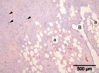
wetenschap
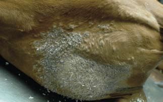
Figure 7. Macroscopic view of the affected skin in the pectoral region of
a 7-year-old Dutch Warmblood gelding (case B) at postmortem exami-
nation. Note not only the extensive hyperkeratosis and exfoliation of
Figure 6. Seven-year-old Dutch Warmblood gelding (case B) with several the skin but also the multiple small prominent subcutaneous nodules
firm not painful nodules varying in diameter between 5 and 15 cm between especially seen surrounding the large greyish hyperkeratotic and scaling
the forelimbs, on the neck, in the lumbar region, and on the abdomen. patch of skin.
were covered with a normal skin showing no alopecia, keratosis was localized multifocally on the right pectoral
crusting, and/or scaling. All nodules seemed to be located region (figure 7) and less severely on both sides of the
subdermally, with the exception of the large nodule in the head, the left cervical region, and the perineum. Small
right pectoral muscle. firm subcutaneous nodules and plaques were found in the
right pectoral region, near the crest, on the back, and on
Further investigations the ventral abdominal wall. These nodules and plaques
Haematology and biochemistry revealed moderate ranged from 2 to 15 cm in diameter. Similar firm multi
anaemia and a slightly raised total protein concentration focal nodular lesions were observed in the left pectoral
with increased ß- and γ-globulin fractions (table 1). musculature, and multifocally within the lungs and the
diaphragm (figure 8).
Diagnosis and treatment
The tentative diagnosis was an immune-mediated reaction
of unknown origin and prolonged corticosteroid treat
ment (prednisolone 1 mg/kg per os s.i.d) was advised. After
the horse was discharged, there was some improvement
and the horse was used again in the riding school. How
ever, the nodules never completely resolved. One month
after the end of prednisolone treatment, the nodules were
even more prominent than before.
Seven weeks after the horse returned home, the
swelling in the pectoral region also enlarged. Findings of
repeated ultrasonographic examination of the pectoral 8a
region raised suspicion about the development of chronic
purulent inflammation, and treatment with laurel
ointmenth was started. The appearance of the overlying
skin changed, and alopecia and severe crusting and scaling
were noticed (figure 7). The nodules all over the body also
increased in size and the horse became more lethargic and
showed exercise intolerance. The owner did not wish any
further examinations and opted to euthanize the horse.
The horse was transported to the Department of Equine
Sciences, euthanized, and postmortem examination was
performed at the Department of Pathobiology. 8b
Figure 8. Macroscopic photographs of the postmortem findings of a
Postmortem examination 7-year-old Dutch Warmblood gelding (case B) depicting circumscript,
Multifocal skin lesions were evident macroscopically. pale, and firm granulomatous nodules (G) measuring 1 to 3 cm in dia
Mildly thickened alopecic skin with extensive hyper meter in the diaphragm (8a), subpleural (top), and within the striated
muscle (bottom). Similar extensive but less circumscript lesions in the
h Laurierzalf®, Virbac Nederland BV, Barneveld, The Netherlands. pulmonary tissue (8b).
658 Tijdschrift voor Diergeneeskunde • Deel 133 • Aflevering 16 • 15 augustus 2008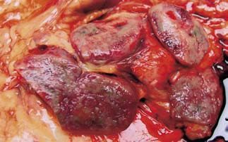
wetenschap
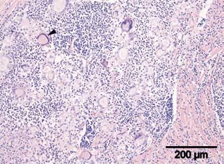
Figure 9. Microscopic examination of the abnormal skin of the pectoral 10a
region of a 7-year-old Dutch Warmblood gelding (case B) revealed an
extensive inflammatory infiltrate within the superficial dermis of the
pectoral region. It is comprised of mainly lymphocytes and histiocytes
with regular presence of multinucleated giant cells (arrowheads). Note
the thickened hyperplastic epidermis with superficial orthokeratotic
hyperkeratosis (H) (haematoxylin-eosin stain).
The lung lesions consisted of two large (measuring
approximately 3 x 10 cm) firm pale areas of dorsal sub
pleural parenchymal consolidation in the left and right
main lobes. A third focus of approximately 3 cm in
diameter was seen in the left top lung lobe (figure 8b). The
cervical, sternal, left axillary, and prescapular lymph nodes
were diffusely moderately swollen and firm. On incision, 10b
all nodular and plaque-like lesions as well as the affected Figure 10. Histology of the lung of a 7-year-old Dutch Warmblood gelding
lymph nodes were circumscribed but non-encapsulated, (case B) showed large multiple areas of atelectatic pulmonary paren-
firm and solid with a pale to greyish discoloration. Other chyma composed of mainly fibrosis and mononuclear inflammatory
histologically evaluated organs were the pituitary gland, infiltrates containing multinucleated giant cells (arrowheads). Several
parotid salivary gland, thyroid glands, heart, spleen, inflated and distended alveoli (a) and a bronchiole (B) are also depicted
pancreas, kidneys, and adrenal glands; these were all (10a). Higher magnification (10b) shows a neutrophilic exsudate within a
unremarkable. bronchiole (B) and two Langhans giant cells (arrowheads) in a fibrotic and
Histological examination of the affected skin revealed consolidated area of lung tissue (haematoxylin-eosin stain).
similar lesions to those in case A: extensive lymphohistio
cytic inflammatory infiltrates admixed with large multi
nucleated giant cells of the Langhans-type and extensive
epidermal hyperkeratosis (figure 9). The firm nodular
lesions in the affected tissues and organs contained
extensive granulomatous inflammatory infiltrates within
a fibrotic matrix. The pulmonary granulomas were
markedly fibrotic (figure 10a and 10b). The inflammatory
cell population consisted of numerous histiocytes and
lymphocytes admixed with lower numbers of plasma cells
and neutrophilic granulocytes; multinucleated Langhans
giant cells were always present (figures 11, 12 and 13).
Comparable small granulomata were present within the
liver (figure 14) and occasionally Langhans-type giant cells
were seen in the femoral bone marrow. Ziehl-Neelsen
stains for acid-fast mycobateria and periodic acid-Schiff Figure 11. This figure shows a higher magnification of a nodular granu-
stains for fungi and parasites proved consistently negative. lomatous panniculitis of the pectoral region where three large Langhans
Like case A, these findings were consistent with a diagno giant cells (bottom left) are surrounded by histiocytes, lymphocytes
sis of generalized sarcoidosis. and plasma cells. Also notice the increased fibrosis of the subcutis and
the presence of several large spherical optically empty adipocytes (V)
(haematoxylin-eosin stain).
Tijdschrift voor Diergeneeskunde • Deel 133 • Aflevering 16 • 15 augustus 2008 659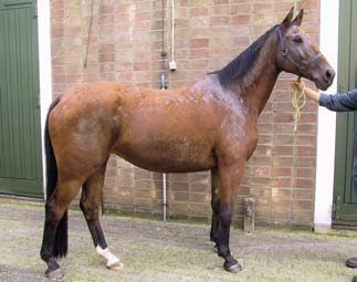
wetenschap
Figure 12. Histology of the nodules in the pectoral striated muscle revea- Figure 14. Small granulomata were also present in the liver containing
led large moderately circumscript granulamatous inflammatory lesions few multinucleated giant cells (arrowheads), in this micrograph affecting
also containing numerous multinucleated giant cells (arrowheads). On a hepatic portal area characterised by bile ducts (G), a small hepatic artery
the top left normal striated muscle is present (haematoxylin-eosin stain). (A) and a large hepatic vein (V) (haematoxylin-eosin stain).
mentioned castration as a possible risk factor because they
found a higher incidence of sarcoidosis in geldings.
In humans, the diagnosis of sarcoidosis is based on
history, clinical appearance, and histopathology of a skin
biopsy (if possible), thereby excluding all other granulo
matous diseases of known cause (6). In horses, the most
typical clinical signs are alopecia, crusting, and scaling,
and the diagnosis is based on this rather typical clinical
appearance and a confirmatory skin biopsy (1, 5, 7, 10, 13,
14, 15, 16). Pruritus has been described in cutaneous
sarcoidosis in humans (4) and in some cases in horses
(7, 16) but not in others (1, 5, 7, 10 and 14). The two horses
presented here showed no signs of pruritus.
In humans, pulmonary sarcoidosis is the most com
Figure 13. A micrograph of an enlarged and affected sternal lymphnode mon form and thoracic radiography findings are abnormal
contains, amongst numerous multinucleated giant cells, an archetypal in more than 90% of affected individuals (6), although
large Langhans type giant cell with abundant finely granular eosinophilic predictions for individual patients based on chest radio
cytoplasm and marginated nuclei in a horse-shoe-shape configuration graph alone are unreliable (6). In horses, thoracic radio
(arrowhead) (haematoxylin-eosin stain). graphs may be helpful in incidental cases with respiratory
signs. The two cases presented here had few respiratory
discussion signs. Moreover, given the pathological outcome, thoracic
In human medicine, sarcoidosis is an idiopathic granu abnormalities would have been detected on radiography
lomatous disease that usually affects the lung, with derma in case B only.
tological problems occurring later or not at all in the Although the literature mentions weight loss as a
course of the disease (6). In horses, most cases start clinical sign of generalized sarcoidosis, both cases present
clinically with dermatological problems (5, 7, 13, 14, 16). ed here had a normal body condition. However, both
The nodular form is quite rare (17, 13). In the cases horses were euthanized relatively early in the course of
presented here, the disease started with skin problems in the disease because of the poor prognosis. In case B typical
one horse (case A) and with nodules in the other (case B). granulomatous inflammatory lesions containing multi
The pathogenesis of the disease is still unknown both in nucleated Langhans type giant cells were also found in the
humans and in horses and is thought to be ‘immune- diaphragm and the bone marrow. To our knowledge, this
mediated’ (6, 13). In both cases presented here, bacterial, has not been described in horses before, although Spiegel
fungal, and yeast involvement was not found in skin et al. (16) mentioned vertebral and femoral involvement
samples or in tissues at postmortem examination. The via radiographs showing lesions of the bone in two horses.
owners of both horses mentioned an event that occurred In equine sarcoidosis blood haematology and biochem
before the onset of clinical problems: colic surgery istry findings as neutrophilia, hyperglobulinaemia, mild
6 months previously in case A and vaccination 3 weeks non-regenerative anaemia, and abnormal kidney and liver
previously in case B. These events are not mentioned as function have been described in equine sarcoidosis (16).
risk factors in the literature, although Spiegel et al. (16) However, only case B had anaemia and hyperglobulinae
660 Tijdschrift voor Diergeneeskunde • Deel 133 • Aflevering 16 • 15 augustus 2008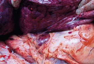
wetenschap
mia. Again, these findings might have changed had the dr. Roel van Nieuwstadt (Department of Equine Sciences)
horses lived longer. Although fine-needle aspiration for clinical help and advice.
biopsy is a minimally invasive technique, punch biopsy of
the skin and/or a tissue biopsy of a subdermal nodule with references
a ‘liver biopsy needle’ are needed to give a reliable ante 1. Axon JE, Robinson P and Lucas J. Generalised granulomatous disease
in a horse. Aust Vet J 2004; 82: 48-51.
mortem diagnosis.
2. English JC, Patel PJ and Greer KE. Sarcoidosis. J Am Acad Dermatol
The prognosis of human sarcoidosis is variable and is 2001; 44: 725-743.
strongly related to the presenting signs (6). In horses, 3. Fadok VA. An overview of equine dermatoses characterized by scaling
Spiegel et al. (16) mentioned a more favourable prognosis and crusting. Vet Clin N Am-Equine 1995; 11: 43-51.
4. Goldberg A, Lang A and Mekori YA. Prolonged generalized pruritus
than other authors (13, 11). The prognosis for localized
associated with selective elevation of IgA as the presenting symptoms
equine sarcoidosis is good for life, but guarded for the of sarcoidosis. Ann Allergy Asthma Immunol 1995; 74: 387-389.
affected skin (15). In humans, treatment for sarcoidosis is 5. Heath SE, Bell RJ, Clark EG et al. Idiopathic granulomatous disease
not always indicated because the disease may spon involving the skin in a horse. J Am Vet Med Assoc 1990; 197:
1033-1036.
taneously resolve and the prolonged use of corticosteroids
6. Judson MA. Sarcoidosis: clinical presentation, diagnosis and approach
has severe side-effects (6). However, corticosteroid therapy to treatment. Am J Med Sci 2008; 335: 26-33.
is indicated in life-threatening disease, if there is cardiac, 7. Loewenstein C, Bettenay SV and Mueller RS. A retrospective study of
neurological, or eye involvement, and should also be equine sarcoidosis. Vet Derm 2004; 15: 67.
8. Newman LS, Rose CS and Maier LA. Sarcoidosis. N Engl J Med 1997;
considered if the disease is progressive (6). Although some
336: 1224-1234.
equine cases may stay unaltered without any treatment, 9. Pascoe RRR and Knottenbelt DC. Sarcoidosis. Immune mediated/
treatment of both generalized and localized sarcoidosis is allergic diseases. In: Manual of Equine Dermatology. London: Harcourt
limited to prolonged (month to years) systemic corti Brace, 1999: 169-170.
10. Rose JF, Littlewood JD, Smith K et al. A series of four cases of
costeroids (13, 15, 16). generalized granulomatous disease in the horse. In: proceedings of 3rd
In conclusion, equine generalized sarcoidosis is a rare World Congress of Veterinary Dermatology 1998: 562-563.
disease with varying clinical syndromes. In the two cases 11. Schlipf JW. Dermatological conditions associated with crusts and
discussed here, one horse first had dermatological prob scales: Sarcoidosis. In: Robinson NE ed. Current therapy in Equine
Medicine, 4th edn. Philadelphia: W.B. Saunders, 1997: 384.
lems (severe crusting) and the other horse initially showed 12. Scott DW. Large Animal Dermatology. Philadelphia: W.B. Saunders,
subdermal nodules with a completely normal macro 1988: 326-328.
scopic appearance of the overlying skin. Generalized 13. Scott DW and Miller WH. Sarcoidosis. In: Equine dermatology.
equine sarcoidosis has a poor prognosis, in contrast to Saunders, St. Louis, 2nd edition, 2003: 675-680.
14. Sellers RS, Toribio RE and Blomme EAG. Idiopathic systemic
localized equine sarcoidosis, which has a favourable granulomatous disease and macrophage expression of pthrp in a
prognosis for life and a guarded prognosis for the localized miniature pony. J Comp Pathol 2001; 125: 214-218.
skin problem. 15. Sloet van Oldruitenborgh-Oosterbaan MM and Koeman JP. Recogni-
tion and therapy of sarcoidosis. In: Proceedings of the 45th beva
Congress. Birmingham, 2006: 184-185.
acknowledgements 16. Spiegel IB, White SD, Foley JE et al. A retrospective study of cutaneous
The authors thank dr. Pieter Jacobs (Equine Practice West equine sarcoidosis and its potential infectious aetiological agents.
Brabant) and dr. Hans Coster (Equine Practice Zwaans Vet Dermatol 2006; 17: 51-62.
hoek) for referring the patients to the Equine Clinic of the 17. Stannard AA. Generalized granulomatous disease. In: Robinson NE ed.
Current therapy in equine medicine, 2nd edn. Philadelphia: W.B.
Department of Equine Sciences and their contributions to Saunders, 1987: 645-646.
the manuscript. They also are grateful to dr. Lidwien
Verdegaal (at that time Department of Equine Sciences, Artikel ingediend: 4 mei 2008
now Bait al Arab Equine Hospital, Kuwait City) and Artikel geaccepteerd: 20 mei 2008
Tijdschrift voor Diergeneeskunde • Deel 133 • Aflevering 16 • 15 augustus 2008 661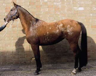
You can also read