Therapeutic Study of Thermosensitive Hydrogel Loaded with Super-Activated Platelet Lysate Combined with Core Decompression Technology for the ...
←
→
Page content transcription
If your browser does not render page correctly, please read the page content below
Hindawi
Stem Cells International
Volume 2021, Article ID 7951616, 7 pages
https://doi.org/10.1155/2021/7951616

Research Article
Therapeutic Study of Thermosensitive Hydrogel Loaded with
Super-Activated Platelet Lysate Combined with Core
Decompression Technology for the Treatment of Femoral
Head Necrosis

 Zhipeng Huang,1 Zhe Zhao,2 Jun Lang,1 Wantao Wang,1 Yinsheng Fu,3
 and Wenbo Wang 1,2
 1
 The First Affiliated Hospital of Harbin Medical University, 23 You Zheng Street, Harbin 150001, China
 2
 Southern University of Science and Technology Hospital, 6019 Liuxian Avenue, Xili, Nanshan District, Shenzhen 518000, China
 3
 Tianqing Stem Cell Co., Ltd., Jubao Second Road, Science and Technology Innovation City, Songbei District, Harbin 150000, China

 Correspondence should be addressed to Wenbo Wang; wenbowang1967@163.com

 Received 16 April 2021; Accepted 1 June 2021; Published 28 June 2021

 Academic Editor: Kun Zhang

 Copyright © 2021 Zhipeng Huang et al. This is an open access article distributed under the Creative Commons Attribution License,
 which permits unrestricted use, distribution, and reproduction in any medium, provided the original work is properly cited.

 Super activated platelet lysate (sPL) is a derivative of platelet-rich plasma (PRP) that contains high levels of several growth factors.
 In this study, we synthesized a temperature-sensitive hydrogel that contained temperature-sensitive Poly(DL-lactide-glycolide-
 glycolide acid) (PLGA), SrCl2, and HA, and loaded it with different concentrations of sPL. The hydrogel showed satisfactory
 encapsulation efficiency and release of the growth factors in a sustained manner, indicating its suitability as a drug carrier. The
 sPL-loaded hydrogel was inserted into the necrotic femoral head of a rat model and core decompression was applied and
 resulted in significantly accelerated bone repair and regeneration. Therefore, encapsulation of sPL in a hydrogel scaffolding may
 be an effective strategy for treating femoral head necrosis.

1. Introduction of patients within 1-3 years. Subchondral bone collapse
 causes resulting in considerable pain and impaired hip joint
Femoral head necrosis (ONFH) is characterized by osteocytic function and will eventually require total hip arthroplasty
necrosis and bone marrow necrosis as a result of insufficient (THA). However, since THA is not the best option for youn-
or a complete lack of blood supply to the subchondral bone. ger patients, action needs to be taken as early as possible to
There are currently 30 million diagnosed cases of ONFH slow down the progression of ONFH and delay the age of
worldwide, of which 8.12 million are in China alone, and joint replacement.
the rate of incidence has been increasing annually [1]. A Core decompression (CD) is a joint-preserving surgery
recent multicenter study conducted on ONFH patients in [3] that is suitable for the early stage ONFH patients with
China found that steroid use was identified as the causative an intact joint surface [4]. It can reduce intramedullary pres-
factor in 26.35% of males patients and 55.75% of female sure on the femoral head, accelerate bone regeneration that
patients [2]. Long-term use of high-dose glucocorticoids may form a cavity after core decompression, and reverse fem-
(GC) may affect the differentiation of cells in the femoral oral head necrosis [5], thereby delaying the progression of the
head and alter bone metabolism, thereby decreasing the disease and preventing femoral head collapse. However, 37%
angiogenic activity of the femoral head and triggering ische- of patients that undergo core decompression therapy will
mic hypoxia. progress to femoral head collapse [4].
 In the absence of an effective method of treatment, Platelet-rich plasma (PRP) can accelerate bone formation
ONFH can progress into subchondral bone collapse in 80% by restoring osteoblast proliferation and activating pathways
2 Stem Cells International

that promote angiogenesis and osteogenesis [6], which is a In brief, PRP was extracted by centrifuging fresh whole
strategy that can be used to the pathological progression of blood. The PRP was ultralow-frozen using melt preparation
ONFH. Growth factors in PRP can promote cartilage forma- and patented cytokine culture technologies, sPL can be effi-
tion [7] and osteogenesis [8] and can therefore alleviate ciently induced, activated, and cultivated.
ONFH [9]. In addition, platelet lysate (PL) growth factors
also promote the chemotactic migration of various cells [10, 2.3. Synthesis and Characterization of Hydrogels. The
11]. PL can be incorporated into biological scaffolds that temperature-sensitive PLGA\SrCl2 and HA were mixed at a
can retain its beneficial effects for a longer period [7, 12] ratio of 94 : 5 : 1, and the mixture was added to 0, 250, and
based on the preparation method, activation, initial platelet 500 μl of sPL placed in a magnetic stirrer. The resulting
concentration, and donor [12, 13]. Super-active platelet PLS0, PLS1, and PLS2 hydrogels were lyophilized and
lysate (sPL) is prepared from PRP via ultralow temperature sprayed with gold, and their morphology was observed under
freeze-thawing. It is enriched in bioactive factors that can a scanning electron microscope (SEM) (Japan Electronics
promote tissue regeneration and vascular remodeling. How- Co. Ltd.).
ever, the short half-life of the growth factors in sPL limits To measure the sustained release of bioactive factors
their biological effects in vivo. In addition, high dosageses from the PLS hydrogels created, the latter were placed in
and/or the frequent administration of sPL is not economi- clean vials (n = 4 per group) and completely submerged in
cally viable, and may lead to toxic side effects. The controlled 4 ml of simulated body fluid (SBF). The vials were then sealed
release of sPL loaded with hydrophilic macromolecules such and incubated in a water bath, and the aliquots in the
as growth factors can significantly improve its efficacy in vivo. medium were collected on days 3, 6, 9, 12, 15, 18, 21, 24,
 Polymers with a high molecular weight [14], such as 27, and 30. The concentration of VEGF and TGF-β was mea-
poly lactic-co-glycolic acid (PLGA), can be used as effec- sured using specific ELISA kits (Jingmei, Jiangsu).
tive in vivo drug delivery systems due to their biocompat- Preweighed lyophilized gels were incubated in deionized
ibility and biodegradability. The polymer material can water at 30°C, 34°C, 38°C, and 42°C for 1 hour to measure
protect sPL from the tissue microenvironment, leading to hydrogel swelling. The swollen gels were retrieved, blotted
controlled release. Bone is a natural organic-inorganic- to remove excess water, and weighed. The colloidal water
inorganic composite material that is mainly composed of content (SR) was calculated using the formula: ðW1 − W0Þ/
collagen and hydroxyapatite (HA, Ca10(PO4)6(OH)2)). A W0, where W0 and W1 indicate dry weight and temperature
variety of organic-inorganic composite materials that can weight, respectively.
mimic the composition and structure of bones have been
developed. Strontium (Sr) is a trace element found in the 2.4. In Vivo Experiments
human body that promotes bone formation and the heal-
ing of osteoporotic tissues. Therefore, we designed a com- 2.4.1. Establishment of an ONFH Model and Treatment. All
posite hydrogel that consisted temperature-sensitive PLGA, animal experiments were conducted in accordance with the
the bone mineral hydroxyapatite (HA), and strontium [15, guidelines on the humane use and care of animals formulated
16]. However, the hydrogel can flow into other parts of by the National Institutes of Health, all experimental animals
the defect, and the properties of the hydrogel need to be are taken care of, and all operations on animals are approved
changed to prevent the hydrogel from flowing out. by the Experimental Animal Use and Welfare Ethics Com-
 Temperature-sensitive PLGA/HA/SrCl2 hydrogel loaded mittee of the First Affiliated Hospital of Harbin Medical Uni-
with sPL was used to reconstruct the degenerated bone tissue versity (Ethical approval number:2019029).
in combination with CD surgery. sPL was slowly released Male SD rats weighing 280-300 g were reared at the ani-
from the hydrogel in a temperature-sensitive manner and mal center of the First Affiliated Hospital of Harbin Medical
resulted in accelerated bone repair after core decompression University and fed ad libitum on a standard laboratory diet
of the femoral head and ONFH. Therefore, our study has laid and water. A total of 27 rats were intravenously injected with
the foundation for the clinical application of heat-sensitive 10 μg/kg lipopolysaccharide (LPS), followed by 24 h later
hydrogel materials, and also for a considerable reduction in with three intramuscular injections of 20 mg/kg methylsulfo-
the cost of treating ONFH patients. nate (MPS) then at 24 h intervals. Osteonecrotic lesions first
 appeared two weeks after the procedure and continued to
2. Materials and Methods appear until 6 weeks after the procedure [18]. Three of the
 rats were euthanized, and tissue samples were collected for
2.1. Materials. Temperature-sensitive PLGA was purchased CT and histopathological examination. Once a diagnosis of
from Jinan Daigang Biomaterial Co., Ltd, SrCl2 was pur- ONFH was confirmed, the other rats were randomly assigned
chased from Sinopharm Chemical Reagent Co. Ltd., HA to the PLS0, PLS1, and PLS2 groups (n = 8). The rats were
was purchased from Aladdin, lipopolysaccharide was pur- anesthetized using 3% pentobarbital sodium, and the right
chased from Sigma-Aldrich Inc. (USA), while Methylsulfonate hip was exposed using an anterior and posterior approach
was purchased from Pfizer Pharmaceuticals (Hangzhou, while preserving the main blood vessels in the femoral head.
China). The hip joint was prevented from shifting by cutting the
 switch capsule, and the femoral head and neck were exposed.
2.2. sPL Preparation. sPL was isolated from human blood via Decompression was performed from the greater trochanter
ultralow temperature freezing, as previously described [17]. to the core of the femoral head using a drill, and the lesion
Stem Cells International 3

 PLS0 PLS1 PLS2

 10 m 10 m 10 m

 (a)
 TGF- (pg/ml) VEGF (pg/ml) SR

 VEGF concentration
 6000
 TGF- concentration

 10

 Water content of gel
 15000
 8
 10000 4000
 6
 2000 4
 5000
 2
 0 0 0
 30 34 38 42
 0
 3
 6
 9
 12
 15
 18
 21
 24
 27
 30

 0
 3
 6
 9
 12
 15
 18
 21
 24
 27
 30
 Time (days) Time (days) Temperature

 PLS0
 PLS1
 PLS2
 (b) (c) (d)

Figure 1: (a) Representative SEM images showing the structure of lyophilized thermosensitive hydrogel. (b, c) Release of growth factors from
the loaded sPL hydrogel. (d) Temperature-dependent change in water content.

was filled with PLS. The implanted region was covered with 2.4.5. Immunohistochemistry. The tissue sections were
bone wax, and the wound was closed. All animals were probed using primary antibodies against type I collagen and
intramuscularly injected with gentamicin (4 mg/kg) before CD31, followed by a secondary antibody with/out Cy5 conju-
and after the operation to prevent any wound infections. gate. After counterstaining with hematoxylin, the slides were
 observed under a fluorescence microscope (Leica, Mannheim,
2.4.2. Radiological Analysis. Four animals from each group Germany) or a light microscope (Leica, Mannheim, Germany).
were sacrificed to be used for the Micro-CT analysis on the
4th and 12th weeks postsurgery. Changes in the femoral head 2.5. Statistical Analysis. Data are expressed as mean ±
were monitored using a QuantumGX CT imaging system standard deviation (SD). One-way analysis of variance
(PerkinElmer, USA). An isotropic 20 mm voxel pitch dataset (ANOVA) was used to compare data between the groups.
was obtained using a total rotation of 360° and step length of A P value of
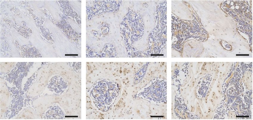
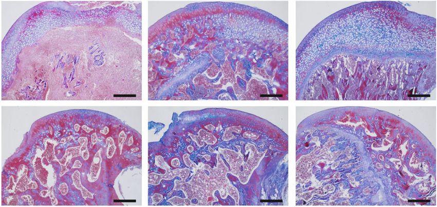
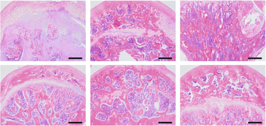
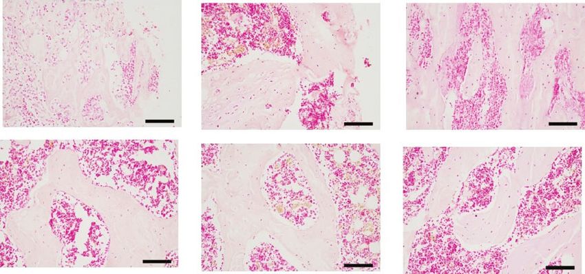
4 Stem Cells International

 PLS0 PLS1 PLS2

 4w

 12w

 Figure 2: Representative coronal, transverse and sagittal micro-CT scans of the different treatment groups.

 PLS0 PLS1 PLS2 PLS0 PLS1 PLS2

 4w 4w

 12w
12w

Figure 3: HE staining after treatment for femoral head necrosis Figure 5: ALP staining of the histopathological osteogenic marker
(40x). (200x).

 PLS0 PLS1 PLS2 PLS0 PLS1 PLS2

4w 4w

12w 12w

Figure 4: Histopathology Masson staining conducted 4 weeks and Figure 6: Histopathological type I staining demonstrated the
12 weeks after ONFH treatment (40x). composition of bone tissue (200x).

 PLS0 PLS1 PLS2
pressure of the femoral head. The core decompression chan-
nel for femoral head necrosis was well constructed, and the
hydrogel was accurately implanted without any damage to 4w
the muscles, nerves, or blood vessels. All animals were
healthy and no obvious signs of infection were observed. Sev-
eral studies have reported the presence of osteogenic growth
factors in PL [10, 20], and encapsulation of sPL in a hydrogel
 12w
scaffolding enhances the retention capacity of constituent
growth factors. As shown in Figure 2, the cross-sectional
and longitudinal CT images show that PLS1 was partially
repaired in the necrotic region, 4 weeks after the implanta- Figure 7: Histopathological CD31 staining demonstrating the
tion, compared with the PLS0 group, whereas PLS2 showed formation of blood vessels in the femoral head (200x).
a stronger therapeutic effect. Necrotic areas were still visible
even after 12 weeks in the PLS0 groups but had been mostly bone marrow cavity with large fat cells. Implantation of the
reconstituted in rats treated with PLS1 and PLS2. hydrogels did not trigger any localized inflammatory reaction
 Histological examination of the femoral head necrosis or fibrosis. PLS2 resulted in the formation of new bone and
showed that ONFH induction resulted in sparse cavities, dif- trabeculae by the 4th week after implantation with causing
fuse spot-like bone marrow necrosis, and a sparse trabecular osteonecrosis and hematopoietic necrosis. Likewise, PLS0
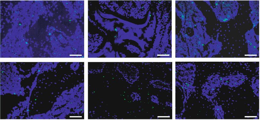
Stem Cells International 5

 PLS0 PLS1 PLS2
 ⁎
 ⁎⁎⁎⁎
 40 ⁎⁎⁎⁎ ⁎
 4w
 30

 20

 12w 10

 0
 4 12 4 12 4 12
 Time (weeks)
 PLS0
 PLS1
 PLS2
 (a) (b)

Figure 8: (a) Tunel staining conducted on the 4th week and 12th week after ONFH treatment (200x). (b) The apoptotic rate was calculated
using the Tunel-staining results.

and PLS1 administration caused the formation of new bones respectively, after 12 weeks (Figure 8(b)). In the 4th week,
but did not induce a trabecular arrangement (Figures 3 and compared with the PLS0 group, the apoptosis rate of the
4). Consistent with the observations mentioned above, bone PLS1 and PLS2 groups was significantly reduced (P < 0:05).
tissues expressing the osteogenic factor ALP and collagen I With the addition of sPL, the apoptosis rate in the tissues
were observed in all groups, and the degree of positive was also significantly reduced (P < 0:05). In the 12th week,
staining increased in the PLS-implanted groups in a sPL compared with the PLS0 group, the apoptosis rate of PLS1
concentration-dependent manner (Figures 5 and 6). and PLS2 groups was significantly reduced (P < 0:05). With
 the addition of sPL, the apoptosis rate between PLS0 and
3.3. The sPL-Loaded Hydrogel Promoted Angiogenesis. In PLS1 groups, and PLS1 and PLS2 groups had no significant
addition to, the growth factors in sPL promote the adherence statistical significance. In the PLS0 group, PLS1 group, and
and proliferation of osteoblasts, while also facilitating angio- PLS2 group, compared with 4 weeks, the apoptosis rate in tis-
genesis via the proliferation of vascular endothelial cells. sues at 12 weeks after operation was significantly reduced
Platelet endothelial cell adhesion molecule-1 (CD31), which (P < 0:05).
is typical for the endothelial lineage [21]. As shown in ER stress-induced apoptosis can be inhibited by PRP
Figure 7, compared with the PLS0 group, CD31 staining exosomes in a manner independent of the PERK/CHOP
increased in the PLS1 and PLS2 treatment groups during pathway [24]. Bone mesenchymal stem cells (BMSCs)
the 4th week of treatment. The number of CD31-positive cells treated with PRP exosomes and dexamethasone showed
increased in a time and sPL concentration-dependent man- enhanced phosphorylated Akt (protein kinase B) and Bad
ner and peaked in the PLS2 treatment group, 12 weeks after (Bcl-2 related death promoter) levels, as well as enhanced
surgery. Vadasz et al. [22] found that the decrease in VEGF Bcl-2 expression, indicating that PRP exosomes can inhibit
(vascular endothelial growth factor, promote angiogenesis) apoptosis by activating the Akt/Bad/Bcl-2 signaling path-
levels was correlated with ONFH progression and promoted way [24].
VEGF synthesis induced angiogenesis in the trabecular space Despite the encouraging results, our study has two main
in femoral head necrosis. Bai et al. [23] found that compared limitations. First, we established an ONFH rat model in rats,
to BMP-2 alone, the combination of VEGF and BMP-2 which did not develop femoral head collapse. However,
increased the formation of the vascular networks and signif- when we performed core decompression, the cartilage sur-
icantly enhanced the formation of ectopic bone. Consistent face was partially destroyed, simulating the destruction of
with previous reports, angiogenic factor CD31 was highly cartilage as a result of the femoral head collapse. The patho-
expressed in the PLS2-treated group (Figure 7). logical condition of ONFH was not consistent with the
 actual clinical situation and also precluded the long-term
3.4. The sPL-Loaded Hydrogel Inhibited Apoptosis in the effects of the use of the hydrogel. Therefore, at this stage, it
Femoral Head Tissue. Numerous Tunel-positive apoptotic is impossible to determine whether the hydrogel can prevent
bone cells were observed in the necrotic femoral head, while collapse. Instead, femoral collapse would have to be estab-
the number of positive cells decreased significantly after lished in a large animal model to demonstrate cartilage
hydrogel implantation. As shown in Figure 8, the rate of apo- repair. Second, we evaluated angiogenesis based only on
ptosis in the PLS0, PLS1, and PLS2 groups were 31:48 ± CD31 expression levels, and microangiography can be used
1:80%, 13:60 ± 1:27%, and 8:05 ± 0:27%, respectively, after to observe the formation of new blood vessels in a more
4 weeks, and 8:59 ± 1:55%, 4:97 ± 0:29%, and 3:08 ± 0:92%, accurate manner.
6 Stem Cells International

4. Conclusion hydrogel on cartilage formation,” Biomaterials, vol. 33,
 no. 14, pp. 3651–3661, 2012.
Compared to thermosensitive hydrogel, the sPL-loaded ther- [8] V. E. Santo, A. R. Duarte, E. G. Popa, M. E. Gomes, J. F. Mano,
mosensitive hydrogel steadily released biological factors that and R. L. Reis, “Enhancement of osteogenic differentiation of
reduced the level of osteoblast apoptosis, promoted osteogen- human adipose derived stem cells by the controlled release of
esis and angiogenesis, and effectively prevented the develop- platelet lysates from hybrid scaffolds produced by supercritical
ment of ONFH in a rat model. Therefore, treatment using fluid foaming,” Journal of Controlled Release, vol. 162, no. 1,
sPL-loaded thermosensitive hydrogel can be used in combi- pp. 19–27, 2012.
nation with core decompression surgery to improve the out- [9] J. Han, F. Gao, Y. Li et al., “The use of platelet-rich plasma for
comes of femoral head necrosis treatment. the treatment of osteonecrosis of the femoral head: a system-
 atic review,” BioMed Research International, vol. 2020, Article
 ID 2642439, 11 pages, 2020.
Data Availability [10] M. C. Phipps, Y. Xu, and S. L. Bellis, “Delivery of platelet-
 derived growth factor as a chemotactic factor for mesenchymal
The data used to support the findings of this study are
 stem cells by bone-mimetic electrospun scaffolds,” PLoS One,
included within the article.
 vol. 7, no. 7, article e40831, 2012.
 [11] Y. Yu, S. Zhu, Y. Hou, J. Li, and S. Guan, “Sulfur contents in
Conflicts of Interest sulfonated hyaluronic acid direct the cardiovascular cells fate,”
 ACS Applied Materials & Interfaces, vol. 12, no. 41, pp. 46827–
The authors have declared that there are no competing 46836, 2020.
interests. [12] P. S. Babo, R. L. Pires, L. Santos et al., “Platelet lysate-loaded
 photocrosslinkable hyaluronic acid hydrogels for periodontal
Acknowledgments endogenous regenerative technology,” ACS Biomaterials Sci-
 ence & Engineering, vol. 3, no. 7, pp. 1359–1369, 2017.
This study was supported by the Postgraduate Research & [13] J. M. de Leon, V. R. Driver, C. P. Fylling et al., “The clinical rel-
Practice Innovation Program of Harbin Medical University evance of treating chronic wounds with an enhanced near-
(Grant No. YJSKYCX2019-39HYD). physiological concentration of platelet-rich plasma gel,”
 Advances in Skin & Wound Care, vol. 24, no. 8, pp. 357–368,
 2011.
References [14] R. Xu, K. Zhang, J. Liang, F. Gao, J. Li, and F. Guan, “Hyaluro-
 nic acid/polyethyleneimine nanoparticles loaded with copper
 [1] D. W. Zhao, M. Yu, K. Hu et al., “Prevalence of nontraumatic ion and disulfiram for esophageal cancer,” Carbohydrate Poly-
 osteonecrosis of the femoral head and its associated risk factors mers, vol. 261, article 117846, 2021.
 in the Chinese population: results from a nationally represen-
 tative survey,” Chinese Medical Journal, vol. 128, no. 21, [15] S. C. Verberckmoes, M. E. De Broe, and P. C. D'Haese, “Dose-
 pp. 2843–2850, 2015. dependent effects of strontium on osteoblast function and
 mineralization,” Kidney International, vol. 64, no. 2, pp. 534–
 [2] D. Li, X. Xie, Z. Yang, C. Wang, Z. Wei, and P. Kang,
 543, 2003.
 “Enhanced bone defect repairing effects in glucocorticoid-
 induced osteonecrosis of the femoral head using a porous [16] B. Zhao, X. Li, H. Xu, Y. Jiang, D. Wang, and R. Liu, “Influence
 nano-lithium-hydroxyapatite/gelatin microsphere/erythro- of simvastatin-strontium-hydroxyapatite coated implant
 poietin composite scaffold,” Biomaterials Science, vol. 6, formed by micro-arc oxidation and immersion method on
 no. 3, pp. 519–537, 2018. osteointegration in osteoporotic rabbits,” International Jour-
 nal of Nanomedicine, vol. 15, pp. 1797–1807, 2020.
 [3] M. Chughtai, N. S. Piuzzi, A. Khlopas, L. C. Jones, S. B. Good-
 man, and M. A. Mont, “An evidence-based guide to the treat- [17] Z. Huang, W. Wang, Q. Wang et al., “Coaxial nanofiber scaf-
 ment of osteonecrosis of the femoral head,” The Bone & Joint fold with super-active platelet lysate to accelerate the repair
 Journal, vol. 99-b, no. 10, pp. 1267–1279, 2017. of bone defects,” RSC Advances, vol. 10, no. 59, pp. 35776–
 [4] D. R. Marker, T. M. Seyler, S. D. Ulrich, S. Srivastava, and 35786, 2020.
 M. A. Mont, “Do modern techniques improve core decom- [18] L. Qin, G. Zhang, H. Sheng et al., “Multiple bioimaging modal-
 pression outcomes for hip osteonecrosis?,” Clinical Orthopae- ities in evaluation of an experimental osteonecrosis induced by
 dics and Related Research, vol. 466, no. 5, pp. 1093–1103, 2008. a combination of lipopolysaccharide and methylpredniso-
 [5] J. R. Lieberman, S. M. Engstrom, R. M. Meneghini, and N. F. lone,” Bone, vol. 39, no. 4, pp. 863–871, 2006.
 SooHoo, “Which factors influence preservation of the osteone- [19] X. Wu, S. Yang, D. Duan, Y. Zhang, and J. Wang, “Experimen-
 crotic femoral head?,” Clinical Orthopaedics and Related tal osteonecrosis induced by a combination of low-dose lipo-
 Research, vol. 470, no. 2, pp. 525–534, 2012. polysaccharide and high-dose methylprednisolone in
 [6] V. T. Nguyen, M. Nardini, A. Ruggiu, R. Cancedda, rabbits,” Joint, Bone, Spine, vol. 75, no. 5, pp. 573–578, 2008.
 F. Descalzi, and M. Mastrogiacomo, “Platelet Lysate Induces [20] E. Jain, S. Sheth, A. Dunn, S. P. Zustiak, and S. A. Sell, “Sus-
 in Human Osteoblasts Resumption of Cell Proliferation and tained release of multicomponent platelet-rich plasma pro-
 Activation of Pathways Relevant for Revascularization and teins from hydrolytically degradable PEG hydrogels,” Journal
 Regeneration of Damaged Bone,” International Journal of of Biomedical Materials Research. Part A, vol. 105, no. 12,
 Molecular Sciences, vol. 21, no. 14, p. 5123, 2020. pp. 3304–3314, 2017.
 [7] L. S. Moreira Teixeira, J. C. Leijten, J. W. Wennink et al., “The [21] M. Hristov, W. Erl, and P. C. Weber, “Endothelial progenitor
 effect of platelet lysate supplementation of a dextran-based cells: mobilization, differentiation, and homing,” Arteriosclerosis,
Stem Cells International 7

 Thrombosis, and Vascular Biology, vol. 23, no. 7, pp. 1185–1189,
 2003.
[22] Z. Vadasz, I. Misselevich, D. Norman, E. Peled, and J. H. Boss,
 “Localization of vascular endothelial growth factor during the
 early reparative phase of the rats' vessels deprivation-induced
 osteonecrosis of the femoral heads,” Experimental and Molec-
 ular Pathology, vol. 77, no. 2, pp. 145–148, 2004.
[23] Y. Bai, Y. Leng, G. Yin et al., “Effects of combinations of BMP-
 2 with FGF-2 and/or VEGF on HUVECs angiogenesis in vitro
 and CAM angiogenesis in vivo,” Cell and Tissue Research,
 vol. 356, no. 1, pp. 109–121, 2014.
[24] S. C. Tao, T. Yuan, B. Y. Rui, Z. Z. Zhu, S. C. Guo, and C. Q.
 Zhang, “Exosomes derived from human platelet-rich plasma
 prevent apoptosis induced by glucocorticoid-associated endo-
 plasmic reticulum stress in rat osteonecrosis of the femoral
 head via the Akt/Bad/Bcl-2 signal pathway,” Theranostics,
 vol. 7, no. 3, pp. 733–750, 2017.
You can also read