Case Report Suspected Spontaneous Aqueous Humor Misdirection Syndrome in a Boston Terrier
←
→
Page content transcription
If your browser does not render page correctly, please read the page content below
Hindawi
Case Reports in Veterinary Medicine
Volume 2020, Article ID 1092562, 6 pages
https://doi.org/10.1155/2020/1092562

Case Report
Suspected Spontaneous Aqueous Humor Misdirection
Syndrome in a Boston Terrier

          Ashley E. Zibura , Michael G. Davidson, and Hans D. Westermeyer
          Comparative Ophthalmology, Department of Clinical Sciences, College of Veterinary Medicine, North Carolina State University,
          1060 William Moore Drive, Raleigh, NC 27607, USA

          Correspondence should be addressed to Hans D. Westermeyer; hdwester@ncsu.edu

          Received 11 January 2020; Accepted 10 March 2020; Published 29 May 2020

          Academic Editor: Sheila C. Rahal

          Copyright © 2020 Ashley E. Zibura et al. This is an open access article distributed under the Creative Commons Attribution
          License, which permits unrestricted use, distribution, and reproduction in any medium, provided the original work is
          properly cited.

          An eight-year-old female spayed Boston Terrier presented to the North Carolina Veterinary Hospital with glaucoma in the
          left eye (OS). Initial ophthalmic examination revealed moderate ocular hypertension, a diffusely and markedly shallow
          anterior chamber with anteriorly displaced iris and lens, vitreal prolapse, and a normal iridocorneal angle (ICA)
          morphology. The patient displayed a paradoxical response to topical latanoprost with an increase in intraocular pressure.
          These examination findings led to a putative diagnosis of spontaneous aqueous humor misdirection syndrome (AHMS).
          The patient was successfully managed with topical carbonic anhydrase inhibitors (CAIs) and apraclonidine for eight
          months until progressive ulcerative keratitis necessitated enucleation of the affected globe. Histopathology and high-field
          magnetic resonance imaging (MRI) of the enucleated globe did not identify an underlying cause for the glaucoma. This
          case suggests that AHMS should be considered in dogs presenting with a shallow anterior chamber, vitreal prolapse,
          increased intraocular pressure, and no other causes of glaucoma.

1. Introduction                                                       penetrating keratoplasty [5], phacoemulsification [6], and
                                                                      neodymium: YAG laser posterior capsulotomy [7]. However,
Aqueous humor misdirection syndrome (AHMS) is a rare                  spontaneous AHMS has not yet been described in dogs.
form of secondary glaucoma thought to result from posteri-               This report describes a suspected case of spontaneous
orly misdirected aqueous humor which becomes trapped in               AHMS in a canine patient.
the vitreous chamber [1, 2]. This leads to the anterior dis-
placement of the iris and lens, resulting in progressive pupil-
lary block and increased intraocular pressure (IOP) [1, 2].           2. Case Presentation
    Also known as malignant glaucoma, AHMS is relatively
rare. Spontaneous AHMS has been described primarily in                2.1. Signalment and History. An eight-year-old female spayed
geriatric cats. These cats typically present with a uniformly         Boston Terrier was presented for the evaluation of acute
shallow anterior chamber, anterior displacement of vitreous,          glaucoma OS. An IOP of 38 mmHg OS (13 mmHg in the
and modest increases in IOP [1, 2]. Initially, the iridocorneal       right eye (OD)) was noted 24 hours earlier by the referring
angle (ICA) is open [2], but with progressive anterior                veterinarian who prescribed three times daily topical dorzo-
displacement of the iris-lens diaphragm, angle narrowing              lamide OS (dorzolamide hydrochloride 2% ophthalmic solu-
and ciliary cleft collapse can develop [1, 2].                        tion, Sandoz Inc., Princeton, NJ, USA). Three years prior, a
    Induced aqueous humor misdirection syndrome                       conjunctival pedicle graft had been placed to treat a deep
(AHMS) has been reported in a dog [3] and a llama [4]                 stromal corneal ulcer OS. The owners reported no other past
following phacoemulsification, and in humans following                 health problems.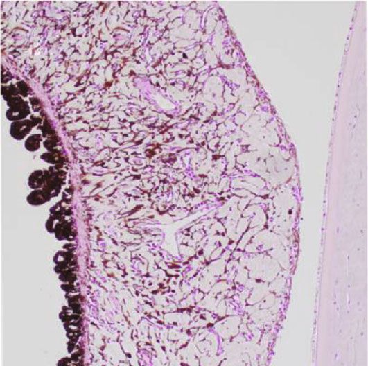
2                                                                                              Case Reports in Veterinary Medicine

                                            (a)                                          (b)

                                           (c)                                           (d)

Figure 1: Gonioscopy images of both eyes with an ICA Grade 2-3 and sparse areas of fibrae latae. (a) View of one quadrant OS. (b) View of
one quadrant OD. (c) View of a different quadrant OD. (d) Magnified view of a sector of the ICA OD showing foci of pigment (white arrow)
within the trabecular meshwork and small foci of fibrae latae (white arrowhead) at the opening of the ICA.

2.2. Initial Ophthalmic Examination. On presentation, neu-            ter, NY, USA) and latanoprost 0.005% ophthalmic solution
roophthalmic examination was unremarkable with intact                 (Bausch & Lomb). After 30 minutes of therapy, the IOP OS
palpebral reflex, menace response, direct and indirect pupil-          increased to 42 mmHg and moderate miosis was noted. A
lary light reflexes (PLR), and dazzle reflex. On slit lamp bio-         drop of tropicamide 1% (Bausch & Lomb) was administered
microscopy (Kowa SL-15, Tokyo, Japan) of the left eye,                OS in an attempt to relieve the suspected pupillary block
very mild faint corneal fibrosis was noted as the remnant of           component of the patient’s ocular hypertension. Thirty
the patient’s previous conjunctival pedicle graft. The anterior       minutes later, the IOP OS was 40 mmHg with no significant
chamber was uniformly, markedly shallow such that the iris            changes in pupil size noted. Four additional doses of Cosopt
nearly contacted the corneal endothelium. The pupil was               and three additional doses of latanoprost were administered
slightly larger than midrange. No iridodonesis or phacodon-           over the next 90 minutes in an attempt at achieving ocular
esis was noted. Vitreal strands extended through the pupil            hypotension; however, this resulted in an IOP of 48 mmHg.
into the shallow anterior chamber, filling the ventral 1/3 of          At that time, two doses of mannitol (20%, 1 gram/kg slow
the pupil opening. An early immature anterior cortical cata-          IV push through an in-line filter over 30 min, Hospira Inc.,
ract was also noted, affecting roughly 20% of the lens. IOP            Lake Forest, IL, USA) were administered 90 minutes apart.
(Tono-Vet, iCare, Vantaa, Finland) measured 32 mmHg                   Four hours later, the IOP OS had decreased to 10 mmHg.
OS, compared to 8 mmHg OD. Binocular indirect ophthal-                Cosopt (q8h OS) was continued overnight. The following
moscopy (Vantage LED Plus, Keeler, Malvern, PA, USA,                  morning, the IOP OS was 8 mmHg with a notably deeper
and 20D or 28D condensing lenses, Volk Optical Inc., Men-             anterior chamber depth. The dog was discharged with
tor, OH, USA) OD was unremarkable and was not performed               Cosopt (q8h OS) and tramadol (2.5 mg/kg PO q8-12h PRN
OS. The gonioscopic evaluation (RetCam, Clarity Medical               for 5 days, Amneal Pharmaceuticals, Bridgewater, NJ, USA).
Systems, Pleasanton, CA, USA) of the right eye revealed a
normal iridocorneal angle opening, ranging from grade 2 to            2.4. Subsequent Ophthalmic Examination and Treatment.
3 throughout the eye’s circumference [8], and minimal pecti-          Three days later, the IOP OS was 17 mmHg OS (10 mmHg
nate ligament dysplasia characterized by a few small sectors          OD) with normal anterior chamber depth, and similar find-
of fibrae latae (Figure 1). Direct gonioscopy of the left eye          ings to those noted at discharge. Indirect ophthalmoscopy
was not performed at this initial examination due to the              revealed a normal fundus and gonioscopy of the left eye par-
markedly shallow anterior chamber.                                    alleled that of the right eye with a normal iridocorneal angle
                                                                      opening ranging from grade 2 to 3 throughout the eye’s cir-
2.3. Initial Response to Therapy. The left eye was treated with       cumference and minimal pectinate ligament dysplasia char-
several doses of alternating dorzolamide hydrochloride                acterized by a few small sectors of fibrae latae (Figure 1).
2%/timolol maleate 0.5% (Cosopt, Bausch & Lomb, Roches-               Sixteen days later, the IOP OS was 19 mmHg (11 mmHg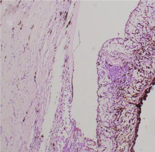
Case Reports in Veterinary Medicine                                                                                                       3

                                      ⁎
                                                                                         ⁎

                                           (a)                                               (b)

Figure 2: (a) Axial and (b) peripheral transverse sections of the enucleated canine globe (OS) imaged with high field 9.4 T MRI. Note the
corneal stromal loss consistent with the patient’s bacterial keratitis, the shallow anterior chamber with notable loss of aqueous humor, and
the multifocal undulation of the neurosensory retina (white arrow) consistent with multifocal retinal detachment which occurred
artifactually, subsequent to globe fixation and processing. The white asterisks denote the plastic drinking straw used for globe positioning
during image acquisition.

OD) with a slightly shallow anterior chamber and vitreous               remained static with an IOP of 19 mmHg OS (13 mmHg
strands filling the 50% of the pupil opening. Cosopt (q8h                OD), and a 3-4 mm diameter midstromal corneal ulcer with
OS) was continued, and apraclonidine (0.5% ophthalmic solu-             50% stromal loss. An aerobic culture of the ulceration identi-
tion, 2 drops 2 minutes apart twice daily OS, Akorn Inc., Lake          fied a coagulase-negative staphylococcus species and Coryne-
Forest, IL, USA) was added for adjunctive therapy. Two weeks            bacterium species. Five days later, the ulcer had progressed to
and 5 months later, the IOP OS was 10 mmHg and 13 mmHg,                 70% stromal loss and the owners elected to enucleate OS. At
respectively (10 mmHg and 14 mmHg OD, respectively).                    the time of globe removal, performed via a subconjunctival
     Eight months after initial evaluation, the dog’s left eye          approach, normal retrobulbar and extraocular anatomy was
became acutely red, cloudy, and painful. Menace response,               noted. In an effort to try to further investigate the ocular
direct pupillary light reflex (PLR), and indirect PLR (from              pathology, the excised globe was submitted for high-field
OD to OS) were absent OS. Indirect PLR (from OS to OD)                  magnetic resonance imaging and histopathologic analysis.
and dazzle reflex OS were intact. Biomicroscopic examina-
tion OS revealed moderate conjunctival hyperemia with                   2.5. MRI Methods. The enucleated left globe was fixed in 10%
scleral injection, superficial corneal neovascularization, mul-          formalin (Azer Scientific, Morgantown, PA, USA) for twenty
tifocal areas of corneal edema with corneal bullae, and multi-          days. The eye was then carefully transferred to a formalin-
focal punctate areas of superficial corneal ulceration. The              filled 60 milliliter syringe (Becton Dickinson, Franklin Lakes,
anterior chamber was uniformly shallow, mirroring the                   NJ, USA). Imaging was performed using a Bruker BioSpec
chamber depth noted at the time of initial glaucoma diagno-             94/30USR 9.4 T horizontal bore scanner MRI (Bruker Cor-
sis. The iris leaflets were uniformly anteriorly displaced.              poration, Billerica, MA, USA). The eye was imaged in a
Anterior uveitis with a concurrent hypermature cataract                 transverse plane overnight, and images were acquired with
was noted with a miotic, nonresponsive pupil and 2+ aque-               the ParaVision software (Bruker Corporation).
ous flare. An incipient cortical cataract was noted OD with
no other changes. The IOP OS was 34 mmHg (10 mmHg                       2.6. MRI Findings. Focal disruption in axial corneal epithelium
OD). The patient was treated with 1 drop of Cosopt ophthal-             was noted with moderate focal stromal loss. The corneal curva-
mic solution OS every 5 minutes for a total of 30 minutes.              ture had a diffusely undulating appearance. The anterior
Subsequent IOP reading OS was 24 mmHg. The patient was                  chamber was markedly shallow secondary to dehydration in
discharged on continued Cosopt therapy three times daily                formalin. However, the ciliary cleft appeared open. The lens
OS, apraclonidine ophthalmic therapy increased to three                 appeared slightly posteriorly subluxated. The vitreous chamber
times daily OS, hypertonic saline 5% ophthalmic solution                appeared intact with multifocal undulation of the neurosen-
(Muro-128, Bausch & Lomb) four times daily OS, flurbipro-                sory retina consistent with multifocal retinal detachment. The
fen sodium 0.03% ophthalmic solution (Bausch & Lomb)                    optic nerve head appeared intact and unremarkable (Figure 2).
four times daily OS, ofloxacin 0.3% ophthalmic solution (Ris-
ing Pharmaceuticals Inc., Saddle Brook, NJ, USA) every 2                2.7. Histopathology Findings. The sections of the globe were
hours during waking hours, and oral carprofen (2.2 mg/kg,               routinely stained with hematoxylin and eosin. Grossly, the
Rimadyl, Zoetis, Kalamazoo, MI, USA) by mouth twice daily.              lens was posteriorly luxated. There was moderate keratiniza-
Three days later, the patient’s neuroophthalmic examination             tion of the corneal epithelium. The peripheral superior aspect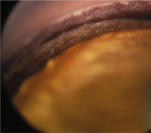
4 Case Reports in Veterinary Medicine
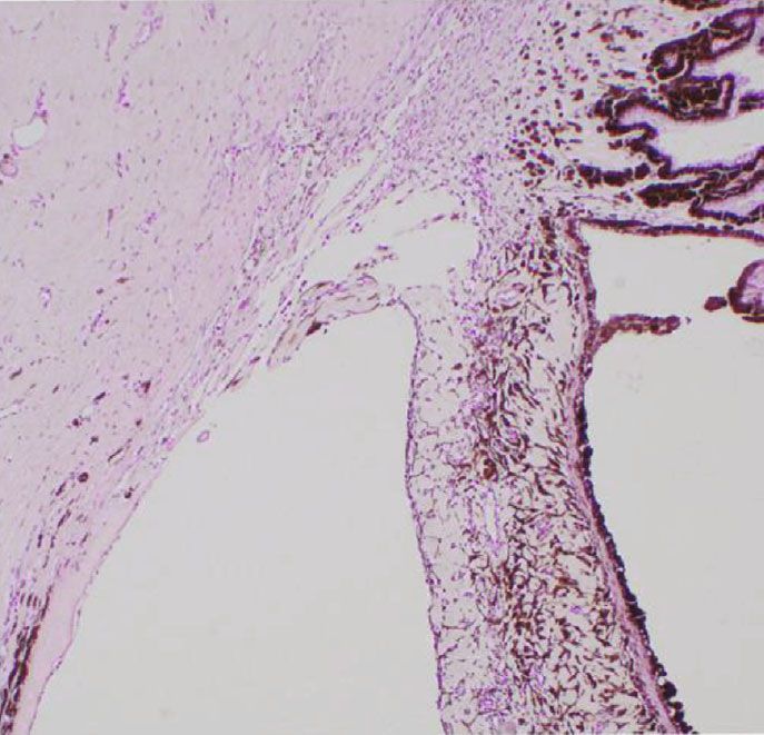
(a) (b) (c)
Figure 3: Histopathology images of the enucleated canine globe (OS) with hematoxylin and eosin (H&E) stain. (a) Image of the
nondependent ICA demonstrating an open angle and lack of goniodysgenesis at 4x magnification, thus ruling out primary glaucoma as
the cause of this patient’s glaucoma. (b) Image of the dependent ICA demonstrating an open angle, lack of goniodysgeneis, moderate
lymphoplasmacytic infiltrate in the iris, ciliary body, and trabecular meshwork, and mild preiridal fibrovascular membrane (PIFM)
formation, viewed at 4x magnification. This very mild degree of inflammation is incongruent with an 8 month history of uveitis-driven
glaucoma, thus supporting a differential cause for this patient’s glaucoma, such as AHMS. (c) Image of the iris leaflet with scant
lymphoplasmacytic infiltration and scant PIFM.
of the cornea had marked fibrosis and vascularization of the anterior chamber, anterior displacement of vitreous, a nor-
superficial corneal stroma. There was a marked loss of the mal iridocorneal angle, and a modest increase in IOP. Addi-
axial corneal stroma associated with moderate edema and tionally, iridocorneal anomalies associated with primary
mild fibroplasia, in addition to focally extensive doubling of angle closure glaucoma (PACG) were ruled out gonioscopi-
Descemet’s membrane with the formation of an axial retro- cally and histopathologically, and no other findings associ-
corneal membrane. There was moderate lymphoplasmacytic ated with secondary glaucoma were identified. Moreover,
infiltrate in the iris, ciliary body, and trabecular meshwork, the rise in IOP in response to latanoprost implicates pupillary
with a mild preiridal fibrovascular membrane (PIFM). Even block as at least one of the likely mechanisms generating
after multiple deeper sections of the block, the iridocorneal increased IOP in this patient, possibly as a component of
angles appeared normal, and no lesions consistent with gonio- ciliovitreolenticular block as previously described in cases
dysgenesis were identified (Figure 3). There was moderate liq- of feline AHMS [2]. The dog of this report also manifested
uefaction of the cortical lens fibers with the formation of signs of vitreous humor degeneration with anterior chamber
morgagnian globules, multifocal areas of mineralization, and presentation of vitreous. This is thought to contribute to the
posterior migration of the lens epithelium. Alcian blue stain pathogenesis of AHMS in humans [10]. A marked increase in
revealed the moderate degeneration of the vitreous with accu- IOP, from 32 to 42 mmHg, after latanoprost administration
mulation of dense aggregates of vitreal matrix adjacent to and was noted in the present case. This 31% IOP increase is sig-
associated with the equatorial lens capsule. The number of nificantly outside the range of potential variation from
ganglion cells was mildly decreased, and the optic nerve pre- tonometry [11–13] and is likely due to exacerbation of pupil-
sented moderate gliosis. The retinal and optic nerve lesions lary block from the shallow anterior chamber and iris-lens-
were very mild, and thus definitive confirmation of the pres- vitreous anterior displacement. The IOP decreased, albeit
ence of glaucoma was not possible. very slightly (from 42 to 40 mmHg), following the adminis-
tration of tropicamide which was given with the aim of
2.8. Follow-up. At recheck examination 7 days following enu- releasing the patient’s suspected pupillary block. Tropica-
cleation, the patient’s surgical incision was intact and healing mide was unsuccessful at relieving the patient’s ocular hyper-
well, and her right eye remained comfortable and visual. On tension and the pupil remained miotic. Thus, in this patient,
follow-up communication with her owners 10 months follow- the effects of tropicamide were likely overridden by the
ing surgery, her right eye remained comfortable. Her cataract miotic effects of latanoprost. It should also be noted that sub-
OD had progressed, causing moderate visual impairment; sequent doses of latanoprost resulted in progressive IOP ele-
however, no clinical signs of glaucoma were observed. vation, reaching 48 mmHg prior to mannitol treatment. This
provides further evidence to support the occurrence of pupil-
3. Discussion lary block in this patient as a likely primary mechanism driv-
ing glaucoma.
The patient’s clinical presentation and ophthalmic examina- Other mechanisms by which the flow of aqueous humor
tion findings mirror those reported in cases of feline sponta- can be blocked at the level of the ciliary body, anterior vitre-
neous AHMS [1, 2, 9]. Specifically, a distinctly shallow ous face, and/or pupil include: iris bombe, iridociliary cysts,
Case Reports in Veterinary Medicine                                                                                               5

intraocular neoplasia, lens sub/luxation, intumescent lens, and     traction of the globe, leading to artefactual lens subluxation
spherophakia [1]; however, all of these disease processes were      and multifocal retinal detachments noted during ex vivo
definitively ruled out in this case through ophthalmic examina-      analysis. The vitreal condensation initially described in cats
tion, histopathologic globe analysis, and high-field MRI of the      with AHMS [2, 16] was not seen histologically in the current
enucleated globe. A block of aqueous flow distal to the level of     report either. However, the last episode of elevated intraocu-
the ICA is highly unlikely in this case based on the absence of     lar pressure due to suspected AHMS occurred more than 8
overt uveitis at initial presentation, histopathologically normal   months prior to enucleation. It is possible that these bands
trabecular meshwork and ciliary cleft, and normal retrobulbar       were present during the episodes of elevated intraocular pres-
anatomy noted at the time of enucleation. While gonioscopy          sure but resolved over time.
cannot rule out the possibility of primary open angle glaucoma           Additional diagnostic tests which should be considered
(POAG), this form of glaucoma has neither been reported in          for thorough evaluation of cases of suspected AHMS include
the Boston Terrier breed nor has it been reported in association    streak retinoscopy to evaluate for potential refractive errors
with anterior iris displacement and vitreal prolapse [14]. More-    subsequent to anterior displacement of the iris-lens dia-
over, an elevated intraocular pressure was not recorded in the      phragm and vitreous body [6, 17, 18], high-resolution ocular
opposite eye as of the last follow up—approximately 1.5 years       ultrasonography (HRUS) or ultrasound biomicroscopy
after initial pressure elevation in the first eye.                   (UBM) to better quantify the degree of anterior chamber nar-
     It seems unlikely uveitis was the cause of the initial pres-   rowing and evaluate for posterior segment foci of aqueous
sure elevations, even though clinical uveitis was observed          humor fluid [6, 18, 19], and optical coherence tomography
prior to enucleation and histologic evidence of uveitis was         (OCT) to further quantify anterior and posterior segment
noted. There were no clinical signs of uveitis noted at the         abnormalities [19, 20]. These tests were not performed in the
time of glaucoma diagnosis, or at any point over the follow-        presented case; however, they may have provided insight to
ing eight months. Additionally, if uveitis were the driving fac-    further support a diagnosis of spontaneous AHMS and should
tor for glaucoma for eight months, more robust uveal                be considered for workup of future cases. Clinical B-scan ocu-
inflammation and PIFM would be expected. Thus, the very              lar ultrasonography with a 10 or 12 MHz probe was not per-
mild inflammatory infiltrate of the dependent iridocorneal            formed due to the assessed low clinical yield of identifying
angle and the thin PIFM noted on histopathologic examina-           pools of aqueous fluid in the vitreal cavity with this probe fre-
tion is incongruent with an 8-month history of uveitis-driven       quency. HRUS or UBM provides higher resolution which is
glaucoma. Therefore, AHMS is a more plausible differential           valuable for diagnosing more subtle lesions [21]. As a result,
for the initial glaucoma. It is much more likely the uveitis        HRUS and UBM are the preferred ultrasonic imaging modal-
noted prior to enucleation was unrelated to the initial glau-       ities for AHMS diagnosis in humans [6, 17–19] and have been
coma and was a separate process driven by a combination             used successfully in the diagnosis of feline AHMS [2].
of ulcerative keratitis and a hypermature cataract.                      It is important to note that although detecting pockets of
     Treatment with dorzolamide/timolol and apraclonidine           aqueous humor is considered a positive diagnostic finding in
adequately controlled the patient’s IOP for eight months in         AHMS, the lack of this finding on HRUS or the absence of
the case presented here. However, the patient’s uveitis and         this test does not preclude diagnosis. In fact, only 9 of the
concurrent ocular changes towards the end of the disease            32 cats in the seminal publication describing spontaneous
course were likely driving glaucoma through different second-        feline AHMS [2] underwent HRUS. Additionally, the authors
ary mechanisms. Outside of the patient’s complicating end-          of that study suggest aqueous humor may become diffusely
stage factors, it is unclear what the true duration of response     distributed throughout the vitreous. Having HRUS showing
to AHMS therapy the patient would have experienced. Con-            pockets of aqueous humor within the vitreous would have
servative management with topical carbonic anhydrase inhib-         helped confirm the etiology of pressure elevations in the pre-
itor medications is the mainstay of therapy for feline              sented case. However, based on the present literature avail-
spontaneous AHMS [1, 2], with phacoemulsification, anterior          able on the subject [4–7, 17, 18, 20, 22–24], the negative
vitrectomy, posterior capsulotomy, and/or endocyclophoto-           predictive value of HRUS in diagnosing AHMS is low. In
coagulation considered in refractory cases [1, 2, 9]. In feline     other words, an ultrasound had been performed and no
patients, AHMS tends to progress slowly, although most cases        aqueous humor pockets were observed, AHMS could not be
ultimately become refractory to treatment with a guarded to         ruled out. Thus, the lack of ultrasound examination in this
poor overall prognosis for vision or IOP control [1, 2].            case report, although a weakness, does not represent a failure
     While systematic clinical exclusion of other differentials      to positively diagnose this syndrome in a dog.
validates a diagnosis of spontaneous AHMS in the presented               This report describes a presumptive case of spontaneous
case, definitive confirmation was not possible. AHMS is typ-          AHMS in a dog. While not previously reported in dogs, this
ically confirmed through observation of pools of aqueous             syndrome should be considered as a differential diagnosis
humor within the vitreous chamber or between the vitreous           in cases of glaucoma with atypical clinical findings, open
and the retina [1, 2]. However, the majority of the aqueous         ICA morphology, and paradoxical response to standard
fluid in the globe diffused out during processing with forma-         medical therapy.
lin as a result of its hyperosmolar properties [15], thereby
preventing observation of pools of aqueous fluid in the poste-       Conflicts of Interest
rior segment on histopathologic or MRI analysis. This loss of
fluid also appears to have contributed to diffuse volume con-         The authors declare that they have no conflicts of interest.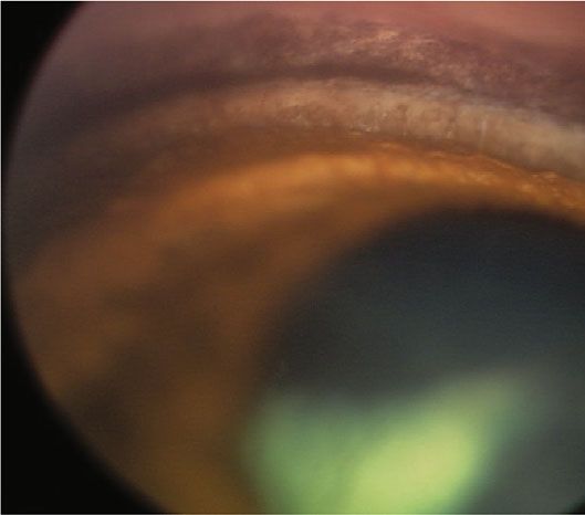
6                                                                                                   Case Reports in Veterinary Medicine

References                                                                [16] G. J. McLellan and L. B. C. Teixeira, “Feline glaucoma,” Veter-
                                                                               inary Clinics of North America - Small Animal Practice, vol. 45,
 [1] G. J. McLellan and P. E. Miller, “Feline glaucoma-a compre-               no. 6, pp. 1307–1333, 2015.
     hensive review,” Veterinary Ophthalmology, vol. 14, pp. 15–          [17] A. Grzybowski and P. Kanclerz, “Acute and chronic fluid mis-
     29, 2011.                                                                 direction syndrome: pathophysiology and treatment,” Graefe’s
 [2] J. M. C. Czederpiltz, N. C. la Croix, A. . . Woerdt et al., “Puta-        Archive for Clinical and Experimental Ophthalmology, vol. 256,
     tive aqueous humor misdirection syndrome as a cause of glau-              no. 1, pp. 135–154, 2018.
     coma in cats: 32 cases (1997-2003),” Journal of the American         [18] H. Shahid and J. F. Salmon, “Malignant glaucoma: a review of
     Veterinary Medical Association, vol. 227, no. 9, pp. 1434–                the modern literature,” Journal of Ophthalmology, vol. 2012,
     1441, 2005.                                                               Article ID 852659, 6 pages, 2012.
 [3] H. M. Denis, “anterior lens capsule disruption and suspected         [19] J. Foreman-Larkin, P. A. Netland, and S. Salim, “Clinical man-
     malignant glaucoma in a dog,” Veterinary Ophthalmology,                   agement of malignant glaucoma,” Journal of Ophthalmology,
     vol. 5, no. 2, pp. 79–83, 2002.                                           vol. 2015, Article ID 283707, 6 pages, 2015.
 [4] C. C. Powell, T. M. Nuhsbaum, and J. R. Gionfriddo, “Aqueous         [20] C. Wirbelauer, A. Karandish, H. Häberle, and D. T. Pham,
     misdirection and ciliary block (malignant) glaucoma after cat-            “Optical coherence tomography in malignant glaucoma fol-
     aract removal in a llama,” Veterinary Ophthalmology, vol. 5,              lowing filtration surgery,” British Journal of Ophthalmology,
     no. 2, pp. 99–101, 2002.                                                  vol. 87, no. 8, pp. 952–955, 2003.
 [5] I. Gama, W. M. Rodrigues, H. P. Filipe, M. Y. Faria, and L. D.       [21] C. E. Plummer, A. Regnier, and K. N. Gelatt, “The canine
     Almeida, “Síndrome de dirección inadecuada del humor                      Glaucomas,” in Veterinary Ophthalmology, K. N. Gelatt, B.
     acuoso refractario: una posible complicación de la querato-               C. Gilger, and T. J. Kern, Eds., pp. 1050–1145, Wiley-Black-
     plastia penetrante,” Archivos de La Sociedad Espanola de Oftal-           well, Ames, Iowa, 5th edition, 2013.
     mologia, vol. 92, no. 8, pp. 390–393, 2017.
                                                                          [22] V. Debrouwere, P. Stalmans, J. Van Calster, W. Spileers,
 [6] P. Moinul and C. M. L. Hutnik, “Aqueous misdirection syn-                 T. Zeyen, and I. Stalmans, “Outcomes of different manage-
     drome: an interesting case presentation,” Clinical Ophthalmol-            ment options for malignant glaucoma: a retrospective study,”
     ogy, vol. 9, pp. 183–186, 2015.                                           Graefe’s Archive for Clinical and Experimental Ophthalmology,
 [7] L. Mastropasqua, M. Ciancaglini, P. Carpineto, L. Lobefalo,               vol. 250, no. 1, pp. 131–141, 2012.
     and P. E. Gallenga, “Aqueous misdirection syndrome: a com-           [23] D. Tabibian, F. Hoogewoud, N. Mavrakanas, and J. S. Schutz,
     plication of neodymium: YAG posterior capsulotomy,” Jour-                 “Misdirected aqueous flow in rhegmatogenous retinal detach-
     nal of Cataract and Refractive Surgery, vol. 20, no. 5,                   ment: a pathophysiology update,” Survey of Ophthalmology,
     pp. 563–565, 1994.                                                        vol. 60, no. 1, pp. 51–59, 2015.
 [8] B. Ekesten and K. Narfstrom, “Correlation of morphologic fea-        [24] A. Sharma, F. Sii, P. Shah, and G. R. Kirkby, “Vitrectomy–pha-
     tures of the iridocorneal angle to intraocular pressure in Sam-           coemulsification–vitrectomy for the management of aqueous
     oyeds,” American Journal of Veterinary Research, vol. 52,                 misdirection syndromes in phakic eyes,” Ophthalmology,
     no. 11, pp. 1875–1878, 1991.                                              vol. 113, no. 11, pp. 1968–1973, 2006.
 [9] R. M. Atkins, M. D. Armour, and J. A. Hyman, “Surgical out-
     come of cats treated for aqueous humor misdirection syn-
     drome: a case series,” Veterinary Ophthalmology, vol. 19,
     pp. 136–142, 2016.
[10] F. Stringa, T. Iqbal, A. Makuloluwa, and V. Shankar, “Clinical
     biomarker in aqueous misdirection syndrome: the pupillary
     snap sign,” JCRS Online Case Reports, vol. 7, no. 1, article
     S2214167718300516, pp. 6–8, 2019.
[11] J.-T. Ahn, M. B. Jeong, Y. W. Park et al., “Accuracy of intraoc-
     ular pressure measurements in dogs using two different
     tonometers and plano therapeutic soft contact lenses,” Veteri-
     nary Ophthalmology, vol. 15, pp. 83–88, 2012.
[12] C. Görig, R. T. I. Coenen, F. C. Stades, S. C. Djajadiningrat-
     Laanen, and M. H. Boevé, “Comparison of the use of new
     handheld tonometers and established applanation tonometers
     in dogs,” American Journal of Veterinary Research, vol. 67,
     no. 1, pp. 134–144, 2006.
[13] Icare TonoVet, icare Tonovet User’s and Maintenance Manual,
     vol. 2, ICare Finland Oy, Vantaa, Finland, 2013.
[14] P. E. Miller and E. Bentley, “Clinical signs and diagnosis of the
     canine primary glaucomas,” Veterinary Clinics of North Amer-
     ica - Small Animal Practice, vol. 45, no. 6, pp. 1183–1212, 2015.
[15] R. Thavarajah, V. Mudimbaimannar, U. Rao, K. Ranganathan,
     and J. Elizabeth, “Chemical and physical basics of routine
     formaldehyde fixation,” Journal of Oral and Maxillofacial
     Pathology, vol. 16, no. 3, p. 400, 2012.
You can also read