Keratin - based materials in Biomedical engineering
←
→
Page content transcription
If your browser does not render page correctly, please read the page content below

IOP Conference Series: Materials Science and Engineering

PAPER • OPEN ACCESS

Keratin - based materials in Biomedical engineering
To cite this article: Sonia Singh 2021 IOP Conf. Ser.: Mater. Sci. Eng. 1116 012024

View the article online for updates and enhancements.

                               This content was downloaded from IP address 46.4.80.155 on 22/09/2021 at 13:18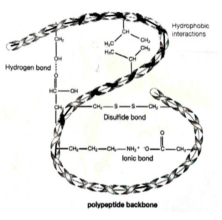
FSAET 2020                                                                                                               IOP Publishing
IOP Conf. Series: Materials Science and Engineering              1116 (2021) 012024           doi:10.1088/1757-899X/1116/1/012024

Keratin - based materials in Biomedical engineering
                    Sonia Singh
                    Institute of Pharmaceutical Research
                   GLA University
                   17km Stone, NH-2, Mathura-Delhi Road Mathura,
                    Chaumuhan, Uttar Pradesh 281406

                     sonia.singh@gla.ac.in

                     Abstract. A biomaterial is used to replace tissue or its function within the living body. Many
                     natural occurring polymers like collagen, fibrin, elastin, gelatin, silk fibroin, hyaluronic acid
                     and chitosan, that have been broadly utilized as in biomaterial applications. In addition to this,
                     proteins are known to be used as one of the popular biomaterials because of their capability to
                     work as synthetic ECM. Among this, keratin is a protein used as effective biopolymers in the
                     fabrication of many new biomaterial(s). Various new techniques have been made for their
                     extraction and structural characterization. Keratin is being characterized as repetitive sequences
                     of amino acid that led in the production of self-assembly. The self-assemble character of
                     keratin has attained to develop into many physical appearances such as sponges, nano-particles
                     and films, found helpful in many drug deliveries and biomedical tissue engineering. This
                     manuscript detailed the advanced utilisation of keratin biomaterials in the area of tissue
                     engineering; wound healing, drug delivery, and so on.
                     Keywords: tissue engineering, wound, fibre, biomaterial, self-assembly

1. Introduction
Keratin-based materials are the biodegradable natural polymers, which have brought a great revolution
in the field of biomaterial field because of their definite properties such as biocompatibility, durability
and biodegradability. Although, such materials would be utilized in the form of hydrogels, films, and
also even as sponge forms in the applications of biomedical engineering [1]. Keratin plays a critical
role in the constitution of major parts of wool, nails, hairs, feathers and hairs [2]. This current
manuscript will give more insights on different sources, structural and functional properties, the
history involved in the development of keratin-based biomaterial(s), their extraction method and
application(s) in the directions of biomedical field, respectively.

2. Historical background of keratin research
As per the literature survey, the keratin was reported for their therapeutical applications in the 16th
century by Li Shi –Zhen, chinese herbalism practitioner [3]. The term “keratin” was first introduced in
literature to outline the substances obtained from animal horns, hooves and even also hard tissues in
1850. Although in the early days of the 20th century, many researchers have focused on various
methods of keratin extraction. In 1920s, the main objective of research was to understand the
structural, functional and extraction method of keratin protein [4]. During the periodical year of World

              Content from this work may be used under the terms of the Creative Commons Attribution 3.0 licence. Any further distribution
              of this work must maintain attribution to the author(s) and the title of the work, journal citation and DOI.
Published under licence by IOP Publishing Ltd                          1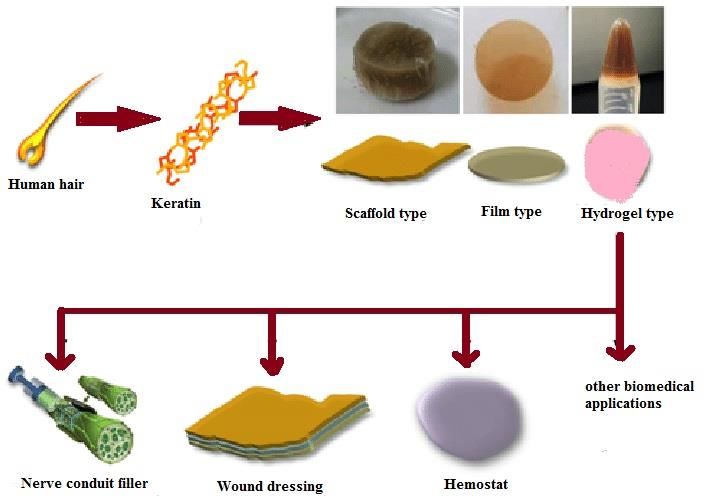
FSAET 2020                                                                                      IOP Publishing
IOP Conf. Series: Materials Science and Engineering   1116 (2021) 012024   doi:10.1088/1757-899X/1116/1/012024

War, many research projects have utilized keratin for the production of textile. In 1940, the CSIR
called as Council for Scientific and Industrial Research, has bought department of protein chemistry
called as Division of Protein Chemistry, situated in Australia to expand the applications of wool and
keratin. Moreover, the main objective of such a department was to understand the structural properties
of keratin fibre [5]. Another research has been conducted through the University of Leeds and the
Wool Industries Research Association in the UK, which has suggested the structure of keratin based
fibre; as it has consist of a cortex followed by a layer of cuticle. Additionally, in the Netherlands, the
investigators have patented a method of fabricating the keratin films and the textile fibre obtained
from hooves [4]. In other words, many experimental works have been conducted between 1940 and
1970 on Keratin, which had established a new platform for the fundamentals of biomaterial(s) [6-8].
                In 1970s, it has been found an exponential achievement in the techniques, explored to
extract and characterize keratin(s) and their products. Several researchers have worked on the proper
utilization of extracted keratin in different forms like films, gels, foams, coatings and fibres [9-11].
Additionally in past decades, the application of keratin and their derivatives for various biomedical
applications such as drug delivery wound healing, tissue engineering continued to become the topic of
interest. During the year of 1980s, chitosan, alginate, hyaluronic acid and collagen has become the
most widely utilized biomaterials in different medical applications [12,13].

3. Keratin: structure
Keratin comprises of cysteine, as a fibrous protein associated mainly with the intermediate filaments,
which are important in the formation of cytoskeleton and skin appendageal structures including wool,
nails, horns, hairs and feathers [1]. Such biopolymers are widely present as depending on their
variations in structure and properties; classified as hard and soft keratins on the presence of sulfur
content. The hard keratin contains a higher percentage of crosslinks attached with sulfur. It also
contributes to epidermal structure and mainly consists of filaments that are arranged in ordered arrays
embedded within a crosslinked matrix [14,15]. Soft keratins, with low sulfur content, prepared from
the bundles of cytoplasmic filaments that are loosely packed. It also provides resilience towards
epithelial tissues [14].
              In literature, the structure of keratins has been studied into alpha-keratins (α-keratins) and
beta-keratins (β-keratins), respectively [16]. Among these distributions, the α-keratin is used as a
primary constituent in the formation of nails, wool, horns, hairs and even of outermost layer of
epidermis. However, on the other hand, the beta-keratin is employed as a primary component of avian
and reptilian claws, beaks, scales and feathers [14,15].
               In α-keratin, the polypeptide chains are mainly comprised in the form of α-helices;
whereas, in case of β- keratin, they have been arranged as pleated β-sheets [17]. In a few decades, the
wool has been investigated as a representative material of α-keratin [18-20]. The α-keratin filament
has a diameter of 6–10 nm i-e in nanoscale and 2–4 nm for β-keratin [21,22]. Wool keratin has a 3D-
structure; consists of approximate proteins (94.9%), minerals (0.5%) and some traces amount of lipid
content [23,24]. Keratin consists of a polypeptide chain containing different amino acids, served as a
backbone having inter-molecular as well as intra-molecular binding (Figure 1). Despite this, the
disulfide, hydrogen, and ionic bonds enhanced the stability and the strength of keratin [25,26]. Such
proteins are insoluble in a polar solvent, alkali, weak acids organic solvents, and protein-digesting
enzymes including pepsin or trypsin. In higher concentration, these biopolymers contain amino acids
such as proline, serine, cysteine and glycine; and methionine, histidine and lysine are present in a
lower concentration. [27]. Three-dimensional mesh structures, resistance towards dissolution in
different solvents and even high stability of keratin is mainly because of disulphide bonds. The
solubilization of wool structure mostly takes place through the disarranging of keratin-complex
structure. [28].

                                                         2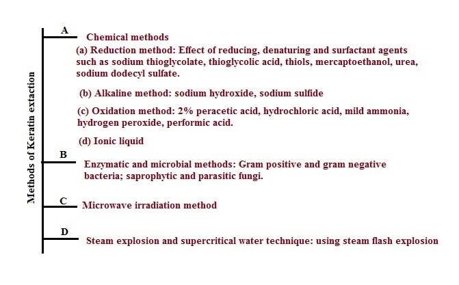
FSAET 2020                                                                                        IOP Publishing
IOP Conf. Series: Materials Science and Engineering   1116 (2021) 012024     doi:10.1088/1757-899X/1116/1/012024

                                      Figure 1: Structure of Keratin [50].

4. Methods used in Keratin based extraction
Initially, keratins are detached from cortex using some chemicals which are used to break
down the disulphide bonds that extensively present in the keratinized tissue(s). The α-keratin
and even gamma-keratin are then converted into non-crosslinked forms via the oxidation or
reduction process. Moreover, during these processes, cystine gets converted into cysteic acid
or cysteine. Free proteins are extracted out in the presence of denaturing solvents using
filtration as well as the dialysis process. In oxidative extraction technique, keratins generally
called as “keratoses” are produced by the application of hydrogen peroxide or peracetic acid.
Keratoses are non-disulphide, hygroscopic and crosslinks. These are polar soluble, and highly
susceptible to hydrolysis at extremely high pH, because of polarization generated by electron-
withdrawing properties of cysteic acid. Although such characteristics properties can lead the
biomaterials to undergo degradation relatively very fast as in vivo [29].
                 Reductive process of extraction can be carried out using dithiothreitol,
thiolglycolic acid and 2-mercaptoethanol. Such extraction technique may form reduced
keratin called as “kerateines" which are less water-soluble, stable at pH values and re-
crosslinked. And these methods would remain in vivo for about weeks to months,
respectively. Most employed techniques for the keratin extraction from keratin-rich
biomaterials including oxidation, reduction, alkali treatment, steam explosion, microwave
irradiation techniques, or by the applications of ionic liquids [30-35]. Figure 2 detailed
various methods employed in the extraction and solubilization of keratin.

                                                         3
FSAET 2020 IOP Publishing
IOP Conf. Series: Materials Science and Engineering 1116 (2021) 012024 doi:10.1088/1757-899X/1116/1/012024
Figure 2: Methods of Keratin extraction [50]
5. Keratin biomaterials - Biomedical applications
Various properties of keratins, concerning physical, chemical and biological aspects led to the
evolution of several keratin biomaterials utilized in the biomedical application(s). The extracted
keratin has a great intrinsic ability to get polymerize into porous and fibrous scaffolds which have
been studied exclusively. The occurrence of self –assembly is greatly found in highly conserved
structure of hair fibre; even when it has been processed correctly it is responsible for architecture,
porosity and dimensionality of keratin-derived materials. Additionally, keratin based biomaterials
obtained from human hair as well as wool revealed to contain cell-binding motifs like glutamic acid-
aspartic acid-serine binding residues, responsible for supporting cellular binding. Though, all these
properties together would create three-dimensional matrixes responsible for cellular infiltration
attachment and proliferation. Keratin biomaterials play an important role in the regulatory functions
found helpful in mediating the cellular behaviour of the body. Many investigators have searched
various methods that modulate the physical as well as mechanical properties of keratins and their
application of interest [29]. Some of the applications of keratin-based biomaterials (Table 1) are
mentioned below:
• Keratin based films: used as a bone morphogenic protein carrier and even in ocular surface
reconstruction
• Keratin based scaffolds employed in the urinary tract tissue engineering
• Keratin hydrogels utilized in drug delivery system and as dynamic matrices in the treatment of
wound healing.
• Keratin powder employed as an absorbent in wound dressing showed the promotion of skin
healing. Such application is because of the release of keratin derivative peptides to the wound
[29].
4FSAET 2020 IOP Publishing
IOP Conf. Series: Materials Science and Engineering 1116 (2021) 012024 doi:10.1088/1757-899X/1116/1/012024
Table.1: Keratin based biomaterial composition and their applications
Sl.no. Biomaterial composition Applications References
Aqueous Keratin dialysate In-vitro as in wound healing of
1 incorporated with alkaline based corneal epithelial. [36]
keratin dialysate
2 Photo active keratin derived films Wound healing, tissue engineering [37]
Improve stability in artificial
Keratin film cross-linked with trans-
3 gastric juice environment. [38]
glutaminase
Photo cross-linkable keratin- 2-D & 3-D cell culture substrates,
4 polyethene glycol hydrogels via the microfabrication techniques. [39]
thiol-norbornene "click" reaction
5 Keratin film Reconstruction of ocular surface [40]
6 Keratin, chitosan/gelatin Soft tissue engineering [41]
7 Keratin –chitosan Wound dressing material [42]
Facilitates osteoblast attachment
8 PLA/chitosan/keratin composites [43]
and proliferation.
9 Keratin wound dressing Used as hemostatic material. [44]
Used as a substrate for cellular
10 Keratin gel attachment and proliferation, [45]
delivery of therapeutic agents.
For repairing cardiac tissue after
11 Hydrogels in injectable forms [46]
myocardial infarction
12 Recombinant keratin proteins Dermal wound healing. [47]
Keratin based therapeutic dermal Wound healing.
13 [48]
patches
Keratin/poly (vinyl alcohol) Tissue engineering.
14 [49]
nanofiber
Keratin has emerged as a strong and effective biomaterial. They are bioactive and
abundantly formed as a bio-sourced of autologous protein(s). Many experimental works have
conducted to examine the application of keratin-based materials in the field of biomedical (Fig 2).
In recent studies, keratin films were utilized in the reconstruction of the ocular surface which uses
amniotic membrane (AM) of humans. Such studies revealed the fact that keratin films can be used
as a replacement of Am in the ophthalmology; such films are transparent and much stiffer when
compared with AM in relevant levels of attachment with the corneal epithelial cells. Keratin
hydrogels derived from human hair were introduced into hearts resulting angiogenesis without
producing inflammation and elevated adverse effects on the heart. Researchers revealed the
potential effect of keratin hydrogels on animal models by showing the development of peripheral
nerves regeneration. Such studies suggested that the keratin-based biomaterial is considered as a
neuroconductive nature that helps in regulating the molecules to enhance the regeneration of nerve
tissue followed by elevated Schwann cells activities [29]. Keratin powder used in wound healing
dressing as an absorbent; thereby it releases its derivative i-e peptides to the wounded area which
may promote the wound healing. Additionally, some polar soluble keratin peptides obtained from
an oxidative method demonstrated to enhance the proliferation of fibroblasts in human skin [51-
52].
5FSAET 2020 IOP Publishing
IOP Conf. Series: Materials Science and Engineering 1116 (2021) 012024 doi:10.1088/1757-899X/1116/1/012024
Figure 2 Keratin based biomaterials [55]
Keratin derivatives were experimented on animals to treat burn wounds where it
showed a significant reduction in the size of wounded area and eventually enhanced the rate of
wound healing. Similarly, the other forms of keratin-based biomaterials such as cross-linked powder,
hydrogels and films possessed potential proliferation of wound healing cell line areas. Another
research suggested that keratin material such as keratose showed beneficial effects in acute ischemic,
due to its improved viscosity as well as colloid oncotic properties [53-54]. Human hair derived keratin
scaffolds were tested for skin disorders in rodents via subcutaneously. Results demonstrated the
potential bio-compatibility and wound healing property of keratin-based materials [29], [54].
Conclusions
Several keratin biomaterials have been counterfeited and also investigated extensively their
application in biomedical sciences as fibres, films, scaffolds, sponges and hydrogels. Keratin based
biomaterials showed biocompatibility, specific chemical property, and also biodegradability. Despite
all these properties, only a few of these biomaterials progressed to clinical trials and possessed a small
share in the commercial market when compared to other biomaterials. Therefore, accompanied with a
novel development of reproducible methods for keratin extraction, along with adequate mechanical
and biological properties for biomedical application (s) would ultimately produce vital steps toward
the tissue regenerations.
References
[1] McLellan, J., Thornhill, S. G., Shelton, S., & Kumar, M. (2019). Keratin-based
biofilms, hydrogels, and biofibers. In Keratin as a Protein Biopolymer (pp. 187-200).
Springer, Cham.
[2] Costa, F., Silva, R., & Boccaccini, A. R. (2018). Fibrous protein-based biomaterials
(silk, keratin, elastin, and resilin proteins) for tissue regeneration and repair. In Peptides and
proteins as biomaterials for tissue regeneration and repair (pp. 175-204). Woodhead
Publishing.
[3] Zhen, L. S., & Mu, B. C. G. (2005). The Time Literature & Art Press:
Changchun. Jilin, China.
[4] Rouse, J. G., & Van Dyke, M. E. (2010). A review of keratin-based biomaterials for
biomedical applications. Materials, 3(2), 999-1014.
6FSAET 2020 IOP Publishing
IOP Conf. Series: Materials Science and Engineering 1116 (2021) 012024 doi:10.1088/1757-899X/1116/1/012024
[5] Rivett, D. E., Ward, S. W., Belkin, L. M., Ramshaw, J. A. M., & Wilshire, J. F. K.
(1996). Keratin and wool research. The Lennox Legacy; CSIRO Publishing: Collingwood,
Australia.
[6] Rogers, G. E. (1959). Electron microscope studies of hair and wool. Annals of the
New York Academy of Sciences, 83(3), 378-399.
[7] Parry, D. A., & Creamer, L. K. (1979). Fibrous proteins, scientific, industrial, and
medical aspects. In International Conference on Fibrous Proteins 1979: Massey University).
Academic Presss.
[8] Earland, C., & Knight, C. S. (1956). Studies on the structure of keratin II. The amino
acid content of fractions isolated from oxidized wool. Biochimica et biophysica acta, 22(3),
405-411.
[9] Anker, C. A. (1972). U.S. Patent No. 3,642,498. Washington, DC: U.S. Patent and
Trademark Office.
[10] Kawano, Y., & Okamoto, S. (1975). Film and gel of keratins. Kagaku to
Seibutsu, 13(5), 291-2.
[11] Okamoto, S. (1977). On the formation of films from some proteins. Nippon Shokuhin
Kogyo Gakkaishi, 24(1), 40-50.
[12] Noishiki, Y., Ito, H., Miyamoto, T., & Inagaki, H. (1982). Application of denatured
wool keratin derivatives to an antithrombogenic biomaterial-vascular graft coated with a
heparinized keratin derivative. Kobunshi Ronbunshu, 39(4), 221-227.
[13] Miyamoto, T., Takahashi, S. I., Ito, H., Inagaki, H., & Noishiki, Y. (1989). Tissue
biocompatibility of cellulose and its derivatives. Journal of biomedical materials
research, 23(1), 125-133.
[14] Fraser, R. D. B., MacRae, T. P., & Rogers, G. E. (1972). Keratins: their composition,
structure and biosynthesis.
[15] Schweizer, J., Bowden, P. E., Coulombe, P. A., Langbein, L., Lane, E. B., Magin, T.
M., & Wright, M. W. (2006). New consensus nomenclature for mammalian keratins. The
Journal of cell biology, 174(2), 169-174.
[16] Spearman, R. I. C. (1967). On the nature of the horny scales of the
pangolin. Zoological Journal of the Linnean Society, 46(310), 267-273.
[17] Jones, L. N., Simon, M., Watts, N. R., Booy, F. P., Steven, A. C., & Parry, D. A. D.
(1997). Intermediate filament structure: hard α-keratin. Biophysical chemistry, 68(1-3), 83-93.
[18] Kumawat, T. K., Sharma, A., Sharma, V., & Chandra, S. (2018). Keratin Waste: The
Biodegradable Polymers. In Keratin. IntechOpen.
[19] Patrucco, A., Visai, L., Fassina, L., Magenes, G., & Tonin, C. (2019). Keratin-based
matrices from wool fibers and human hair. In Materials for Biomedical Engineering (pp. 375-
403). Elsevier.
[20] Rajabinejad, H., Bucişcanu, I. I., & Maier, S. S. (2019). Current approaches for raw
wool waste management and unconventional valorization : a review. Environmental
Engineering & Management Journal (EEMJ), 18(7).
[21] Squire, J., & Vibert, P. J. (1987). Fibrous protein structure. Academic Press.
[22] Fraser, R. B., & Parry, D. A. (2011). The structural basis of the filament-matrix
texture in the avian/reptilian group of hard β-keratins. Journal of structural biology, 173(2),
391-405.
[23] Ayutthaya, S. I. N., Tanpichai, S., & Wootthikanokkhan, J. (2015). Keratin extracted
from chicken feather waste: extraction, preparation, and structural characterization of the
7FSAET 2020 IOP Publishing
IOP Conf. Series: Materials Science and Engineering 1116 (2021) 012024 doi:10.1088/1757-899X/1116/1/012024
keratin and keratin/biopolymer films and electrospuns. Journal of Polymers and the
Environment, 23(4), 506-516.
[24] Fortunati, E., Aluigi, A., Armentano, I., Morena, F., Emiliani, C., Martino, S., ... &
Puglia, D. (2015). Keratins extracted from Merino wool and Brown Alpaca fibres: Thermal,
mechanical and biological properties of PLLA based biocomposites. Materials Science and
Engineering: C, 47, 394-406.
[25] Park, M., Kim, B. S., Shin, H. K., Park, S. J., & Kim, H. Y. (2013). Preparation and
characterization of keratin-based biocomposite hydrogels prepared by electron beam
irradiation. Materials Science and Engineering: C, 33(8), 5051-5057.
[26] Poole, A. J., Lyons, R. E., & Church, J. S. (2011). Dissolving feather keratin using
sodium sulfide for bio-polymer applications. Journal of Polymers and the
Environment, 19(4), 995-1004.
[27] Korniłłowicz-Kowalska, T., & Bohacz, J. (2011). Biodegradation of keratin waste:
theory and practical aspects. Waste management, 31(8), 1689-1701.
[28] Tonin, C., Aluigi, A., Varesano, A., & Vineis, C. (2010). Keratin-based
nanofibres. nanofibers. InTech, 139-158.
[29] Vasconcelos, A., & Cavaco-Paulo, A. (2013). The use of keratin in biomedical
applications. Current drug targets, 14(5), 612-619.
[30] Buchanan, J. H. (1977). A cystine-rich protein fraction from oxidized α-
keratin. Biochemical Journal, 167(2), 489-491.
[31] Yamauchi, K., Yamauchi, A., Kusunoki, T., Kohda, A., & Konishi, Y. (1996).
Preparation of stable aqueous solution of keratins, and physiochemical and biodegradational
properties of films. Journal of Biomedical Materials Research: An Official Journal of The
Society for Biomaterials and The Japanese Society for Biomaterials, 31(4), 439-444.
[32] Tsuda, Y., & Nomura, Y. (2014). Properties of alkaline‐hydrolyzed waterfowl
feather keratin. Animal Science Journal, 85(2), 180-185.
[33] Tonin, C., Zoccola, M., Aluigi, A., Varesano, A., Montarsolo, A., Vineis, C., &
Zimbardi, F. (2006). Study on the conversion of wool keratin by steam
explosion. Biomacromolecules, 7(12), 3499-3504.
[34] Zoccola, M., Aluigi, A., Patrucco, A., Vineis, C., Forlini, F., Locatelli, P., ... &
Tonin, C. (2012). Microwave-assisted chemical-free hydrolysis of wool keratin. Textile
Research Journal, 82(19), 2006-2018.
[35] Xie, H., Li, S., & Zhang, S. (2005). Ionic liquids as novel solvents for the
dissolution and blending of wool keratin fibers. Green Chemistry, 7(8), 606-608.
[36] Feng, Y., Borrelli, M., Meyer-ter-Vehn, T., Reichl, S., Schrader, S., & Geerling, G.
(2014). Epithelial wound healing on keratin film, amniotic membrane and polystyrene in
vitro. Current Eye Research, 39(6), 561-570.
[37] Aluigi, A., Sotgiu, G., Torreggiani, A., Guerrini, A., Orlandi, V. T., Corticelli, F., &
Varchi, G. (2015). Methylene blue doped films of wool keratin with antimicrobial
photodynamic activity. ACS applied materials & interfaces, 7(31), 17416-17424.
[38] Cui, L., Gong, J., Fan, X., Wang, P., Wang, Q., & Qiu, Y. (2013).
Transglutaminase‐modified wool keratin film and its potential application in tissue
engineering. Engineering in Life Sciences, 13(2), 149-155.
[39] Yue, K., Liu, Y., Byambaa, B., Singh, V., Liu, W., Li, X., ... & Ng, K. W. (2018).
Visible light crosslinkable human hair keratin hydrogels. Bioengineering & translational
medicine, 3(1), 37-48.
8FSAET 2020 IOP Publishing
IOP Conf. Series: Materials Science and Engineering 1116 (2021) 012024 doi:10.1088/1757-899X/1116/1/012024
[40] Reichl, S., Borrelli, M., & Geerling, G. (2011). Keratin films for ocular surface
reconstruction. Biomaterials, 32(13), 3375-3386.
[41] Kakkar, P., Verma, S., Manjubala, I., & Madhan, B. (2014). Development of
keratin–chitosan–gelatin composite scaffold for soft tissue engineering. Materials Science and
Engineering: C, 45, 343-347.
[42] Singaravelu, S., Ramanathan, G., Raja, M. D., Barge, S., & Sivagnanam, U. T.
(2015). Preparation and characterization of keratin-based biosheet from bovine horn waste as
wound dressing material. Materials letters, 152, 90-93.
[43] Tanase, C. E., & Spiridon, I. (2014). PLA/chitosan/keratin composites for
biomedical applications. Materials Science and Engineering: C, 40, 242-247.
[44] Burnett, L. R., Richter, J. G., Rahmany, M. B., Soler, R., Steen, J. A., Orlando, G.,
& Van Dyke, M. E. (2014). Novel keratin (KeraStat™) and polyurethane (Nanosan®-Sorb)
biomaterials are hemostatic in a porcine lethal extremity hemorrhage model. Journal of
Biomaterials Applications, 28(6), 869-879.
[45] Han, S., Ham, T. R., Haque, S., Sparks, J. L., & Saul, J. M. (2015). Alkylation of
human hair keratin for tunable hydrogel erosion and drug delivery in tissue engineering
applications. Acta biomaterialia, 23, 201-213.
[46] Hasan, A., Khattab, A., Islam, M. A., Hweij, K. A., Zeitouny, J., Waters, R., ... &
Paul, A. (2015). Injectable hydrogels for cardiac tissue repair after myocardial
infarction. Advanced Science, 2(11), 1500122.
[47] Veerasubramanian, P. K., Thangavel, P., Kannan, R., Chakraborty, S.,
Ramachandran, B., Suguna, L., & Muthuvijayan, V. (2018). An investigation of konjac
glucomannan-keratin hydrogel scaffold loaded with Avena sativa extracts for diabetic wound
healing. Colloids and Surfaces B: Biointerfaces, 165, 92-102.
[48] Nayak, K. K., & Gupta, P. (2017). Study of the keratin-based therapeutic dermal
patches for the delivery of bioactive molecules for wound treatment. Materials Science and
Engineering: C, 77, 1088-1097.
[49] Wang, J., Hao, S., Luo, T., Zhou, T., Yang, X., & Wang, B. (2017). Keratose/poly
(vinyl alcohol) blended nanofibers: fabrication and biocompatibility assessment. Materials
Science and Engineering: C, 72, 212-219.
[50] Feroz, S., Muhammad, N., Ranayake, J., & Dias, G. (2020). Keratin-Based materials
for biomedical applications. Bioactive Materials, 5(3), 496-509.
[51] Van Dyke, M. E., Timmons, S. F., Blanchard, C. R., Siller-Jackson, A. J., & Smith,
R. A. (2001). U.S. Patent No. 6,270,793. Washington, DC: U.S. Patent and Trademark Office.
[52] Nunez, F., Trach, S., Burnett, L., Handa, R., Van Dyke, M., Callahan, M., & Smith,
T. (2011). Vasoactive Properties of Keratin‐Derived Compounds. Microcirculation, 18(8),
663-669.
[53] Xu, S., Sang, L., Zhang, Y., Wang, X., & Li, X. (2013). Biological evaluation of
human hair keratin scaffolds for skin wound repair and regeneration. Materials Science and
Engineering: C, 33(2), 648-655.
[54] Chen, Y. H., Dong, W. R., Xiao, Y. Q., Zhao, B. L., Hu, G. D., & An, L. B. (2006).
Preparation and bioactivity of human hair keratin-collagen sponge, a new type of dermal
analogue. Nan fang yi ke da xue xue bao= Journal of Southern Medical University, 26(2),
131-138.
[55] Lee, H., Noh, K., Lee, S. C., Kwon, I. K., Han, D. W., Lee, I. S., & Hwang, Y. S. (2014).
Human hair keratin and its-based biomaterials for biomedical applications. Tissue Engineering and
Regenerative Medicine, 11(4), 255-265.
9FSAET 2020 IOP Publishing
IOP Conf. Series: Materials Science and Engineering 1116 (2021) 012024 doi:10.1088/1757-899X/1116/1/012024
10You can also read