Characterization of bacterial communities associated with blood fed and starved tropical bed bugs, Cimex hemipterus (F.) (Hemiptera): a high ...
←
→
Page content transcription
If your browser does not render page correctly, please read the page content below
www.nature.com/scientificreports
OPEN Characterization of bacterial
communities associated
with blood‑fed and starved tropical
bed bugs, Cimex hemipterus (F.)
(Hemiptera): a high throughput
metabarcoding analysis
Li Lim & Abdul Hafiz Ab Majid*
With the development of new metagenomic techniques, the microbial community structure of
common bed bugs, Cimex lectularius, is well-studied, while information regarding the constituents
of the bacterial communities associated with tropical bed bugs, Cimex hemipterus, is lacking. In this
study, the bacteria communities in the blood-fed and starved tropical bed bugs were analysed and
characterized by amplifying the v3-v4 hypervariable region of the 16S rRNA gene region, followed
by MiSeq Illumina sequencing. Across all samples, Proteobacteria made up more than 99% of the
microbial community. An alpha-proteobacterium Wolbachia and gamma-proteobacterium, including
Dickeya chrysanthemi and Pseudomonas, were the dominant OTUs at the genus level. Although the
dominant OTUs of bacterial communities of blood-fed and starved bed bugs were the same, bacterial
genera present in lower numbers were varied. The bacteria load in starved bed bugs was also higher
than blood-fed bed bugs.
Cimex hemipterus Fabricus (Hemiptera), also known as tropical bed bugs, is an obligate blood-feeding insect
throughout their entire developmental cycle, has made a recent resurgence probably due to increased worldwide
travel, climate change, and resistance to insecticides1–3. Distribution of tropical bed bugs is inclined to tropical
regions, and infestation usually occurs in human dwellings such as dormitories and h otels1,2. Bed bugs are a
nuisance pest to humans as people that are bitten by this insect may experience allergic reactions, iron deficiency,
and secondary bacterial infection from bite s ores4,5.
Eradication of bed bugs from an infested area is challenging not only because they are resistant to insecticides
but also because they can crawl and hide into tight crevices, making them difficult to discover4,6. Moreover,
bed bugs could survive for many months without feeding, causing more problems for the residents4. The vari-
ous mechanisms, including metabolic, behavioural, and physiological of bed bugs, could have contributed to
maintaining wellness4,5. Nevertheless, the role played by microbiota may be as well crucial in bed bugs’ survival.
Insects are commonly possessing symbiotic microorganisms that essential in host’s growth, development,
nutrient supplement, protection, insecticides resistance, and r eproduction5,7. For example, Buchnera, an obligate
symbiont of aphids, is essential in maintaining their h ost8. Due to a restricted diet, bed bugs are not unusual
to maintain symbiotic microorganisms that played roles in their survival1,9. Wolbachia is indeed a confirmed
endosymbiont in common bed bugs, Cimex lectularius, which is essential in reproduction and provides the B
vitamins necessary for g rowth10.
Metagenomics is the study to acquire a general view of the microbial community composition in certain eco-
environments via sequence the whole genetic material of the microbial community11. With the development of
next-generation sequencing (NGS) and bioinformatics, metagenomics study becomes more convenient, efficient,
cost-effective and overcome the problem of which most of the microorganism cannot be isolated and maintained
in pure culture12,13. Many metagenomic studies of arthropods including mosquito14, tick15, assassin bugs16, tsetse
Household and Structural Urban Entomology Laboratory, Vector Control Research Unit, School of Biological
Sciences, Universiti Sains Malaysia, 11800 Minden, Penang, Malaysia. *email: abdhafiz@usm.my
Scientific Reports | (2021) 11:8465 | https://doi.org/10.1038/s41598-021-87946-w 1
Vol.:(0123456789)
www.nature.com/scientificreports/
flies17 as well as common bed bugs18 had used the sequencing of 16S rRNA gene to characterize the microbiota.
However, the microbiome of tropical bed bugs, C. hemipterus, remains mostly unexplored, and the application of
high-throughput sequencing in the characterization of bacteria community in tropical bed bugs is not available.
In order to understand how endosymbiotic bacteria in contributing to their hosts’ wellness, characterization
of the bacterial community in C. hemipterus is a preliminary step before further investigations. As the limita-
tion of blood meals or starvation faced by bed bugs may affect the structure and relative abundance of bacteria
community that reside within their host due to nutrients d epletion19, characterizing the bacterial composition
of bed bugs during starvation may as well imperative, given that these bacteria may play an essential role in
their host survival. Thus, both bacterial compositions in blood-fed and starved C. hemipterus were analysed and
characterized using 16S rRNA PCR amplification followed by Miseq Illumina sequencing.
Materials and methods
Bed bugs. Cimex hemipterus that used in this study was originated from specimens collected in Kuala Lum-
pur International Airport (KLIA) back in 2014 and maintained since that time at the Household and Struc-
tural Urban Entomology Laboratory, Vector Control Research Unit, School of Biological Sciences, Universiti
Sains Malaysia3. The bed bugs colony was maintained in plastic containers (200 ml) covered with cloth mesh at
23 ± 1 °C and fed on the volunteer arm directly once every two weeks. The feeding of bed bugs on human blood
followed the protocol with code, USM/JEPeM/19120868, approved by the Human Research Ethics Committee
USM (HREC).
DNA extraction and sequencing. Male, adult stage bed bugs were used in this experiment. Two tested
treatments: bed bugs under blood-fed state (bed bugs were euthanized and preserved after feeding) and bed
bugs under a starved state (bed bugs were euthanized and preserved 45 days after feeding). There were three
biological replications for each treatment with only one sample (one individual male adult tropical bed bug) in
each replication. The samples were preserved in absolute ethanol in the freezer under − 20 °C. Before DNA iso-
lation, each bed bug preserved in absolute ethanol was surface sterilized with 70% ethanol for 15 min and then
washed with sterile distilled water. The bed bug sample was then placed in a new 1.5 ml microcentrifuge tube
and homogenized with a micro pestle.
Microbial DNA was extracted from the homogenized bed bug sample using the HiYield Genomic DNA isola-
tion kit (Real Biotech Corporation, Taiwan) following the manufacturer’s standard protocol. DNA concentration
and quality were assessed using Quawell Micro Volume Spectrophotometer (Labgene Scientific, Switzerland).
DNA extracts’ integrity and purity were assessed through visualized on 1% (w/v) agarose gels. 16S v3-v4 amplicon
metagenomics paired-end sequencing was conducted on the Illumina MiSeq platform following the standard
protocol. The raw sequence data is accessible at the NCBI Sequence Read Archive (SRA) database under the
bioproject number PRJNA600667.
Bioinformatics analysis. The quality of generated sequences or raw fastq files was assessed using QIIME
(version 1.9.0)20. Trimmomatic21 was subsequently used to trim the sequences at any site receiving an aver-
age quality score less than 20 and discard those shorter than 50 bp. Sequences that overlap longer than ten bp
were assembled according to their overlap sequence using F LASH22. Operational taxonomic units (OTUs) were
clustered with 97% similarity cut-off using U PARSE23 while UCHIME24 was used to identified and removed
chimeric sequences. The taxonomy of each 16S rRNA gene sequence was analysed using RDP C lassifier25 against
the Silva 16S rRNA d atabase26. The taxon identity from kingdom to species level was then assigned to the rep-
resentative OTU sequences. 16S rRNA gene sequences were also aligned to the Parallel-META 327 reference
database for OTU picking and taxonomical annotation. The rarefaction curve was plotted using R software28 to
determine the sequencing depth sufficiency. Heatmap showed that the percentage of microorganisms in a matrix
was also derived using R software28.
Shannon, Simpson (Simpson’s Index of Diversity), and CHAO1 estimators were used to measure the alpha-
diversity of blood-fed and starved bed bugs using the Parallel-Meta 3 pipeline. The results were presented through
boxplots by R software. Significant differences (P < 0.05) in diversity indexes were determined using the pairwise
Wilcoxon test. The observed number of OTUs also used as an indicator of species richness.
Beta-diversity differences were calculated based on the Bray–Curtis, Weighted-, and Unweighted-UniFrac dis-
tance matrices. Principal coordinates analysis (PCoA) plots were generated based on Weighted- and Unweighted-
UniFrac distance matrices using R s oftware28. Mantel test with 999 permutations was used to compare the dis-
similarity of distance matrices between blood-fed and starved bed bugs and determine the R statistics, and the
P-value with P-values less than 0.05 were considered statistically significant.
The number of sequences at the genus level between blood-fed and starved bed bugs was also compared and
screened the significantly different bacterial taxa using a paired sample T-test. Taxa that present in both blood-
fed and starved bed bugs were selected for this analysis.
Ethical approval. Household and Structural Urban Entomology laboratory research mainly focus on urban
pests, including household ants, termites, cockroaches, and bed bugs. However, to rear and breed the bed bugs
sufficient for different experiments, including physiology, molecular, and insecticide resistance studies, human
blood-feeding for bed bugs is unavoidable since studies have shown bed bugs with human blood-feeding have a
higher percentage in profuse breeding.
The participant that takes part in bed bugs blood feeding is on his freewill. The participant is well-educated
and well-aware of the possible risks regarding direct blood-feeding. We then obtained informed consent from
the participant. No allergy or any medical issues have arisen of bed bugs being direct feed on the volunteer’s arm.
Scientific Reports | (2021) 11:8465 | https://doi.org/10.1038/s41598-021-87946-w 2
Vol:.(1234567890)
www.nature.com/scientificreports/
State Sample Number of reads Average length of reads Number of OTUs
BB1 32,816 413.50 29
Blood fed BB2 43,211 411.61 18
BB3 32,171 413.13 28
BB1 138,298 409.79 154
Starved BB2 109,834 409.41 52
BB3 125,804 408.98 34
Table 1. General characteristics of sequencing data for each bed bug sample. *BB1-BB3 were replicates.
Our research protocol has been reviewed and approved for implementation by The Human Research Eth-
ics Committee of USM or Jawatankuasa Etika Penyelidikan Manusia Universiti Sains Malaysia (JEPeM) with
assigned study protocol code USM/JEPeM/19120868. JEPeM-USM complies with the Declaration of Helsinki,
International Conference on Harmonization (ICH) Guidelines, Good Clinical Practice (GCP) Standards, Council
for International Organizations of Medical Sciences (CIOMS) guidelines, World Health Organization (WHO)
Standards and Operational Guidance for Ethics Review of Health-Related Research and Surveying and Evaluating
Ethical Review Practices, EC/IRB Standard Operating Procedures (SOPs), and Local Regulations and Standards
in Ethical Review. Statement regarding the participant’s informed consent and JEPeM approval has been added
in the manuscript’s method section.
Results
Data summary. The general characteristics of the sequencing datasets were shown in Table 1. For bed bugs
samples in the blood-fed state, the dataset consisted of 32,816, 43,211, and 32,171 sequences with an average
read lengths of 413.50, 411.61, and 413.13 from individuals BB1, BB2, and BB3, respectively. A total of 138,298,
109,834, and 125,804 reads were generated with average read lengths of 409.79, 409.41, and 408.98 from starved
bed bugs samples BB1, BB2, and BB3, respectively. The higher number of OTUs in starved bed bugs indicated
higher species richness (Table 1).
Both Shannon and Simpson indices indicate higher species richness in blood-fed bed bugs. However, the Chao
1 indices estimated greater species richness for starved bed bugs. As a whole, although there is variability in the
richness of the bacterial assemblages between blood-fed and starved bed bugs, comparisons of the sequences
using several alpha diversity metrics including Shannon, Simpson, and CHAO1, demonstrated in the boxplot
revealed no significant differences (Shannon, p = 1; Simpson, p = 0.4; CHAO1, p = 0.4) between blood-fed and
starved bed bugs of which supported by pairwise Wilcoxon test (Fig. 1).
Nevertheless, the rarefaction curves generated by plotting the number of observed taxa against the number of
sequences derived through Parallel-META 3 showed starved bed bugs generally had steeper and higher curves
than blood-fed bed bugs indicated higher taxa richness (Fig. 2). Except for curves of samples fed.BB2 and fed.
BB3, others in Fig. 2 showed plateau or approached saturation, indicating that adequate sequencing depth was
achieved13. The curves that have not reached a plateau indicating more undetected OTUs may exist in these
samples, which could probably affect the Shannon and Simpson indices.
The principal coordinates analysis (PCoA) plots shown beta-diversity differences between each blood-fed
and starved bed bug sample based on Weighted-UniFrac distances matrix (Fig. 3). The first three PCoA axes
with PC1-78.4%, PC2-12.9%, and PC3-4.19% based on Weighted-UniFrac explain the bacterial community vari-
ance among blood-fed and starved bed bugs. Points that are ordinated closer together in the plots indicate more
miniature dissimilarity community composition than those ordinated farther apart. According to tropical bed
bugs’ physiological status, clustering was observed (Fig. 3), indicating dissimilarity and difference in the bacte-
rial community between samples of blood-fed and starved bed bugs. Nevertheless, the Mantel test reveal that
the dissimilarity of bacterial community had no significant difference between blood-fed and starved bed bugs
based on Weighted-Unifrac (r = − 0.100; p = 0.156) and Unweighted-UniFrac (r = − 0.579; p = 0.702) measures.
This pattern also held with Bray–Curtis community dissimilarity matrices (r = − 0.224; p = 1.000).
The rather far apart of each point also presented inter-individual variability within both treatment groups
(Fig. 3). However, the variability between individuals also has no significant difference (data not shown).
Bacterial community. Heatmap revealed that Proteobacteria was the dominant phylum with relative
abundances more than 99% across all samples from both blood-fed (Fig. 4) and starved (Fig. 5) bed bugs. For
blood-fed bed bugs, the remaining OTUs (less than 1%) were classified into the phyla Firmicutes, Actinobac-
teria, Acidobacteria, Chloroflexi, Deinococcus-Thermus, Bacteroidetes, Tenericutes or were unassigned at the
phylum level (Fig. 4).
The assigned phylum for remaining OTUs of starved bed bugs was slightly different from blood-fed bed bugs
and more diversified. After Proteobacteria, Cyanobacteria was the second-highest abundance in starved bed
bugs, followed by Bacteroidetes, Chloroflexi, Actinobacteria, Unclassified phylum, Firmicutes, Patescibacteria,
Omnitrophicaeota, Gemmatimonadetes, Planctomycetes, WOR-1, Deinococcus-Thermus, WS2, Latescibacteria,
and Calditrichaeota (Fig. 5).
The bacterial genera shared between the three samples, BB1, BB2, and BB3 of blood-fed and starved bed bugs
was shown in Table 2. The relative abundance of bacterial taxa identified refers to the proportion of reads for
Scientific Reports | (2021) 11:8465 | https://doi.org/10.1038/s41598-021-87946-w 3
Vol.:(0123456789)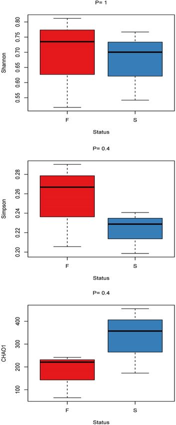
www.nature.com/scientificreports/

                                            Figure 1.  Alpha-diversity boxplot of blood-fed (F, red) and starved (S, blue) bed bugs.

                                            each taxon relative to the total number of reads for all taxa. Alpha-proteobacteria and gamma-proteobacteria
                                            were dominant at the class level. There were seven bacterial genera, including Wolbachia, Dickeya chrysanthemi,
                                            Pseudomonas, Enhydrobacter, Paracoccus, Methylobacterium, and Curvibacter were present in both blood-fed
                                            and staved bed bugs. All blood-fed samples shared four bacterial genera: Shimwellia, Skermanella, Pelomonas,

          Scientific Reports |   (2021) 11:8465 |                https://doi.org/10.1038/s41598-021-87946-w                                              4

Vol:.(1234567890)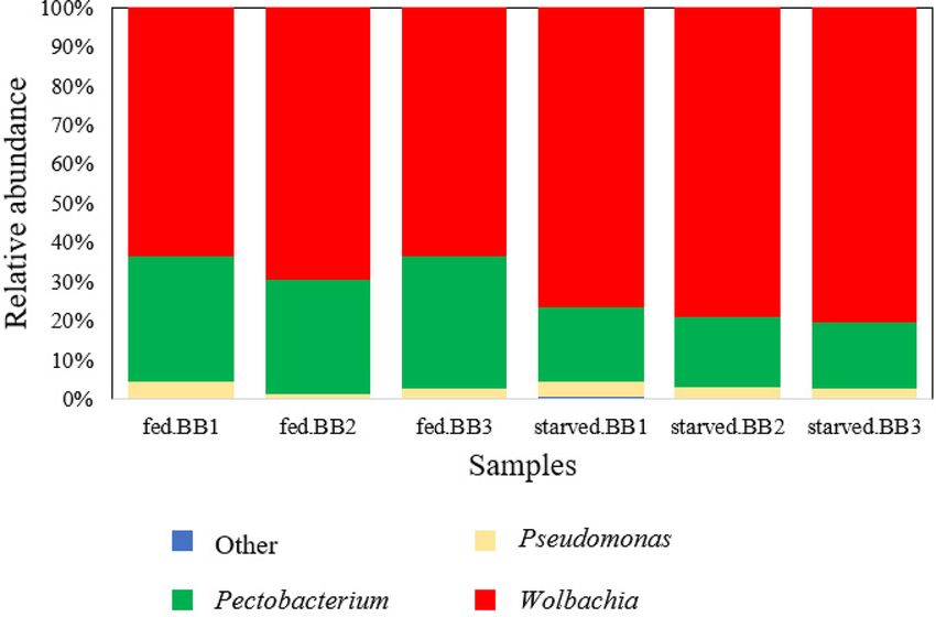
www.nature.com/scientificreports/

                                  Figure 2.  Rarefaction curves of the three samples, fed.BB1 (blue line), fed.BB2 (yellow line) and fed.BB3 (green
                                  line) from blood-fed bed bugs and three samples, BB1 (purple line), BB2 (red line) and BB3 (turquoise line)
                                  from starved bed bugs.

                                  Figure 3.  Principal coordinates analysis (PCoA) using Weighted-UniFrac distances of bacterial communities of
                                  blood-fed (red points) and starved (turquoise points) bed bugs.

                                  and Mesorhizobium, which exhibited a low relative abundance (less than 100 of total sequences). Eleven bacteria
                                  genera, including Pectobacterium, Bradyrhizobium, Phyllobacterium, Ralstonia, Sphingomonas, Acinetobacter
                                  indicus, Stenotrophomonas, Brevundimonas, Achromobacter, Staphylococcus, and Bacillus were found exclusively
                                  in the starved bed bugs samples (Table 2).

Scientific Reports |   (2021) 11:8465 |               https://doi.org/10.1038/s41598-021-87946-w                                                  5

                                                                                                                                             Vol.:(0123456789)
www.nature.com/scientificreports/
Figure 4. Heatmap analysis of three samples, BB1, BB2 and BB3 from blood-fed bed bugs.
The genera, Wolbachia, and Dickeya chrysanthemi were tremendously dominated the sequence data, with
Wolbachia containing 19,988, 29,877, and 20,038 sequences and the other, D. chrysanthemi containing 10,733,
12,555, and 10,791 sequences for three individuals of blood-fed bed bugs. The number of sequences of these two
OTUs obtained from the three starved bed bugs was even higher than the number of sequences acquired from
blood-fed bed bugs with 82,041, 82,848, and 86,363 sequences Wolbachia and 20,496, 18,216, 18,317 sequences
for D. chrysanthemi (Table 2). The cumulative sequences from these two OTUs constitute approximately 97%
and 96% of all sequences in blood-fed and starved bed bugs (Fig. 6).
Paired sample T-test was performed to determine the significant differences in the number of sequences of
bacterial genera present in both blood-fed and starved bed bugs (Table 3). The results showed that the number
of Wolbachia sequences in starved bed bugs was significantly higher than in blood-fed bed bugs (p = 0.004).
Dickeya chrysanthemi (p = 0.023) and Pseudomonas (p = 0.005) in starved bed bugs were also significantly higher
than in blood-fed bed bugs. The number of sequences of Enhydrobacter (p = 0.329), Paracoccus (p = 0.869),
Methylobacterium (p = 0.071), and Curvibacter (p = 0.108) showed no significant differences between blood-fed
and starved bed bugs (Table 3).
Although the number of sequences of dominant genera in starved bed bugs is significantly higher than blood-
fed bed bugs, the abundance ratio of the top three most abundant genera of which, Wolbachia, D. chrysanthemi
Scientific Reports | (2021) 11:8465 | https://doi.org/10.1038/s41598-021-87946-w 6
Vol:.(1234567890)www.nature.com/scientificreports/
Figure 5. Heatmap analysis of three samples, BB1, BB2 and BB3 from starved bed bugs.
followed by Pseudomonas, accounted for more than 99% of the total sequences between blood-fed and starved
bed bugs were similar (Fig. 6).
Discussion
This study is the first study that characterizes the bacterial community found in blood-fed and starved tropical
bed bugs, Cimex hemipterus, using high-throughput sequencing. Based on the bacterial community analysis,
Proteobacteria, which mainly documented to display antiparasitic activity in insects’ guts, was the phylum that
predominates in both blood-fed and starved bed bugs29.
Despite inter-individual variability (Fig. 3), the variability between individuals is less than 0.01% abundance
with no significant difference, and most of the OTUs contain only averaged one to three sequences (data not
shown). Since all bed bug samples were fed on the same blood source, taxa variability may occur due to indi-
vidual variation30.
The alpha-diversity in both blood-fed and starved tropical bed bugs was relatively low (Fig. 1)30,31. The low
bacterial diversity observed may due to the samples used were laboratory strains. Boissière et al. (2012) also
reported that bacterial richness and diversity are low in laboratory mosquitoes30. Other reasons, such as selective
pressures, including microbial interactions or insect immune systems, may also affect the bacterial community’s
composition30.
Beta-diversity estimated based on Bray–Curtis, Weighted- and Unweighted-UniFrac distance matrices showed
no significant differences between blood-fed and starved bed bugs when compared using the Mantel test. No
significant differences may be due to the dominant genera (more than 99%), of which Wolbachia, D. chrysanthemi,
and Pseudomonas were the same between both blood-fed and starved bed bugs. The result suggests that blood-
feeding has a limited influence on the bacterial community’s overall diversity within tropical bed bugs. Zolnik
et al. (2016) and Swei and Kwan (2017) reported similar findings in ticks of which blood-feeding induce limited
changes in microbial communities’ composition or even reduced the microbiome d iversity32,33. Although there
Scientific Reports | (2021) 11:8465 | https://doi.org/10.1038/s41598-021-87946-w 7
Vol.:(0123456789)www.nature.com/scientificreports/
Number of sequences
Blood fed Starved
Taxonomy (Phylum; class; family; genus) BB1 BB2 BB3 BB1 BB2 BB3
Proteobacteria; Alpha-proteobacteria; Anaplasmataceae; Wolbachia 19,988 29,877 20,038 82,041 82,848 86,363
Proteobacteria; Gamma-proteobacteria; Enterobacteriacese; Dickeya chrysan-
10,733 12,555 10,791 20,496 18,216 18,317
themi
Proteobacteria; Gamma-proteobacteria; Pseudomonadaceae; Pseudomonas 1389 596 808 4000 3164 2907
Proteobacteria; Gamma-proteobacteria; Enterobacteriaceae; Shimwellia 95 1 4 – – –
Proteobacteria; Gamma-proteobacteria; Moraxellaceae; Enhydrobacter 54 2 24 8 4 13
Proteobacteria; Alpha-proteobacteria; Azospirillaceae Skermanella 47 2 1 – – –
Proteobacteria; Alpha-proteobacteria; Rhodobacteraceae; Paracoccus 25 3 2 4 2 18
Proteobacteria; Alpha-proteobacteria; Beijerinckiaceae; Methylobacterium 8 3 4 69 55 24
Proteobacteria; Gamma-proteobacteria; Burkholderiaceae; Curvibacter 1 1 10 39 64 25
Proteobacteria; Gamma-proteobacteria; Burkholderiaceae; Pelomonas 2 1 3 – – –
Proteobacteria; Alpha-proteobacteria; Rhizobiaceae; Mesorhizobium 3 2 1 – – –
Proteobacteria; Gamma-proteobacteria; Enterobacteriaceae; Pectobacterium – – – 46 534 92
Proteobacteria; Alpha-proteobacteria; Xanthobacteraceae; Bradyrhizobium – – – 9 358 9
Proteobacteria; Alpha-proteobacteria; Rhizobiaceae; Phyllobacterium – – – 6 248 4
Proteobacteria; Gamma-proteobacteria; Burkholderiaceae; Ralstonia – – – 12 154 1
Proteobacteria; Alpha-proteobacteria; Sphingomonadaceae; Sphingomonas – – – 14 94 12
Proteobacteria; Gamma-proteobacteria; Moraxellaceae; Acinetobacter indicus – – – 17 13 7
Proteobacteria; Gamma-proteobacteria; Xanthomonadaceae; Stenotrophomonas – – – 10 7 1
Proteobacteria; Alpha-proteobacteria; Caulobacteraceae; Brevundimonas – – – 6 10 1
Proteobacteria; Gamma-proteobacteria; Burkholderiaceae; Achromobacter – – – 1 3 4
Firmicutes; Bacilli; Staphylococcaceae; Staphylococcus – – – 12 13 7
Firmicutes; Bacilli; Bacillaceae; Bacillus – – – 8 1 16
Table 2. Taxonomic classification of bacterial sequences (OTUs) that shared in all three samples either from
blood fed or starved bed bugs. “–” indicated not found.
were no significant differences in alpha and beta-diversity measures between blood-fed and starved bed bugs,
the number of bacterial species (the number of OTUs) was lower in blood-fed bed bugs than starved bed bugs
(Table 1). The result is also similar to the study by Sontowski and Van Dam (2020) showed a blood meal in A.
aegypti reduces the diversity and number of OTUs34.
There are several bacterial taxa present in both blood-fed and starved bed bugs. The bacterial taxa shared
across all samples suggested they probably vertically transmitted, and their presence is probably essential to their
host by providing supplementation and maintenance of insects’ homeostasis and immune system35,36. Among the
shared bacterial taxa of blood-fed and starved bed bugs, two OTUs of Proteobacteria dominate the bacterial com-
munity of C. hemipterus, the alpha-proteobacteria, Wolbachia, and gamma-proteobacterium, D. chrysanthemi.
The identification of Wolbachia and a gamma-proteobacteria as the predominant genera in C. hemipterus was in
accordance with the finding of Hypša and A ksoy37, Hosokawa et al.10, and Meriweather et al.18 in C. lectularius,
which also reported Wolbachia and a gamma-proteobacteria were predominant in common bed bugs. However,
the gamma-proteobacterium reported from these studies was unclassified.
Wolbachia infections are common in the Cimicidae38. As an obligate endosymbiont of bed bugs, Wolbachia
are vertically transmitted, could be found in many cell t ypes10,37,39–41, and play an essential role in host repro-
duction as well as the production of B vitamins, including biotin and r iboflavin10,41,42. Thus, the high frequency
of Wolbachia found in C. hemipterus is not surprising but provides evidence that the 16S rRNA assessment is
mainly congruent with expectations.
Dickeya chrysanthemi (Pectobacterium) was the second most bacteria found in C. hemipterus. It is a com-
mon plant pathogen and could synthesize siderophores, an iron-chelating agent essential for iron acquiring
under iron-limited conditions43,44. This characteristic implies their importance in bed bugs and may explain the
increased relative abundance of this species in starved bed bugs. The presence of both Wolbachia and gamma-
proteobacteria and their possible functions in common bed bugs, C. lectularius, have been reported by Goodman
(2016) of which both Wolbachia and the gamma-proteobacteria was essential for bed bugs growth45. Moreover,
the absence of Dickeya leads to reductions in egg production in C. lectularius, suggesting that removing the
gamma-proteobacteria can reduce f ertility18.
Other than Wolbachia and Dickeya, genera present in both blood-fed and starved bed bugs, including Pseu-
domonas, Methylobacterium, Paracoccus, Enhydrobacter, and Curvibacter (Table 2). Pseudomonas is a generalist,
which could be found from the different broad environments. It is also a common pollutant degrader46 and can
produce siderophore47,48. Although Pseudomonas has been reported as a contaminant in metagenome studies49,
Pseudomonas found in this study is unlikely a contaminant based on loads of Pseudomonas in three starved bed
bugs was significantly higher than the blood-fed bed bugs (Table 3). If the Pseudomonas was the contaminant,
Scientific Reports | (2021) 11:8465 | https://doi.org/10.1038/s41598-021-87946-w 8
Vol:.(1234567890)www.nature.com/scientificreports/
Figure 6. The relative abundance of bacteria genera in each sample from blood-fed bed bugs (start from left,
fed.BB1–fed.BB3) and starved bed bugs (starved.BB1–starved.BB3).
the loads from bed bugs under both blood-fed and starved states should be similar. Pseudomonas also could be
found in many insects, including m osquitoes34,50, beetles51, and scolitines52, and always linked to detoxifying
activities in insect’s guts. Infections of Pseudomonas are beneficial in these cases, but targeted studies are needed
to investigate Pseudomonas’s function in tropical bed bugs.
Methylobacterium is a common plant growth promoter and could be found in the natural environment
ubiquitously53,54. The presence of Methylobacterium in bed bugs may be necessary as this bacterium is also one
Number of sequences (Mean ± SD)
Genera Blood-fed Starved P-value
Wolbachia 23,301.00 ± 5695.04 83,750.67 ± 2298.05 0.004
Dickeya chrysanthemi 11,359.67 ± 1035.60 19,009.67 ± 1288.19 0.023
Pseudomonas 931.00 ± 410.56 3357.00 ± 571.49 0.005
Enhydrobacter 26.67 ± 26.10 8.33 ± 4.51 0.329
Paracoccus 10.00 ± 13.00 8.00 ± 8.72 0.869
Methylobacterium 5.00 ± 2.65 49.33 ± 23.03 0.071
Curvibacter 4.00 ± 5.20 42.67 ± 19.76 0.108
Table 3. The number of sequences between blood-fed and starved bed bugs with P-value determined using
paired samples T-test.
of the ten most abundant OTUs characterized by C. lectularius18. Paracoccus has been isolated from industrial
effluent, Cayenne ticks, and Tsetse fly29. There is no information regarding Enhydrobacter and Curvibacter in
blood-feeding insects, and the role of these bacteria in the gut of blood-feeding insects is not explicit. Neverthe-
less, bacterial groups consistently present and shared among all the bed bug samples suggest it could serve the
host’s essential functions.
There were exclusive OTUs in blood-fed and starved bed bugs, even in extremely low abundance (less than
1%) in the overall bacterial community. Genera that only detected in blood-fed bed bugs, including Shimwellia,
Mesorhizobium, Skermanella, and Pelomonas. According to Brzuszkiewicz et al. (2012), Shimwellia can synthe-
size coenzyme B12 or vitamin B12 de n ovo55. Mesorhizobium and Skermanella could be found from various
ecosystems29,56, while Pelomonas has been isolated from the assassin bug, Triatoma maculate16. However, their
role in blood-feeding insects is not elucidated.
The bacterial community in starved bed bugs is more diverse and abundant than blood-fed bed bugs. Genera
Acinetobacter, Phyllobacterium, Achromobacter, Bradyrhizobium, Staphylococcus, Stenotrophomonas, Ralstonia,
Bacillus, Sphingomonas, and Brevundimonas were only characterized in starved bed bugs (Table 2). The changes
in the bacterial community structure of bed bugs in the starved state could be due to host-driven mechanisms.
For instance, the host may produce different antimicrobial proteins, less secretion of mucus on the gut lining,
pH changes, and reduced intestines’ size during the starved period may alter the bacterial c ommunity19.
Based on Fig. 6, the abundance ratio of Wolbachia, D. chrysanthemi, and Pseudomonas between the two
physiological statuses is similar. However, the number of sequences of these dominant species is significantly
Scientific Reports | (2021) 11:8465 | https://doi.org/10.1038/s41598-021-87946-w 9
Vol.:(0123456789)www.nature.com/scientificreports/
higher in starved bed bugs than blood-fed bed bugs (Table 3). The change of bacteria’s load may be due to the
selection pressure, of which in this study, starvation forced onto the beg bugs. Selection pressure could induce
changes in loads of bacteria, and the loads are always higher of samples in the critical s tage57. The result is in
line with the study by Rodgers et al. (2017) on the microbiota of An. coluzzii, of which mosquitoes restore their
gut homeostasis after the blood meals by excreting bacteria with the blood bolus, result in a 98% reduction in
bacterial loads31. Tchioffo et al. (2016) also showed blood-fed mosquitoes (Anopheles) induced a drop in bacterial
diversity and abundance at first and increased later after prolonged s tarvation58.
The decrease of the concentration of symbiotic bacteria may due to the digestive processes that various
digestive enzymes killed the symbiotic bacteria. After processing the blood meal, the nutrient supply within
the ingested blood may increase the symbiotic bacteria concentration59. Besides, the increased concentration of
symbiotic bacteria during the starved period could strengthen the host’s immune system19,40.
Nevertheless, there are also study showing the contrasting result. A study by Fisher et al. (2018) on com-
mon bed bugs, Cimex lectularius, showed the Wolbachia titer declined significantly with prolonged starvation
at 40 d after feeding60. These studies31,58,60 showed that bacterial community composition was affected mainly
by arthropod s pecies61.
Conclusion
This study is the first metabarcoding analysis of bacterial communities’ characterization from blood-fed and
starved tropical bed bugs, C. hemipterus. Overall, the bacterial community in both blood-fed and starved bed
bugs was dominated by Proteobacteria with dominant genera or species, Wolbachia, D. chrysanthemi, and Pseu-
domonas. The findings also indicate starvation has a limited effect on the bacterial composition of bed bugs
but significantly affects the concentrations of dominant bacterial species. Furthermore, less abundance specific
bacterial genera were isolated either from blood-fed or starved bed bugs. It provides information regarding
the microbial community that may associate with blood meal digestion and provides fundamentals for future
research on the bacterial members’ functional roles except for Wolbachia.
Received: 21 January 2021; Accepted: 7 April 2021
References
1. Beard, B. Kissing bugs and bedbugs, the Heteroptera. In Biology of Disease (ed. Marquardt, W. H.) 57–65 (Elsevier, 2004).
2. Doggett, S. L., & Russell, R. C. (2008). The resurgence of bed bugs, Cimex spp. (Hemiptera: Cimicidae) in Australia. In Proceedings
of the Sixth International Conference on Urban Pests (pp. 407–425). Hungary: Veszprém.
3. Zulaikha, Z., Hafis, A. R. A. & Hassan, A. A. A survey on the infestation levels of tropical bed bugs in Peninsular Malaysia: current
updates and status on resurgence of Cimex hemipterus (Hemiptera: Cimicidae). Asian Pac. J. Trop. Dis. 6(1), 40–45 (2016).
4. Benoit, J. B., del Grosso, N. A., Yoder, J. A. & Denlinger, D. L. Resistance to dehydration between bouts of blood feeding in the bed
bug, Cimex lectularius, is enhanced by water conservation, aggregation, and quiescence. Am. J. Trop. Med. Hyg. 76(5), 987–993
(2007).
5. Dang, K., Doggett, S. L., Singham, G. V. & Lee, C. Y. Insecticide resistance and resistance mechanisms in bed bugs, Cimex spp.
(Hemiptera: Cimicidae). Parasit. Vectors 10(1), 318 (2017).
6. Karunaratne, S. H. P. P., Damayanthi, B. T., Fareena, M. H. J., Imbuldeniya, V. & Hemingway, J. Insecticide resistance in the tropical
bedbug Cimex hemipterus. Pestic. Biochem. Physiol. 88(1), 102–107 (2007).
7. De Gaio, A. O. et al. Contribution of midgut bacteria to blood digestion and egg production in Aedes aegypti (Diptera: Culicidae)
(L.). Parasit. Vectors 4(1), 105 (2011).
8. Douglas, A. E. Nutritional interactions in insect-microbial symbioses: aphids and their symbiotic bacteria Buchnera. Annu. Rev.
Entomol. 43(1), 17–37 (1998).
9. Zhou, G., Scaraffia, P. Y. & Wells, M. A. Vector nutrition and energy metabolism. In Biology of Disease Vectors (ed. Marquardt, W.
H.) 311–315 (Elsevier, 2004).
10. Hosokawa, T., Koga, R., Kikuchi, Y., Meng, X.-Y. & Fukatsu, T. Wolbachia as a bacteriocyte-associated nutritional mutualist. Proc.
Natl. Acad. Sci. 107(2), 769–774 (2010).
11. Shi, W., Syrenne, R., Sun, J. Z. & Yuan, J. S. Molecular approaches to study the insect gut symbiotic microbiota at the ‘omics’ age.
Insect Sci. 17(3), 199–219 (2010).
12. Palavesam, A. et al. Pyrosequencing-based analysis of the microbiome associated with the horn fly, Haematobia irritans. PLoS
ONE 7(9), e44390 (2012).
13. Greay, T. L. et al. Recent insights into the tick microbiome gained through next-generation sequencing. Parasit. Vectors 11(1), 12
(2018).
14. Wang, Y., Gilbreath, T. M. III., Kukutla, P., Yan, G. & Xu, J. Dynamic gut microbiome across life history of the malaria mosquito
Anopheles gambiae in Kenya. PLoS ONE 6(9), e24767 (2011).
15. Carpi, G. et al. Metagenomic profile of the bacterial communities associated with Ixodes ricinus ticks. PLoS ONE 6(10), e25604
(2011).
16. Montoya-Porras, L. M., Omar, T. C., Alzate, J. F., Moreno-Herrera, C. X. & Cadavid-Restrepo, G. E. 16S rRNA gene amplicon
sequencing reveals dominance of Actinobacteria in Rhodnius pallescens compared to Triatoma maculata midgut microbiota in
natural populations of vector insects from Colombia. Acta Trop. 178, 327–332 (2018).
17. Griffith, B. C. et al. Analysis of the gut-specific microbiome from field-captured tsetse flies, and its potential relevance to host
trypanosome vector competence. BMC Microbiol. 18(1), 55–67 (2018).
18. Meriweather, M., Matthews, S., Rio, R. & Baucom, R. S. A 454 survey reveals the community composition and core microbiome
of the common bed bug (Cimex lectularius) across an urban landscape. PLoS ONE 8(4), e61465 (2013).
19. Kohl, K. D., Amaya, J., Passement, C. A., Dearing, M. D. & McCue, M. D. Unique and shared responses of the gut microbiota to
prolonged fasting: A comparative study across five classes of vertebrate hosts. FEMS Microbiol. Ecol. 90(3), 883–894 (2014).
20. Caporaso, J. G. et al. QIIME allows analysis of high-throughput community sequencing data. Nat. Methods 7(5), 335 (2010).
21. Bolger, A. M., Lohse, M. & Usadel, B. Trimmomatic: A flexible trimmer for Illumina sequence data. Bioinformatics 30(15), 2114–
2120 (2014).
22. Magoč, T. & Salzberg, S. L. FLASH: fast length adjustment of short reads to improve genome assemblies. Bioinformatics 27(21),
2957–2963 (2011).
23. Edgar, R. C. UPARSE: highly accurate OTU sequences from microbial amplicon reads. Nat. Methods 10(10), 996 (2013).
Scientific Reports | (2021) 11:8465 | https://doi.org/10.1038/s41598-021-87946-w 10
Vol:.(1234567890)www.nature.com/scientificreports/
24. Edgar, R. C., Haas, B. J., Clemente, J. C., Quince, C. & Knight, R. UCHIME improves sensitivity and speed of chimera detection.
Bioinformatics 27(16), 2194–2200 (2011).
25. Wang, Q., Garrity, G. M., Tiedje, J. M. & Cole, J. R. Naive Bayesian classifier for rapid assignment of rRNA sequences into the new
bacterial taxonomy. Appl. Environ. Microbiol. 73(16), 5261–5267 (2007).
26. Pruesse, E. et al. SILVA: a comprehensive online resource for quality checked and aligned ribosomal RNA sequence data compat-
ible with ARB. Nucl. Acids Res. 35(21), 7188–7196 (2007).
27. Jing, G. et al. Parallel-META 3: Comprehensive taxonomical and functional analysis platform for efficient comparison of microbial
communities. Sci. Rep. 7(1), 1–11 (2017).
28. R Core Team. R: A language and environment for statistical computing (R Foundation for Statistical Computing, 2015).
29. Malele, I. et al. Bacterial diversity obtained by culturable approaches in the gut of Glossina pallidipes population from a non-sleeping
sickness focus in Tanzania: preliminary results. BMC Microbiol. 18(1), 107–116 (2018).
30. Boissière, A. et al. Midgut microbiota of the malaria mosquito vector Anopheles gambiae and interactions with Plasmodium falci-
parum infection. PLoS Pathog. 8(5), e1002742 (2012).
31. Rodgers, F. H., Gendrin, M., Wyer, C. A. & Christophides, G. K. Microbiota-induced peritrophic matrix regulates midgut homeo-
stasis and prevents systemic infection of malaria vector mosquitoes. PLoS Pathog. 13(5), e1006391 (2017).
32. Zolnik, C. P., Prill, R. J., Falco, R. C., Daniels, T. J. & Kolokotronis, S. O. Microbiome changes through ontogeny of a tick pathogen
vector. Mol. Ecol. 25(19), 4963–4977 (2016).
33. Swei, A. & Kwan, J. Y. Tick microbiome and pathogen acquisition altered by host blood meal. ISME J. 11(3), 813–816 (2017).
34. Sontowski, R. & Van Dam, N. M. Functional variation in Dipteran gut bacterial communities in relation to their diet, life cycle
stage and habitat. Insects 11(8), 543 (2020).
35. Weiss, B. L., Wang, J. & Aksoy, S. Tsetse immune system maturation requires the presence of obligate symbionts in larvae. PLoS
Biol. 9(5), e1000619 (2011).
36. Engel, P. & Moran, N. A. The gut microbiota of insects-diversity in structure and function. FEMS Microbiol. Rev. 37(5), 699–735
(2013).
37. Hypša, V. & Aksoy, S. Phylogenetic characterization of two transovarially transmitted endosymbionts of the bedbug Cimex lectu-
larius (Heteroptera: Cimicidae). Insect Mol. Biol. 6(3), 301–304 (1997).
38. Sakamoto, J. M., Feinstein, J. & Rasgon, J. L. Wolbachia infections in the Cimicidae: Museum specimens as an untapped resource
for endosymbiont surveys. Appl. Environ. Microbiol. 72(5), 3161–3167 (2006).
39. Brownlie, J. C. et al. Evidence for metabolic provisioning by a common invertebrate endosymbiont, Wolbachia pipientis, during
periods of nutritional stress. PLoS Pathog. 5(4), e1000368 (2009).
40. Micieli, M. V. & Glaser, R. L. Somatic Wolbachia (Rickettsiales: Rickettsiaceae) levels in Culex quinquefasciatus and Culex pipiens
(Diptera: Culicidae) and resistance to West Nile virus infection. J. Med. Entomol. 51(1), 189–199 (2014).
41. Bellinvia, S., Johnston, P. R., Reinhardt, K. & Otti, O. Bacterial communities of the reproductive organs of virgin and mated com-
mon bedbugs, Cimex lectularius. Ecol. Entomol. 45(1), 142–154 (2020).
42. Nikoh, N. et al. Evolutionary origin of insect: Wolbachia nutritional mutualism. Proc. Natl. Acad. Sci. 111(28), 10257–10262 (2014).
43. Neilands, J. B. Siderophores: structure and function of microbial iron transport compounds. J. Biol. Chem. 270(45), 26723–26726
(1995).
44. Hugouvieux-Cotte-Pattat, N., Condemine, G., Nasser, W. & Reverchon, S. Regulation of pectinolysis in Erwinia chrysanthemi.
Annu. Rev. Microbiol. 50(1), 213–257 (1996).
45. Goodman, M. H. (2016). Endosymbiotic bacteria in the bed bug, Cimex lectularius L. (Hemiptera: Cimicidae) (Doctoral thesis,
University of Kentucky, US). Retrieved from https://doi.org/10.13023/ETD.2016.527
46. Kumar, M. et al. Enhanced biodegradation of β- and δ-hexachlorocyclohexane in the presence of α- and γ-isomers in contaminated
soils. Environ. Sci. Technol. 39(11), 4005–4011 (2005).
47. Ambrosi, C., Leoni, L., Putignani, L., Orsi, N. & Visca, P. Pseudobactin biogenesis in the plant growth-promoting rhizobacterium
Pseudomonas strain B10: Identification and functional analysis of the L-ornithine N5-oxygenase (psbA) gene. J. Bacteriol. 182(21),
6233–6238 (2000).
48. Mossialos, D. et al. Quinolobactin, a new siderophore of Pseudomonas fluorescens ATCC 17400, the production of which is
repressed by the cognate pyoverdine. Appl. Environ. Microbiol. 66(2), 487–492 (2000).
49. Salter, S. J. et al. Reagent and laboratory contamination can critically impact sequence-based microbiome analyses. BMC Biol.
12(1), 87 (2014).
50. Chavshin, A. R. et al. Malpighian tubules are important determinants of Pseudomonas transstadial transmission and long-time
persistence in Anopheles stephensi. Parasit. Vectors 8(1), 1–7 (2015).
51. Ceja-Navarro, J. A. et al. Gut microbiota mediate caffeine detoxification in the primary insect pest of coffee. Nat. Commun. 6, 7618
(2015).
52. Hernández-García, J. A., Briones-Roblero, C. I., Rivera-Orduña, F. N. & Zúñiga, G. Revealing the gut bacteriome of Dendroctonus
bark beetles (Curculionidae: Scolytinae): Diversity, core members and co-evolutionary patterns. Sci. Rep. 7(1), 1–12 (2017).
53. McDonald, D. P. K. I. R. & Wood, A. P. The family methylobacteriaceae. In The Prokaryotes: Alphaproteobacteria and Betaproteo-
bacteria (ed. Rosenberg, E.) 313–340 (Springer, 2014).
54. Chen, C. et al. Diverse bacterial symbionts of insect-pathogenic fungi and possible impact on the maintenance of virulence during
infection. Symbiosis 69(1), 47–58 (2016).
55. Menchaca, A. C. et al. Preliminary assessment of microbiome changes following blood-feeding and survivorship in the Amblyomma
americanum nymph-to-adult transition using semiconductor sequencing. PLoS ONE 8(6), e67129 (2013).
56. Swenson, C. E. & Sadikot, R. T. Achromobacter respiratory infections. Ann. Am. Thorac. Soc. 12(2), 252–258 (2015).
57. Echaubard, P. et al. Rapid evolution of Wolbachia density in insecticide resistant Culex pipiens. Heredity 104(1), 15–19 (2010).
58. Tchioffo, M. T. et al. Dynamics of bacterial community composition in the malaria mosquito’s epithelia. Front. Microbiol. 6, 1500
(2016).
59. Eichler, S. & Schaub, G. A. Development of symbionts in triatomine bugs and the effects of infections with trypanosomatids. Exp.
Parasitol. 100(1), 17–27 (2002).
60. Fisher, M. L. et al. Growth kinetics of endosymbiont Wolbachia in the common bed bug, Cimex lectularius. Sci. Rep. 8(1), 1–9
(2018).
61. Hawlena, H. et al. The arthropod, but not the vertebrate host or its environment, dictates bacterial community composition of
fleas and ticks. ISME J. 7(1), 221–223 (2013).
Acknowledgements
This research was funded by the Fundamental Research Grant (FRGS) (203/PBIOLOGI/6711681). This research
protocol was approved by the Universiti Sains Malaysia, Human Ethics Committee (USM/JEPeM/19120868).
Scientific Reports | (2021) 11:8465 | https://doi.org/10.1038/s41598-021-87946-w 11
Vol.:(0123456789)www.nature.com/scientificreports/
Author contributions
L.L. and A.H.A.M. designed the experiment; L.L. collected data; analyzed the data and wrote the manuscript,
A.H.A.M. read, corrected, and approved the manuscript. All authors have read and agreed to the published
version of the manuscript.
Competing interests
The authors declare no competing interests.
Additional information
Correspondence and requests for materials should be addressed to A.H.A.M.
Reprints and permissions information is available at www.nature.com/reprints.
Publisher’s note Springer Nature remains neutral with regard to jurisdictional claims in published maps and
institutional affiliations.
Open Access This article is licensed under a Creative Commons Attribution 4.0 International
License, which permits use, sharing, adaptation, distribution and reproduction in any medium or
format, as long as you give appropriate credit to the original author(s) and the source, provide a link to the
Creative Commons licence, and indicate if changes were made. The images or other third party material in this
article are included in the article’s Creative Commons licence, unless indicated otherwise in a credit line to the
material. If material is not included in the article’s Creative Commons licence and your intended use is not
permitted by statutory regulation or exceeds the permitted use, you will need to obtain permission directly from
the copyright holder. To view a copy of this licence, visit http://creativecommons.org/licenses/by/4.0/.
© The Author(s) 2021
Scientific Reports | (2021) 11:8465 | https://doi.org/10.1038/s41598-021-87946-w 12
Vol:.(1234567890)You can also read