I-Nema: A Biological Image Dataset for Nematode Recognition
←
→
Page content transcription
If your browser does not render page correctly, please read the page content below
I-Nema: A Biological Image Dataset for
                                                             Nematode Recognition

                                                      Xuequan Lu1? , Yihao Wang2? , Sheldon Fung1 , and Xue Qing2
                                                                           1
arXiv:2103.08335v1 [q-bio.QM] 15 Mar 2021

                                                                             Deakin University, Australia
                                                                    2
                                                                        Nanjing Agricultural University, China

                                                    Abstract. Nematode worms are one of most abundant metazoan groups
                                                    on the earth, occupying diverse ecological niches. Accurate recognition
                                                    or identification of nematodes are of great importance for pest con-
                                                    trol, soil ecology, bio-geography, habitat conservation and against cli-
                                                    mate changes. Computer vision and image processing have witnessed a
                                                    few successes in species recognition of nematodes; however, it is still in
                                                    great demand. In this paper, we identify two main bottlenecks: (1) the
                                                    lack of a publicly available imaging dataset for diverse species of nema-
                                                    todes (especially the species only found in natural environment) which
                                                    requires considerable human resources in field work and experts in tax-
                                                    onomy, and (2) the lack of a standard benchmark of state-of-the-art deep
                                                    learning techniques on this dataset which demands the discipline back-
                                                    ground in computer science. With these in mind, we propose an image
                                                    dataset consisting of diverse nematodes (both laboratory cultured and
                                                    naturally isolated), which, to our knowledge, is the first time in the com-
                                                    munity. We further set up a species recognition benchmark by employing
                                                    state-of-the-art deep learning networks on this dataset. We discuss the
                                                    experimental results, compare the recognition accuracy of different net-
                                                    works, and show the challenges of our dataset. We make our dataset
                                                    publicly available at: https: // github. com/ xuequanlu/ I-Nema

                                                    Keywords: Nematode image dataset · Nematode recognition · Deep
                                                    learning

                                            1     Introduction

                                            Nematode worms are important, due to the following reasons: (1) parasitic ne-
                                            matodes threaten the health of plants on a global scale, causing at least 80 billion
                                            US dollar’s loss per year [31]; (2) interstitial nematodes pervade sediment and
                                            soil ecosystems in overwhelming numbers [21]; and (3) Caenorhabditis elegans
                                            (C. elegans) is a favourable experimental model system [33]. Accurate recog-
                                            nition or identification of nematodes are of great value for pest control (e.g.
                                            choosing a specific nematicide or rotation crop), soil ecology (e.g. evaluation for
                                            the soil quality), bio-geography, habitat conservation and climate maintenance
                                            ?
                                                Joint first author. Q. Xue is the correponding author. This is a preprint.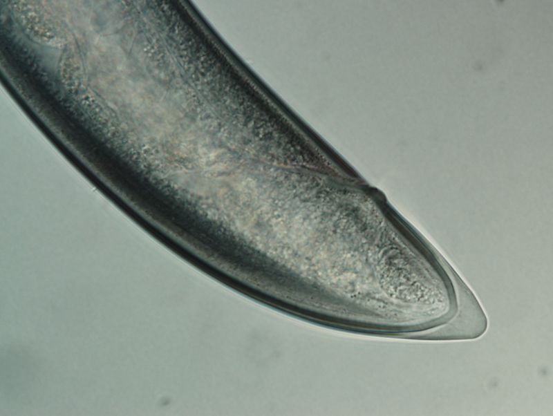
2       X. Lu et al.

Table 1. Datasets for Nematode recognition. #. Samples and #. Classes denote the
numbers of samples and classes, respectively. Our proposed dataset is the first open-
access one which involves both naturally isolated and laboratory cultured nematodes,
covering diverse morphology and life strategies.

              Datasets        #. Samples    #. Classes Open to public
              [25,24]           50              5           NO
              [28]              500             5           NO
              [26]             1,000             5          NO
              [23,22]           500            10           NO
              [43]        480,000 frames        8           NO
              [19]-single  10,476 clips        365          NO
              [19]-multi     308 clips         12           NO
              Our dataset      2,769            19          YES

[11]. However, nematodes recognition is challenging, due to high phenotypic plas-
ticity (i.e. high morphological variations within a single species) [11,29], vague
diagnostic characteristics [13,14], and frequently encountered juveniles [3]. More
importantly, the manual identification is extremely time-consuming and labor-
intensive, especially when dealing with large-scale ecological data. It often re-
quires solid training and well-established experience. As a result, the taxonomy
of nematode species appears to be a significant bottleneck for many relevant
fields.
     Although molecular barcoding [15,5] has been consolidated as a powerful
tool for the species identification and biodiversity investigation, its applications
depend on the availability of molecular biology facilities, sequencing instruments,
as well as the background knowledge. Alternatively, imaging data are often more
accessible and economical for broader users, and have been utilized for nematode
research, such as nematode detection/segmentation [38,30,32,36,35,34,10,42,8],
classification [19,43,25,24,27,23,22,26,28], nematode (eggs) counting [2,17], etc. It
should be noted that the current identification or recognition research are either
for nematode image stacks [25,24,27,23,22,26,28], or predominantly designed for
model organism C. elegans [43,19]. However, the involved issues for identification
tasks are: (1) very few classes involved in the image stacks, e.g. 5 classes in
Table 1; (2) low diversity coverage, i.e. species limited to the laboratory cultured
C. elegans only (Table 1: [43,19]). Importantly, to our knowledge, all relevant
imaging data were used in their own research, and none of them are publicly
available. In summary, an image dataset covering diverse kinds of nematodes
and a standard benchmark using state-of-the-art deep learning techniques is
still missing!
     Provided the above analysis, we are motivated to create an image dataset for
nematodes and complete a benchmark for state-of-the-art deep learning meth-
ods. Despite that laboratory cultured species are much easier to acquire, for ex-
ample, C. elegans complete a reproductive life cycle in 3.5 days at 20◦ and a huge
population can be reproduced, our samples are mostly collected from the natu-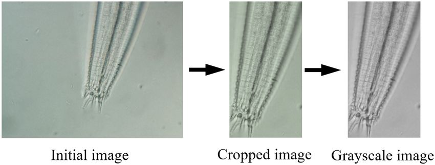
I-Nema: A Biological Image Dataset for Nematode Recognition          3

ral environment (many are not cultivable in laboratory). As a result, the data
collection is more time-consuming and labor-intensive (e.g. sampling in different
ecosystems, manual nematode picking for target species in a mixed population,
etc) than using pure laboratory culture. In particular, we first collect soil from a
wide range of natural environments, including temperate broadleaf, mixed for-
est, crop field, and tundra. Then the nematodes are extracted and further placed
in glass slides. With the aid of the microscope system, manual identification is
performed to determine whether a nematode is needed or not for further image
capturing. If it is needed, we continue image capturing using the microscope
imaging system (a camera, a microscope and a software), and assign the man-
ually identified species label to the involved images. It is discarded otherwise.
We also take images for 2 laboratory cultured species. A single nematode worm
per image is constrained during image capture. The dataset has a total number
of 2, 769 images and 19 different species (17 species from natural environment
and 2 laboratory species). To the best of our knowledge, this proposed dataset is
the first open-access image dataset including diverse nematodes species and life
strategies (plant-parasitic, fungi feeder, bacteria feeder, omnivores, predator).
    In addition to the dataset, we further employ the state-of-the-art deep learn-
ing networks on the species recognition of nematodes. We analyze the results with
regard to different respects: (1) different deep learning networks, (2) pre-training
as initialization versus training from scratch. We also conduct experiments and
analysis on augmentation and discuss the supported research by our work. The
contributions of this work are summarized as follows.
 – We propose an image dataset for diverse species of nematodes. It is, to our
   knowledge, the first open-access biological image dataset for diverse nema-
   todes, representing different evolutionary lineages and life strategies (isolated
   both from natural environment and laboratory).
 – We conduct a benchmark by adjusting and training the state-of-the-art deep
   learning networks on our dataset. We compare and analyze their perfor-
   mance. We also discuss the supported research by our work.

2     Related Work
In this section, we review the research which are most relevant to this work. We
first look back on the nematode detection/segmentation, and then review species
recognition of nematodes. We finally summarize some state-of-the-art CNNs.

2.1   Nematode Detection/Segmentation
An early nematode detection paper presented a computational system which
supports the detection of nematodes in digital images [38]. Nguyen et al. pro-
posed an improved watershed segmentation algorithm using water diffusion and
local shape priors, which has been applied to the segmentation of nematode
worms [30]. Ochoa et al. introduced an approach to extract a considerable num-
ber of individuals even in cluttered regions in population images [32]. Later,
4 X. Lu et al.
novel methods based on skeleton analysis were designed for the detection of in-
dividual nematode worms in population images in presence of worm overlapping
[36,35,34]. Recently, Chou et al. put forwarded an efficient CNN-based regression
network for accurate edge detection [10]. Chen et al. proposed a framework for
detecting worm-shaped objects in microscopic images using convolutional neural
networks (CNNs) [8]. The authors used curved lines as annotation rather than
bounding boxes. Wang et al. introduced a pipeline for automated analysis of C.
elegans imagery [42], which detects, segments the worm, and predicts the age of
individual worms with high accuracy.
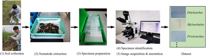
Fig. 1. The pipeline for creating our dataset. Soil samples are first collected (soil
collection), and nematodes are then extracted from soil (extraction). The nematodes
are further picked up and placed in a glass slide (specimen preparation). After manual
identification (identification), we determine which specimens will do image capturing
and annotation (image acquisition and annotation).
2.2 Nematode Recognition
A major proportion of nematode classification research are conducted on image
stacks, with each stack (or each species) consisting of multiple focal planes of the
specimen [25,24,27,26,28,23,22]. Various methods have been developed to handle
the classification based on image stacks, for example, information fusion based
approaches [27,23], 3D X-Ray Transform based method [25], 3D X-Ray Trans-
form based multilinear analysis method [24], projection-based methods [22,26],
deep convolutional neural network (CNN) image fusion based multilinear ap-
proach [28]. Zhou et al. proposed an identification method for mutant types and
other types, based on locomotion patterns [43]. Javer et al. presented a fully
convolutional neural network to discern genetically diverse strains of C. elegans,
based only on their recorded spontaneous activity, and explored how its perfor-
mance changes as different embeddings are used as input [19].
Datasets. [25,24] used 5 species and 10 samples per species for classification.
[28] adopted 500 samples from 5 classes, and [26] utilized 1, 000 samples from 5
species. [23,22] used 500 samples of 10 categories. [43] used a total number of
480, 000 frames from the wild type and 7 other mutant types of C. elegans. [19]I-Nema: A Biological Image Dataset for Nematode Recognition 5 involves two datasets of C. elegans: single-worm dataset and multi-worm dataset. The former includes 10, 476 video clips of individual worms divided between 365 different classes. The latter contains a total number of 308 video clips from 12 strains. We summarize the dataset information in Table 1. It is not surprising to collect a great number of samples for C. elegans, since C. elegans is a common organism with rapid reproduction in laboratory. Also, the data are typically used in their own research and are not released to the public. 2.3 Deep CNNs Nowadays, deep convolutional neural networks (CNNs) are powerful tools for data-driven research. State-of-the-art CNNs are AlexNet [20], VGG [39], ResNet [16], Inception [41], Xception [9], etc. They are well defined and built on image grids, and capable of learning discriminative features from input images. In this work, we adjust AlexNet, VGG-16, VGG-19, ResNet-34, ResNet-50, ResNet-101 and train them on the train set, and test them over the test set of our dataset, in terms of the task of nematode recognition. 3 Our Dataset: I-Nema 3.1 Overview In this section, we explain how we create our dataset, referring to as “I-Nema”. At first, we conduct a country-wide soil sample collection in order to gather specimens representing different lineages in the tree of Nematoda [18]. The indi- vidual nematodes are extracted from soil, fixed, and subsequently processed into glass slide specimens with 10 ∼ 20 nematode worms per glass slide. The species is manually identified and the undesirable taxa are excluded. Images are then captured for each selected specimen using the camera equipped with a micro- scope. The identified species label is simply annotated to the involved images. We finally perform some pre-processing for the initially collected images. Figure 1 shows the pipeline for creating I-Nema. 3.2 Sample Collection Sample collection contains soil collection, nematode extraction and specimen preparation. Soil samples are collected from various ecosystems in Mainland China. Aside from collecting samples in the natural environment, we also use the nematode cultures maintained in laboratory (two species: Pratylenchus sp. and Acrobeloides sp.). For soil samples, nematodes are extracted with a Baer- mann tray after an incubation in room temperature for 24 hours. The nematode extraction are concentrated using a 500 mesh sieve (25µm opening). For the laboratory maintained species, nematodes are directly washed from either car- rot disc (Pratylenchus sp.) or cultural middle (Acrobeloides sp.). After removing water, nematodes are fixed with 4% formalin and gradually transferred to anhy- drous glycerin, following the protocol [40] which is modified based on [37]. The
6      X. Lu et al.

glycerin preserved nematodes are manually picked up and placed in a glass slide.
Each permanent slide contains about 10 ∼ 20 individual worms.

3.3   Specimen Recognition
The species is manually identified before capturing images. This is to collect di-
verse species of nematodes, rather than simply increasing the number of images
in a few species. Nematodes are identified based on the morphological characters
(e.g. the presence of teeth or stylet in buccal cavity, the shape of pharynx and
tail, the type of reproductive system, etc) and morphometric measurements (e.g.
the length of stylet, body width and length, tail length, etc). These characters
can be straightforwardly observed through microscopy and/or measured in a
professional software connected to the camera equipped with the microscope.
The acquired information is subject to the available taxonomic key (e.g. [6,4])
and is further confirmed with the original descriptions. The recovered taxon will
be considered as undesirable and excluded if it is rare in population (difficult to
acquire sufficient specimens) or evolutionary repetitive to a captured species. If
the taxon is abundant in number and represent a different evolutionary lineage,
we continue the following image acquisition step. We selected and identified a
total number of 2 laboratory cultured species (Pratylenchus sp. and Acrobeloides
sp.) and 17 naturally collected species (see Table 2), covering 16 families of com-
mon soil species and all nematode trophic guilds. Figure 1 shows the microscope
setup for the manual specimen identification.

3.4   Image Acquisition and Annotation
We capture images after the step of identifying specimens. The glass slides are
examined and photographed using an Olympus BX51 DIC Microscope (Olym-
pus Optical, produced in Tokyo, Japan), equipped with an camera. The system
(see Figure 1) has also been utilized for specimen identification in Section 3.3.
The procedure for image acquisition are as follows. The nematode in specimen
is moved to the view center of the microscope, to facilitate further image align-
ment. Head regions are given a higher priority, as they involve more informative
taxonomic characters. Tail and middle body regions are also photographed, since
these regions are also informative for species recognition and easy to locate. For
the Mylonchulus sp., Miconchus sp., Pristionchus sp. and Acrobeles sp., 5 ∼ 10
images are taken at different image planes to ensure each of the taxonomic
characters are properly captured. These species have contrasting morphological
structures at different layers. For other species, 3 ∼ 5 images are taken from
the lowest, middle and highest image planes which allow large variations to be
captured. Both juveniles and adult worms are included for capturing images.
    We eventually acquire a total number of 2, 769 images, ranging from 24 to
347 images per species. As for image annotation, since the species of a specimen
has been identified in the last step, the species label can be simply allocated to
the acquired images of corresponding nematodes. Table 2 shows some statistics
of our proposed image dataset (I-Nema). Figure 2 shows some nematode images
I-Nema: A Biological Image Dataset for Nematode Recognition 7
(a) (b) (c)
(d) (e) (f)
(g) (h) (i)
Fig. 2. The example of nematode images used in this study. (a-c): Aporcelaimus sp.;
(d-f): Mylonchulus sp.; (g-i): Pristionchus sp.; (a), (b), (e), (f), (g), (h): head region;
(c,d): tail region; (i): middle body.8 X. Lu et al.
from I-Nema. As shown in the histogram (Figure 3), the data distribution is
diverse. Species with less than 114 images account for more than half of the
whole dataset, while over 324 images are included in three species. Two species
have less than 54 images. Species between 84 images and 294 images have few
variations (except blanks), in terms of the number of classes.
Table 2. Numbers of image samples in each species in our dataset. “sp.” represents
“species”. “#. Samples” denotes the number of samples.
Species #. Samples
Acrobeles sp. 71
Acrobeloides sp. 184
Amplimerlinius sp. 24
Aphelenchoides sp. 347
Aporcelaimus sp. 128
Axonchium sp. 170
Discolimus sp. 64
Ditylenchus sp. 330
Dorylaimus sp. 38
Eudorylaimus sp. 86
Helicotylenchus sp. 77
Mesodorylaimus sp. 96
Miconchus sp. 57
Mylonchulus sp. 139
Panagrolaimus sp. 326
Pratylenchus sp. 286
Pristionchus sp. 196
Rhbiditis sp. 81
Xenocriconema sp. 69
Total 2,769
3.5 Image Pre-processing
The captured images suffer from several issues, for example, nematode worms
existing in different regions on images, various background color. To alleviate
these issues, we first crop the images based on the detected edge information.
Specifically, we use Canny edge detector [7] which is sufficient to detect edges in
each image (i.e. worm area), and calculate the min/max pixels for the detected
edges. We crop each image according to the corresponding min/max pixels. Note
that this is not a worm segmentation, as shown in Figure 4. We then convert
the cropped images to grayscale images. Figure 4 illustrates the procedure for
the image pre-processing step.I-Nema: A Biological Image Dataset for Nematode Recognition 9 Fig. 3. Histogram for our proposed dataset (number of images versus number of classes). Fig. 4. Preprocessing for the initially captured images. We fist crop the less informative regions and then process it into a grayscale image.
10      X. Lu et al.

4     Benchmark

4.1   Experimental Setting

Data. We split our proposed dataset into the train set and test set, with an
approximate ratio of 4 : 1. With respect to data augmentation, we utilize online
random flip (vertical and horizontal) and Gaussian blur during training. In order
to fit the input size of the networks, all images are resized to 224 × 224.
    CNNs. We conduct two types of experiments using 6 state-of-the-art CNNs
which are AlexNet, VGG-16, VGG-19, ResNet-34, ResNet-50 and ResNet-101.
Two types are pre-training initialization and training from scratch, respectively.
All CNNs are imported from Pytorch [1]. In the case of pre-training initializa-
tion, since all networks are pre-trained on the ImageNet [12] which includes 1, 000
classes, we adjust their final output layers to 19 accordingly, i.e. the number of
species in our dataset. For simplicity and consistency, we set the same hyperpa-
rameters for all CNNs, including a learning rate of 1.0 × 10⁻³ , a weight decay of
10−4 , a batch size of 32, a cross-entropy loss function and SGD as the optimizer.
We assign 100 epochs for all CNNs, in terms of the training with pre-training
initialization. We assign 150 epochs for all CNNs, in the case of training from
scratch. In both cases, we select the trained models with the best performance
for fair comparisons. We use a desktop workstation with one NVIDIA TITAN
V and one NVIDIA QUADRO RTX 6000 for training.
    Evaluation metric. To evaluate the classification or recognition outcomes,
we employ two common metrics which are Mean Class Accuracy and Overall Ac-
curacy. We use Mean Cls. Acc. and Overall Acc. to represent them, respectively,
in the tables. In particular, Mean Class Accuracy is to evaluate the average accu-
racy of all classes, while Overall Accuracy is for evaluating the average accuracy
over all sample images. They are defined as
                                              c      ni
                                           1X 1 X
                              accclass =                ai ,
                                           c i=1 ni j=1 j
                                           Pc Pni i                                (1)
                                              i=1       aj
                            accoverall   =     Pc j=1 ,
                                                 i=1 ni

where accclass denotes Mean Class Accuracy, and accoverall indicates Overall
Accuracy. c is the number of classes, i.e. 19 in this work. ni is the number of
images in the i-th class, and aij is the accuracy for the j-th image in the i-th class.
It should be noted that the train set and test set of our dataset are separate for
the metric calculation, and we report the evaluation results of these two metrics
for the test set.

4.2   Species Recognition

We perform species identification (or recognition) of nematodes using the six
above CNNs on our proposed dataset. The classification results are reported
I-Nema: A Biological Image Dataset for Nematode Recognition 11 Table 3. Classification or recognition results of different CNNs on our dataset. Num- bers before / denote the test results of using pre-trained models (on ImageNet) for training CNNs on our proposed dataset. Numbers after / indicate the test results by training CNNs over our dataset from scratch. All numbers are with %. We can see that the ResNet series are usually better than other CNNs over our dataset. “sp.” repre- sents “species”. “Mean Cls. Acc” and “Overall Acc.” denote Mean Class Accuracy and Overall Accuracy, respectively. Species AlexNet VGG16 VGG19 ResNet34 ResNet50 ResNet101 Acrobeles sp. 28.6/0.0 50.0/21.4 57.1/28.6 64.3/28.6 64.3/7.1 71.4/21.4 Acrobeloides sp. 13.9/36.1 33.3/16.7 58.3/16.7 66.7/30.6 77.8/27.8 80.6/8.3 Amplimerlinius sp. 0.0/0.0 0.0/0.0 0.0/0.0 20.0/0.0 20.0/0.0 20.0/0.0 Aphelenchoides sp. 49.3/26.1 84.1/37.7 92.8/36.2 87.0/30.4 85.5/65.2 91.3/55.1 Aporcelaimus sp. 32.0/16.0 16.0/32.0 8.0/44.0 72.0/4.0 76.0/8.0 36.0/0.0 Axonchium sp. 82.4/50.0 82.4/52.9 79.4/64.7 82.4/50.0 85.3/47.1 91.2/61.8 Discolimus sp. 0.0/0.0 0.0/0.0 0.0/0.0 0.0/0.0 7.7/0.0 0.0/0.0 Ditylenchus sp. 73.5/39.7 79.4/77.9 88.2/69.1 72.1/67.7 88.2/64.7 85.3/57.4 Dorylaimus sp. 14.3/0.0 28.6/14.3 42.9/14.3 57.1/14.3 42.9/0.0 42.9/0.0 Eudorylaimus sp. 35.3/0.0 82.3/17.7 58.8/11.8 64.7/23.5 64.7/5.9 58.8/0.0 Helicotylenchus sp. 40.0/0.0 53.3/20.0 93.3/6.7 80.0/26.7 93.3/20.0 93.3/20.0 Mesodorylaimus sp. 10.5/0.0 15.8/0.0 21.1/0.0 47.4/0.0 57.9/0.0 26.3/0.0 Miconchus sp. 45.5/0.0 90.9/45.5 54.6/18.2 90.9/27.3 90.9/9.1 100.0/18.2 Mylonchulus sp. 62.1/0.0 89.7/13.8 75.9/6.9 93.1/0.0 96.6/0.0 96.6/0.0 Panagrolaimus sp. 38.5/23.1 61.5/16.9 73.9/12.3 76.9/18.5 73.9/16.9 83.1/12.3 Pratylenchus sp. 80.0/65.0 90.0/63.3 88.3/65.0 98.3/60.0 96.7/60.0 98.3/61.7 Pristionchus sp. 69.2/7.7 74.4/71.8 64.1/66.7 79.5/59.0 69.2/61.5 71.8/59.0 Rhbiditis sp. 60.0/0.0 66.7/20.0 53.3/26.7 46.7/6.7 46.7/46.7 93.3/26.7 Xenocriconema sp. 36.4/0.0 100.0/27.3 72.7/27.3 100.0/0.0 100.0/0.0 81.8/0.0 Mean Cls. Acc. 40.6/13.9 57.8/28.9 57.0/27.1 68.4/23.5 70.4/23.2 69.6/21.1 Overall Acc. 50.7/24.6 67.0/38.6 69.4/36.8 76.1/33.3 78.6/36.4 79.0/32.8
12      X. Lu et al.

in Table 3. The results consist of the accuracy for recognizing each species of
nematodes, the mean class accuracy as well as the overall accuracy. Note that
the numbers before / are the results by fine-tuning the pre-trained model on our
dataset. The numbers after / are obtained through training each CNN on our
dataset from scratch. It is clear that training from scratch produces inferior test
results to those by the fine-tuning with pre-training. On the one hand, it follows
the general observation, that is, a good weight initialization could help improve
the stability of training and the training outcomes. On the other hand, it also
reveals the challenges of our dataset for these state-of-the-art CNNs, which is
evidenced by the poor classification accuracy (< 40.0%) by training from scratch
as well as the limited recognition accuracy (< 80.0%) by fine-tuning.

Pre-training initialization It can be seen from Table 3 that the ResNet se-
ries often outperform other CNNs in the case of pre-training initialization. This
is probably because the residual concept makes the training of deep networks
easier than other CNNs. The highest accuracy is 79.0%, achieved by fine-tuning
ResNet101. By contrast, AlexNet obtains the lowest recognition accuracy which
is 50.7%. VGG16 and VGG19 achieve 67.0% and 69.4%, respectively.
    With respect to species, we see that the highest test accuracy is 100.0%,
reported in Xenocriconema sp. and Miconchus sp., achieved by ResNet family
and VGG16. By contrast, nearly all CNNs fail to identify Discolimus sp. cor-
rectly. This is probably because the images of this species have few discriminative
features and CNNs mistake them for other species.
    In general, the species with more images are more likely to be correctly
recognized. As shown in Table 3, the classifiers tend to perform well on the
species where sufficient data is provided. Although the number of samples is of
importance to the test accuracy, CNNs may also be able to recognize species
with less training data. For instance, the number of samples for two different
species, Panagrolaimus sp. and Miconchus sp., are 326 and 57 respectively, but
most CNNs produced higher test accuracy in identifying Miconchus. This may
be attributed to high image quality and discriminative features or patterns on
the images, which hence provide useful information for CNNs to learn.

Training from scratch Regarding training from scratch, all CNNs lead to poor
recognition outcomes which are less than 40.0%. Specifically, AlexNet attains a
lowest recognition accuracy which is 24.6%. Other CNNs are a bit better than
AlexNet, achieving over 30.0% accuracy.
    We further noticed that only a few species learned effective features, for exam-
ple, the species of Pristionchus, Pratylenchus, Ditylenchus and Axonchium. We
deduce that this is due to a large number of images in these species. By contrast,
other species have fewer samples and/or less discriminative features/patterns.
We observed the recognition accuracy of some species are even 0%. We suspect
that our dataset involves challenging patterns or features (e.g. twisted worms,
random poses of worms, disturbance information etc) for learning, and a random
weight initialization would be too arbitrary to learn these patterns or features.
I-Nema: A Biological Image Dataset for Nematode Recognition 13
Thanks to the pre-training initialization, we achieve much better recognition
outcomes than training from scratch.
Table 4. Comparisons for different augmentation strategies. “Mean Cls. Acc” and
“Overall Acc.” denote Mean Class Accuracy and Overall Accuracy, respectively.
Accuracy (%) None Flip Flip & blur
Mean Cls. Acc. (ResNet34) 61.3 68.0 68.4
Overall Acc. (ResNet34) 71.9 76.1 76.1
Mean Cls. Acc. (ResNet50) 58.1 65.0 70.4
Overall Acc. (ResNet50) 68.7 74.1 78.6
Mean Cls. Acc. (ResNet101) 68.0 66.1 69.6
Overall Acc. (ResNet101) 76.8 76.8 79.0
4.3 Discussion
Besides the above species recognition results, we also discuss the effects of aug-
mentation, and the supported research of our dataset and benchmark. The for-
mer is for comparisons among none augmentation, a single type of augmentation
(random flip) and two types of augmentations (random flip, Gaussian blur). The
latter explains some crucial research which can be benefited from our work.
Augmentation. As mentioned above, we augmented data during training.
We implemented two types of augmentation which are random flip (vertical and
horizontal) and Gaussian blur. We use the ResNet series in the case of pre-
training initialization for this experiment. We observed from Table 4 that the
augmentation is able to promote the classification accuracy. The complete aug-
mentation (random flip, Gaussian blur) attains a better accuracy than both the
single augmentation (random flip) and none augmentation. The single flip aug-
mentation usually obtains a better accuracy than none augmentation. Though
there is some improvement, it is not significant and the highest accuracy is still
below 80.0%. This further demonstrates the challenges of our proposed dataset,
and that augmentation is difficult to compensate the challenges.
Research to support. Our proposed dataset and benchmark can serve
many relevant research activities, and some are listed as follows.
– Pest control. Crop rotation is the practice of planting different crops sequen-
tially on the same plot of land, in order to improve soil health and control
pest. The success of crop rotation relies on a proper selection of an alterna-
tive non-host crop for the pest. For the control of plant-parasitic nematodes,
the nematode species as prior information is essential, since different species
usually have differing host ranges.
– Soil ecology. Soil nematodes are abundant in number and vary sensitively
to pollutants and environmental disturbance. Therefore the presence and14 X. Lu et al.
the abundance degree of certain soil nematode species is one of the most
important bio-indicators to evaluate soil health, quality, and the physical
or pollution-induced disturbances. In this direction, species recognition is
critical and our work supports it.
– Bio-geography. Many nematode species are cosmopolitan. Their worldwide
distribution are affected by some biological and geographic factors. The un-
derstanding of the involved species and their assemblage (e.g. species recog-
nition) is key to interpret the interactions of these factors, and how they
contribute to the environment. Our work supports this direction as well.
– 3D modeling. 2D image based understanding of nematodes might lose some
important information. 3D reconstruction and rendering of nematodes would
provide more freedom for researchers’ understanding of nematodes. These
can be based on our proposed images dataset (e.g. 3D reconstruction from
2D images).
4.4 Future Work
The above results and discussion reveal the challenges of our dataset, especially
for training a CNN from scratch. It will motivate researchers to analyze the prop-
erties of our data and propose innovative techniques (e.g. CNNs, data balance
methods etc), in order to attain higher classification accuracy. In the future, we
would like to add new data, and invite users to submit their test data, to our
dataset. With more samples and species gathered, this dataset will be expanded
to advance the species recognition of nematodes in the foreseeable future.
5 Conclusion
In this paper, we have presented an open-access imaging dataset for nematode
recognition. The dataset has been collected and annotated manually, with paying
rigorous efforts and time. We have investigated the efficacy of the state-of-the-art
deep learning methods on nematode recognition, a biological computing task. We
have conducted a benchmark of adopting various state-of-the-art Convolutional
Neural Networks (CNNs) on this dataset. We also discussed and analyzed the
results. We found augmentation and pre-training initialization can help boost the
performance of species recognition. However, there is still great space to improve,
given the current highest overall accuracy 79.0%. It is possible to improve the
performance by designing sophisticated deep learning networks. We hope our
dataset and benchmark will be instructive to relevant researchers from different
fields in their future research.
References
1. Pytorch. https://github.com/pytorch (2020)
2. Akintayo, A., Tylka, G.L., Singh, A.K., Ganapathysubramanian, B., Singh, A.,
Sarkar, S.: A deep learning framework to discern and count microscopic nematode
eggs. Scientific reports 8(1), 1–11 (2018)I-Nema: A Biological Image Dataset for Nematode Recognition 15
3. Anderson, R.C.: Nematode parasites of vertebrates: their development and trans-
mission. Cabi (2000)
4. Andrássy, I.: Free-living nematodes of Hungary:(Nematoda errantia). 1. Hungarian
Natural History Museum (2005)
5. Blaxter, M.L.: The promise of a dna taxonomy. Philosophical Transactions of the
Royal Society of London. Series B: Biological Sciences 359(1444), 669–679 (2004)
6. Bongers, T.: De nematoden van Nederland: een identificatietabel voor de in Ned-
erland aangetroffen zoetwater-en bodembewonende nematoden. Koninklijke Ned-
erlandse Natuurhistorische Vereniging Zeist, The Netherlands (1988)
7. Canny, J.: A computational approach to edge detection. IEEE Transactions on
pattern analysis and machine intelligence (6), 679–698 (1986)
8. Chen, L., Strauch, M., Daub, M., Jiang, X., Jansen, M., Luigs, H.G., Schultz-
Kuhlmann, S., Krüssel, S., Merhof, D.: A cnn framework based on line annotations
for detecting nematodes in microscopic images. In: 2020 IEEE 17th International
Symposium on Biomedical Imaging (ISBI). pp. 508–512. IEEE (2020)
9. Chollet, F.: Xception: Deep learning with depthwise separable convolutions. In:
Proceedings of the IEEE conference on computer vision and pattern recognition.
pp. 1251–1258 (2017)
10. Chou, Y., Lee, D.J., Zhang, D.: Edge detection using convolutional neural networks
for nematode development and adaptation analysis. In: International Conference
on Computer Vision Systems. pp. 228–238. Springer (2017)
11. Coomans, A.: Present status and future of nematode systematics. Nematology
4(5), 573–582 (2002)
12. Deng, J., Dong, W., Socher, R., Li, L.J., Li, K., Fei-Fei, L.: ImageNet: A Large-
Scale Hierarchical Image Database. In: CVPR09 (2009)
13. Derycke, S., Fonseca, G., Vierstraete, A., Vanfleteren, J., Vincx, M., Moens, T.:
Disentangling taxonomy within the rhabditis (pellioditis) marina (nematoda, rhab-
ditidae) species complex using molecular and morhological tools. Zoological journal
of the Linnean Society 152(1), 1–15 (2008)
14. Erwin, T.L.: Tropical forests: their richness in coleoptera and other arthropod
species. The Coleopterists Bulletin (1982)
15. Floyd, R., Abebe, E., Papert, A., Blaxter, M.: Molecular barcodes for soil nematode
identification. Molecular ecology 11(4), 839–850 (2002)
16. He, K., Zhang, X., Ren, S., Sun, J.: Deep residual learning for image recognition. In:
Proceedings of the IEEE conference on computer vision and pattern recognition.
pp. 770–778 (2016)
17. Holladay, B.H., Willett, D.S., Stelinski, L.L.: High throughput nematode counting
with automated image processing. BioControl 61(2), 177–183 (2016)
18. Holterman, M., van der Wurff, A., van den Elsen, S., van Megen, H., Bongers,
T., Holovachov, O., Bakker, J., Helder, J.: Phylum-wide analysis of ssu rdna re-
veals deep phylogenetic relationships among nematodes and accelerated evolution
toward crown clades. Molecular biology and evolution 23(9), 1792–1800 (2006)
19. Javer, A., Brown, A.E., Kokkinos, I., Rittscher, J.: Identification of c. elegans
strains using a fully convolutional neural network on behavioural dynamics. In:
Proceedings of the European Conference on Computer Vision (ECCV). pp. 0–0
(2018)
20. Krizhevsky, A., Sutskever, I., Hinton, G.E.: Imagenet classification with deep con-
volutional neural networks. In: Proceedings of the 25th International Conference
on Neural Information Processing Systems - Volume 1. p. 1097–1105. NIPS’12,
Curran Associates Inc., Red Hook, NY, USA (2012)16 X. Lu et al.
21. Lambshead, P.: Recent developments in marine benthic biodiversity reserch. Ocea-
nis 19, 5–24 (1993)
22. Liu, M., Wang, X., Liu, X., Zhang, H.: Classification of multi-focal nematode
image stacks using a projection based multilinear approach. In: 2017 IEEE In-
ternational Conference on Image Processing (ICIP). pp. 595–599 (Sep 2017).
https://doi.org/10.1109/ICIP.2017.8296350
23. Liu, M., Wang, X., Zhang, H.: A multi-direction image fusion based approach
for classification of multi-focal nematode image stacks. In: 2017 IEEE Inter-
national Conference on Image Processing (ICIP). pp. 3835–3839 (Sep 2017).
https://doi.org/10.1109/ICIP.2017.8297000
24. Liu, M., Roy-Chowdhury, A.K.: Multilinear feature extraction and classification
of multi-focal images, with applications in nematode taxonomy. In: 2010 IEEE
Computer Society Conference on Computer Vision and Pattern Recognition. pp.
2823–2830. IEEE (2010)
25. Liu, M., Roy-Chowdhury, A.K., Yoder, M., De Ley, P.: Multi-focal nematode image
classification using the 3d x-ray transform. In: 2010 IEEE International Conference
on Image Processing. pp. 269–272. IEEE (2010)
26. Liu, M., Wang, X., Liu, K., Liu, X.: Multi-focal nematode image stack classification
using a projection-based multi-linear method. Machine Vision and Applications
29(1), 135–144 (2018)
27. Liu, M., Wang, X., Zhang, H.: Classification of nematode image stacks by an
information fusion based multilinear approach. Pattern Recognition Letters 100,
22–28 (2017)
28. Liu, M., Wang, X., Zhang, H.: Taxonomy of multi-focal nematode image stacks by a
cnn based image fusion approach. Computer methods and programs in biomedicine
156, 209–215 (2018)
29. Nadler, S.: Species delimitation and nematode biodiversity: phylogenies rule. Ne-
matology 4(5), 615–625 (2002)
30. Nguyen, H.T., Ji, Q.: Improved watershed segmentation using water diffusion and
local shape priors. In: 2006 IEEE Computer Society Conference on Computer Vi-
sion and Pattern Recognition (CVPR’06). vol. 1, pp. 985–992. IEEE (2006)
31. Nicol, J., Turner, S., Coyne, D., Den Nijs, L., Hockland, S., Maafi, Z.T.: Current
nematode threats to world agriculture. In: Genomics and molecular genetics of
plant-nematode interactions, pp. 21–43. Springer (2011)
32. Ochoa, D., Gautama, S., Vintimilla, B.: Contour energy features for recognition
of biological specimens in population images. In: International Conference Image
Analysis and Recognition. pp. 1061–1070. Springer (2007)
33. Riddle, D.L., Blumenthal, T., Meyer, B.J., Priess, J.R.: Developmental Genetics
of the Germ Line–C. elegans II. Cold spring harbor laboratory press (1997)
34. Rizvandi, N.B., Pizurica, A., Philips, W., Ochoa, D.: Edge linking based method
to detect and separate individual c. elegans worms in culture. In: 2008 Dig-
ital Image Computing: Techniques and Applications. pp. 65–70 (Dec 2008).
https://doi.org/10.1109/DICTA.2008.87
35. Rizvandi, N.B., Pižurica, A., Philips, W.: Automatic individual detection and
separation of multiple overlapped nematode worms using skeleton analysis. In:
Campilho, A., Kamel, M. (eds.) Image Analysis and Recognition. pp. 817–826.
Springer Berlin Heidelberg, Berlin, Heidelberg (2008)
36. Rizvandi, N.B., Pizurica, A., Philips, W.: Machine vision detection of isolated and
overlapped nematode worms using skeleton analysis. In: 2008 15th IEEE Interna-
tional Conference on Image Processing. pp. 2972–2975. IEEE (2008)I-Nema: A Biological Image Dataset for Nematode Recognition 17
37. Seinhorst, J.: On the killing, fixation and transferring to glycerin of nematodes.
Nematologica 8(1), 29–32 (1962)
38. Silva, C., Magalhaes, K., Neto, A.D.: An intelligent system for detection of nema-
todes in digital images. In: Proceedings of the International Joint Conference on
Neural Networks, 2003. vol. 1, pp. 612–615. IEEE (2003)
39. Simonyan, K., Zisserman, A.: Very deep convolutional networks for large-scale
image recognition. In: Bengio, Y., LeCun, Y. (eds.) 3rd International Conference
on Learning Representations, ICLR 2015, San Diego, CA, USA, May 7-9, 2015,
Conference Track Proceedings (2015), http://arxiv.org/abs/1409.1556
40. Sohlenius, B., Sandor, A.: Vertical distribution of nematodes in arable soil under
grass (festuca pratensis) and barley (hordeum distichum). Biology and Fertility of
Soils 3(1-2), 19–25 (1987)
41. Szegedy, C., Liu, W., Jia, Y., Sermanet, P., Reed, S., Anguelov, D., Erhan, D.,
Vanhoucke, V., Rabinovich, A.: Going deeper with convolutions. In: Proceedings
of the IEEE conference on computer vision and pattern recognition. pp. 1–9 (2015)
42. Wang, L., Kong, S., Pincus, Z., Fowlkes, C.: Celeganser: Automated analysis of
nematode morphology and age. In: Proceedings of the IEEE/CVF Conference on
Computer Vision and Pattern Recognition Workshops. pp. 968–969 (2020)
43. Zhou, B.T., Baek, J.H.: An automatic nematode identification method based on
locomotion patterns. In: International Conference on Intelligent Computing. pp.
372–380. Springer (2006)You can also read