Cholangiocarcinoma 2020: the next horizon in mechanisms and management - Nature
←
→
Page content transcription
If your browser does not render page correctly, please read the page content below
CONSENSUS
Statement
Cholangiocarcinoma 2020: the
next horizon in mechanisms and
management
Jesus M. Banales 1,2,3 ✉, Jose J. G. Marin 2,4, Angela Lamarca 5,6,
Pedro M. Rodrigues 1, Shahid A. Khan7, Lewis R. Roberts 8, Vincenzo Cardinale9,
Guido Carpino 10, Jesper B. Andersen 11, Chiara Braconi 12, Diego F. Calvisi13,
Maria J. Perugorria1,2, Luca Fabris 14,15, Luke Boulter 16, Rocio I. R. Macias 2,4,
Eugenio Gaudio17, Domenico Alvaro18, Sergio A. Gradilone19, Mario Strazzabosco 14,15,
Marco Marzioni20, Cédric Coulouarn21, Laura Fouassier 22, Chiara Raggi23,
Pietro Invernizzi 24, Joachim C. Mertens25, Anja Moncsek25, Sumera Rizvi8,
Julie Heimbach26, Bas Groot Koerkamp 27, Jordi Bruix2,28, Alejandro Forner 2,28,
John Bridgewater 29, Juan W. Valle 5,6 and Gregory J. Gores 8
Abstract | Cholangiocarcinoma (CCA) includes a cluster of highly heterogeneous biliary malignant
tumours that can arise at any point of the biliary tree. Their incidence is increasing globally,
currently accounting for ~15% of all primary liver cancers and ~3% of gastrointestinal malignancies.
The silent presentation of these tumours combined with their highly aggressive nature and
refractoriness to chemotherapy contribute to their alarming mortality, representing ~2% of all
cancer-related deaths worldwide yearly. The current diagnosis of CCA by non-invasive approaches
is not accurate enough, and histological confirmation is necessary. Furthermore, the high
heterogeneity of CCAs at the genomic, epigenetic and molecular levels severely compromises
the efficacy of the available therapies. In the past decade, increasing efforts have been made to
understand the complexity of these tumours and to develop new diagnostic tools and therapies
that might help to improve patient outcomes. In this expert Consensus Statement, which is
endorsed by the European Network for the Study of Cholangiocarcinoma, we aim to summarize
and critically discuss the latest advances in CCA, mostly focusing on classification, cells of origin,
genetic and epigenetic abnormalities, molecular alterations, biomarker discovery and treatments.
Furthermore, the horizon of CCA for the next decade from 2020 onwards is highlighted.
Cholangiocarcinoma (CCA) constitutes a diverse group (20–30%) and iCCA (10–20%)1,6,7. CCA is the second
of malignancies emerging in the biliary tree. CCAs are most common primary hepatic malignancy after hepa-
divided into three subtypes depending on their anatom- tocellular carcinoma (HCC), comprising approximately
ical site of origin: intrahepatic (iCCA), perihilar (pCCA) 15% of all primary liver tumours and 3% of gastrointes-
and distal (dCCA) CCA1,2 (Fig. 1). Of note, considered tinal cancers1,6,7. CCAs are usually asymptomatic in early
as an independent entity, mixed HCC–CCA tumours stages and, therefore, often diagnosed when the disease
are a rare type of liver malignancy sharing features of is already in advanced stages, which highly compromises
both iCCA and HCC and presenting an aggressive dis- therapeutic options, resulting in a dismal prognosis1,8.
ease course and poor prognosis3,4. iCCAs arise above the CCA is a rare cancer, but its incidence (0.3–6 per 100,000
second-order bile ducts, whereas the point of anatom- inhabitants per year)1 and mortality (1–6 per 100,000
ical distinction between pCCA and dCCA is the inser- inhabitants per year, globally9, not taking into account
✉e-mail: jesus.banales@ tion of the cystic duct. pCCA and dCCA can also be specific regions with incidence >6 per 100,000 habitants
biodonostia.org collectively referred to as ‘extrahepatic’ (eCCA)5. In the such as South Korea, China and Thailand) have been
https://doi.org/10.1038/ USA, pCCA is the single largest group, accounting for increasing in the past few decades worldwide, repre-
s41575-020-0310-z approximately 50–60% of all CCAs, followed by dCCA senting a global health problem. Despite advances in
NAture RevIewS | GASTROENTEROLOgy & HEPATOLOgy volume 17 | September 2020 | 557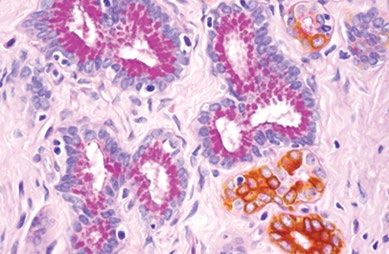
C o n S e n S u S S tat e m e n t
CCA awareness, knowledge, diagnosis and therapies, and what is envisaged on the horizon for CCA, focus-
patient prognosis has not improved substantially in the ing on epidemiology, risk factors, clinical presentation,
past decade, with 5-year survival (7–20%) and tumour diagnosis, genetic and epigenetic landscape, molecular
recurrence rates after resection still disappointing10–17. perturbations, chemoresistance and therapies.
Therefore, a detailed study of these types of cancers is
urgently needed to improve patient welfare and out- Methods
comes. Considering the high heterogeneity of CCAs, This international group of multidisciplinary experts in
individual characterization of these tumours at the CCA (that is, oncologists, surgeons, hepatologists, genet-
genomic, epigenetic and molecular levels is an indispen- icists, immunologists, basic scientists) has been inten-
sable approach to ascertain their pathogenesis, paving sively collaborating within the ENS-CCA since 2015
the path for new therapeutic options and personalized with the main aims of improving our understanding of
medicine. In this expert Consensus Statement, which CCA and the management of patients. In this regard,
is endorsed by the European Network for the Study of this expert consensus is endorsed by the ENS-CCA.
Cholangiocarcinoma (ENS-CCA), we provide a com- The overall goal of this multidisciplinary statement is
prehensive and critical overview of current knowledge to provide a detailed critical overview of the current
knowledge in this field, proposing some expert recom-
Author addresses mendations and highlighting what is envisaged for the
1
Department of Liver and Gastrointestinal Diseases, Biodonostia Health Research
next decade.
Institute – Donostia University Hospital, University of the Basque Country (UPV/EHU), J.M.B. and G.J.G. identified the areas of interest,
San Sebastian, Spain. stratified the consensus statement into the sections pre-
2
National Institute for the Study of Liver and Gastrointestinal Diseases (CIBERehd, sented in the document and assigned them to selected
“Instituto de Salud Carlos III”), San Sebastian, Spain. ENS-CCA members or non-European collaborators
3
Ikerbasque, Basque Foundation for Science, Bilbao, Spain. (L.R.R., S.G., S.R., J.H. and G.J.G.) who are expert in
4
Experimental Hepatology and Drug Targeting (HEVEFARM), IBSAL, University of each field of knowledge and research. To write this docu
Salamanca, Salamanca, Spain. ment, a PubMed search was conducted by combining
5
Department of Medical Oncology, The Christie NHS Foundation Trust, Manchester, UK. the term ‘cholangiocarcinoma’ with the following terms:
6
Division of Cancer Sciences, University of Manchester, Manchester, UK.
‘epidemiology’, ‘risk factors’, ‘classification’, ‘cells of ori-
7
Department of Surgery and Cancer, Imperial College London, Hammersmith Hospital,
London, UK.
gin’, ‘diagnosis’, ‘staging’, ‘genetics’, ‘epigenetics’, ‘signal-
8
Division of Gastroenterology and Hepatology, Mayo Clinic College of Medicine and ling pathways’, ‘epithelial-to-mesenchymal transition’,
Science, Rochester, MN, USA. ‘cancer stem cells’, ‘tumour microenvironment’, ‘immuno
9
Department of Medico-Surgical Sciences and Biotechnologies, Sapienza University biology’, ‘in vitro and in vivo models’, ‘biomarkers’,
of Rome, Rome, Italy. ‘surgery’, ‘liver transplantation’, ‘therapies’, ‘clinical trials’
10
Department of Movement, Human and Health Sciences, Division of Health Sciences, and ‘chemoresistance’. No specific search dates were
University of Rome “Foro Italico”, Rome, Italy. used. All the sections were merged into a first draft by
11
Biotech Research and Innovation Centre (BRIC), Department of Health and Medical P.M.R and J.M.B. and then extensively revised to create
Sciences, University of Copenhagen, Copenhagen, Denmark. the final document that was later circulated among
12
Institute of Cancer Sciences, University of Glasgow, Glasgow, UK.
all the authors for further correction, improvement, dis-
13
Institute of Pathology, University of Regensburg, Regensburg, Germany.
14
Department of Molecular Medicine, University of Padua School of Medicine,
cussion and approval. The data presented in Fig. 2 were
Padua, Italy. obtained by combining the values of mortality rates in
15
Digestive Disease Section, Yale University School of Medicine, New Haven, CT, USA. men and women for both iCCA and eCCA reported
16
MRC-Human Genetics Unit, Institute of Genetics and Molecular Medicine, University in 2019 by Bertuccio et al.9. For the recommendations
of Edinburgh, Edinburgh, UK. on CCA management and research priorities, ideas were
17
Division of Human Anatomy, Department of Anatomical, Histological, Forensic proposed, discussed and approved after final revision by
Medicine and Orthopedics Sciences, Sapienza University of Rome, Rome, Italy. all the authors to reach a consensus.
18
Department of Medicine and Medical Specialties, Sapienza University of Rome,
Rome, Italy. Epidemiology and risk factors
19
The Hormel Institute, University of Minnesota, Austin, MN, USA.
The global mortality for CCA increased worldwide
20
Clinic of Gastroenterology and Hepatology, Universita Politecnica delle Marche,
Ancona, Italy.
during the periods 2000–2004, 2005–2009 and 2010–
21
INSERM, Université de Rennes, Rennes, France. 2014 (Fig. 2), according to the WHO and Pan American
22
Sorbonne Université, INSERM, Centre de Recherche Saint-Antoine (CRSA), Health Organization databases for 32 selected locations
Paris, France. in Europe, America, Asia and Oceania9. Furthermore,
23
Department of Experimental and Clinical Medicine, University of Florence, Florence, CCA mortality was higher in men than in women world-
Italy. wide, and in countries/regions in Asia versus those in
24
Division of Gastroenterology and Center of Autoimmune Liver Diseases, Department the West. Accordingly, Asian individuals were reported
of Medicine and Surgery, San Gerardo Hospital, University of Milano, Bicocca, Italy. to have the highest mortality (2.81 per 100,000 men
25
Department of Gastroenterology and Hepatology, University Hospital Zurich and in Japan). However, in the USA, the more noticeable
University of Zurich, Zürich, Switzerland.
increases in mortality between 2004 and 2014 were
26
Department of Surgery, Mayo Clinic, Rochester, MN, USA.
27
Department of Surgery, Erasmus Medical Center, Rotterdam, Netherlands.
found for African American individuals (45%), followed
28
Barcelona Clinic Liver Cancer (BCLC) group, Liver Unit, Hospital Clínic of Barcelona, by Asian (22%) and white (20%) individuals18. The
Fundació Clínic per a la Recerca Biomédica (FCRB), IDIBAPS, University of Barcelona, age-standardized incidence of CCA shows considerable
Barcelona, Spain. geographical variation, with the highest incidence in
29
Department of Medical Oncology, UCL Cancer Institute, London, UK. Eastern countries/regions; incidence varies from 85 per
558 | September 2020 | volume 17 www.nature.com/nrgastro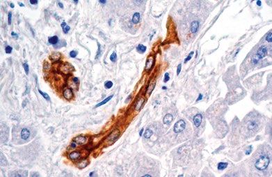
C o n S e n S u S S tat e m e n t
Mass-forming
Periductal-infiltrating
Bile
ductules
iCCA
(10–20%)
Segmental
ducts
Left, right,
common pCCA
Intraductal-growing (50–60%)
hepatic
ducts
Common dCCA
bile (20–30%)
duct
Fig. 1 | Anatomical classification of cholangiocarcinoma. On the basis of the anatomical site of origin,
cholangiocarcinoma (CCA) is classified into intrahepatic CCA (iCCA), perihilar CCA (pCCA) and distal CCA (dCCA).
iCCA is defined as a malignancy located in the periphery of the second-order bile ducts, pCCA arises in the right
and/or left hepatic duct and/or at their junction, and dCCA involves the common bile duct (that is, the choledochus).
Grossly, CCA can show three main patterns of growth: mass-forming, periductal-infiltrating, and intraductal-growing.
Mass-forming CCA is a mass lesion in the hepatic parenchyma. Periductal-infiltrating iCCA grows inside the duct wall
and spreads longitudinally along the wall. Intraductal-growing CCA is a polypoid or papillary tumour growing towards
the duct lumen.
100,000 in northeastern Thailand (the highest reported Furthermore, in the USA, the incidence of iCCA is
value globally) to 0.4 per 100,000 in Canada12. Variations higher in older people (≥45 years old) than in younger
in incidence probably reflect differences in local risk people and in Hispanic individuals than in non-Hispanic
factors and potential genetic predispositions1,2,19. individuals, and is associated with a worse 5-year
The three subtypes of CCA can have different risk survival in both these populations21. Worse overall sur-
factors, pathobiology, clinical presentations, manage- vival (OS) rates have also been reported for African
ment and prognosis, as well as distinct epidemiolog- Americans, followed by American Indians and Alaska
ical trends1,2. Over the past few decades, the reported Native groups21,22. Of note, the hospital charges asso-
age-standardized incidence for iCCA has been stead- ciated with iCCA management almost doubled from
ily increasing in most locations worldwide, whereas 2005 to 2014 in the USA22, and male patients with low
the age-standardized incidence for eCCA has been annual incomes (
C o n S e n S u S S tat e m e n t
Annual mortality rates for CCA (per 100,000 inhabitants) Netherlands Norway Finland Czech Republic Hungary
in the periods 2000-2004 (2002), 2005-2009 (2007) 2002: 1.78 2002: 1.09 2002: 2.65 2002: 1.52 2002: 1.98
2007: 1.83 2007: 1.64 2007: 2.87 2007: 1.65 2007: 1.12
and 2010-2014 (2012) 2012: 2.33 2012: 2.18 2012: 2.42 2012: 1.88 2012: 2.03
4 deaths 2007: 1.49 2007: 2.22 2007: 1.32 2007: 3.79 2007: 2.01
Not applicable 2012: 1.87 2012: 2.49 2012: 1.71 2012: 4.04 2012: 2.75
Canada Belgium
2002: 1.71 2002: 1.74
2007: 2.05 2007: 2.06
2012: 2.71 2012: 2.59
USA UK Japan
2002: 1.56 2002: 1.70 2002: 6.52
2007: 1.71 2007: 2.48 2007: 6.19
2012: 2.05 2012: 3.10 2012: 5.85
Germany South Korea
Mexico
2002: 1.05 2002: 2.16 Incidence >6
2007: 1.14 Puerto Rico 2007: 2.66
2012: 1.17 2002: 1.11 2012: 3.2
2007: 1.00 China
2012: 1.35 Incidence >6
France Switzerland
Venezuela 2002: 1.78 2002: 2.19
2002: 0.85 2007: 2.39 2007: 2.58 Taiwan
2007: 0.75 Colombia 2012: 2.94 2012: 2.85 Incidence >4
2012: 0.83 2002: 1.10
2007: 1.43 Brazil
Portugal Italy Israel
2012: 1.42 2002: 1.31 Hong Kong
2007: 1.43 2002: 2.21 2002: 1.48 2002: 1.36
2007: 1.71 2007: 1.76 2002: 4.33
2012: 1.47 2007: 2.32 2007: 4.31
2012: 2.76 2012: 2.01 2012: 1.92
2012: 4.36
Chile Argentina Spain Australia New Zealand
2002: 0.74 2002: 0.32 2002: 1.65 2002: 2.04 2002: 1.89
2007: 0.96 2007: 0.32 2007: 2.19 Thailand 2007: 2.47 2007: 2.59
2012: 1.13 2012: 0.45 2012: 2.80 Incidence >6 2012: 2.88 2012: 2.43
Fig. 2 | Mortality of cholangiocarcinoma worldwide. Global age-standardized annual mortality rates for
cholangiocarcinoma (CCA) (deaths per 100,000 inhabitants, including intrahepatic CCA, perihilar CCA and distal CCA)
obtained from Bertuccio et al.9. Data refer to the periods 2000–2004 (2002), 2005–2009 (2007) and 2010–2014 (2012).
Yellow indicates countries/regions with low mortality (4 deaths per 100,000 people). Mortality in eastern countries/regions in which CCA is highly prevalent (that is, Thailand,
China, Taiwan and South Korea) have not yet been reported and, therefore, CCA incidence is shown for these countries1.
liver disease)29. In this regard, CCA can be notoriously CCA cases remain sporadic, without any identifiable
difficult to accurately diagnose due to its location often risk factor present. A number of studies are examining
being inaccessible to histology or cytology, a lack of clear the potential influence of commonly used drugs such as
diagnostic imaging criteria, and inaccurate non-invasive aspirin31–33 and lipid-lowering statins34,35 in the preven-
tumour biomarkers1,2. tion of CCA. Notably, post-diagnosis aspirin usage has
Several risk factors, both common and rare, have been found to be associated with a reduced risk of death
been linked to CCA (Table 1). Although some risk factors (HR 0.71) among patients with CCA36. Polymorphisms
are shared by all forms of CCA, others seem to be more of host genes encoding enzymes involved in xeno
specific for one subtype and seem to be more important biotic detoxification, DNA repair, multidrug resistance,
in different regions. A common characteristic amongst immune response and folate metabolism have been
many of these risk factors is that they are associated with linked to CCA development19. There are currently no
chronic inflammation of the biliary epithelium and bile published genome-wide association studies (GWAS)
stasis19. Several recognized risk factors have increased in CCA, but an appropriately powered one is eagerly
globally over recent decades (1990–2016) and could anticipated.
be contributing to increasing CCA rates. For instance,
high alcohol consumption, tobacco smoking and viral Classification and cells of origin
infections (hepatitis B virus (HBV) and hepatitis C virus iCCA can emerge at any point of the intrahepatic biliary
(HCV)) have been reported to increase the risk of CCA tree, ranging from bile ductules to the second-order bile
development30. Moreover, it is also important to high- ducts (segmental bile ducts). In contrast to HCC, iCCA
light the global obesity pandemic, as well as the meta- usually develops in non-cirrhotic liver37. pCCA can arise
bolic syndrome and/or presence of nonalcoholic fatty in the right and/or left hepatic duct and/or at their junc-
liver disease, as risk factors that deserve future central tion (so-called perihilar bile ducts)38, and dCCA involves
attention30. However, in most locations, the majority of the common bile duct39. The current term eCCA is now
560 | September 2020 | volume 17 www.nature.com/nrgastro
C o n S e n S u S S tat e m e n t
discouraged as it combines subtypes with distinct clin- frequently, as intraductal papillary tumours40. CCA can
icopathological features, prognosis and therapeutic be preceded by pre-invasive lesions39. Histologically,
options, and also due to the difficulties in discriminating although the vast majority of pCCA and dCCA are
between intrahepatic and extrahepatic origins of pCCA. conventional mucin-producing adenocarcinomas or
iCCA can show three main patterns of growth: mass- papillary tumours40, iCCA shows several histological
forming, periductal-infiltrating, and intraductal- variants (that is, conventional, cholangiolocarcinoma
growing1,38 (Fig. 1); pCCA and dCCA present as flat and rare variants)41 (Fig. 3; Table 2). Conventional iCCA
or poorly defined nodular sclerosing tumours or, less can be further classified into two main histological
subtypes according to the level or size of the affected
duct42–46 (Fig. 3; Table 2). Small bile duct iCCA presents
Table 1 | Risk factors for cholangiocarcinoma
as a small-sized tubular or acinar adenocarcinoma with
Risk factor Study type OR or RR from nodular growth invading the liver parenchyma, and
selected studies with no or minimal mucin production42–46. Large bile
Choledochal cyst30 Meta-analysis OR 26.71 for iCCA duct iCCA arises in large intrahepatic bile ducts and
OR 34.94 for eCCA comprises mucin-producing columnar tumour cells
Choledocholithiasis30 Meta-analysis OR 10.08 for iCCA arranged in a large duct or papillary architecture38,46–49.
OR 18.58 for eCCA Remarkably, the histological subtyping parallels the high
Cholelithiasis30 Meta-analysis OR 3.38 for iCCA molecular heterogeneity of CCAs and can be ascribed
OR 5.92 for eCCA to different cells of origin and pathogenesis41. Small bile
Cholecystolithiasis30 Meta-analysis OR 1.75 for iCCA duct iCCA can be characterized by isocitrate dehydro-
OR 2.94 for eCCA genase (IDH1, IDH2) mutations or fibroblast growth
Caroli disease396 Population-based study OR 38 for iCCA factor receptor 2 (FGFR2) fusions40,50–55. By contrast,
OR 97 for eCCA large bile duct iCCA, similar to pCCA and dCCA,
Primary sclerosing Population-based study OR 22 for iCCA shows a high frequency of mutations in KRAS and/or
cholangitis396 OR 41 for eCCA TP53 genes51,53,56,57. Interestingly, dCCA is also associated
Cirrhosis30 Meta-analysis OR 15.32 for iCCA with ELF3 mutations58. Growing evidence demonstrates
OR 3.82 for eCCA that distinct cells of origin within an organ can give rise
to different subtypes of cancer, typically tissue-specific
Chronic hepatitis B30 Meta-analysis OR 4.57 for iCCA
OR 2.11 for eCCA stem and progenitor cells59–62. Evidence regarding the
cells of origin of CCA in humans was obtained by phe-
Chronic hepatitis C30 Meta-analysis OR 4.28 for iCCA
OR 1.98 for eCCA notyping the candidate tissues and/or cells of origin with
respect to CCA subtypes through histological and gene
Haemochromatosis396 Population-based study OR 2.1 for iCCA expression analysis38,44,46,63–68, whereas indirect evidence
Inflammatory bowel disease30 Meta-analysis OR 2.68 for iCCA might be derived from risk factors67,68.
OR 2.37 for eCCA Small bile duct and cholangiolocarcinoma iCCA
Chronic pancreatitis396 Population-based study OR 2.7 for iCCA subtypes emerge at the level of smaller intrahepatic
OR 6.6 for eCCA bile ducts, including bile ductules38,44,46,69. In these por-
Liver fluke (Opisthorchis Meta-analysis OR 5 iCCA > eCCA tions of the biliary tree, hepatic stem or progenitor cells
viverrini, Clonorchis sinensis)397 (HpSCs) and cuboidal cholangiocytes represent surface
Type 2 diabetes mellitus398 Meta-analysis OR 1.73 for iCCA epithelium and are the putative cells of origin of these
OR 1.5 for eCCA malignancies38,44,46,69 (Fig. 3; Table 2). Interestingly, HpSCs
Nonalcoholic fatty liver Meta-analysis OR 2.2 for iCCA have been implicated in CK19+ HCC70 and in combined
disease399 OR 1.5 for eCCA HCC–CCA4,64,71. Notably, CCA-like HCC tumours dis-
Obesity30 Meta-analysis OR 1.14 for iCCA play embryonic stem cell-like expression traits, further
OR 1.2 for eCCA substantiating the involvement of bipotent hepatic pro-
Hypertension30 Meta-analysis OR 1.10 for iCCA genitor cells, and in humans display a worse prognosis
OR 1.21 for eCCA than HCC72. In line with these findings, nestin, a maker
of bipotent progenitor oval cells, is greatly increased
Alcohol consumption30 Meta-analysis OR 3.15 for iCCA
OR 1.75 for eCCA in HCC–CCA tumours and is associated with a worse
prognosis, and has been proposed as a new possible
Cigarette smoking30 Meta-analysis OR 1.25 for iCCA
OR 1.69 for eCCA diagnostic and prognostic biomarker3,26. Small bile duct
and cholangiolocarcinoma iCCA usually develop on a
Environmental toxins background of chronic liver disease (such as chronic
Thorotrast (banned 1969)400,401 Retrospective study RR >300 viral hepatitis and cirrhosis)38,46,70, characterized by
1,2-Dichloropropane 402
Retrospective study RR 15 HpSC activation71,73,74.
Large bile duct iCCA, pCCA and dCCA derive
Asbestos403 Case–control study OR 4.8 for iCCA
OR 2.1 for eCCA from columnar mucous cholangiocytes or peribiliary
glands38,44,46,47,49,69 (Fig. 3), which are also implicated in
Asbestos404 Case–control study OR 1.1–1.7 for iCCA
No association with the origin of precursor lesions (such as intraductal pap-
eCCA illary neoplasm)46. These malignancies mainly develop
eCCA, extrahepatic cholangiocarcinoma; iCCA, intrahepatic cholangiocarcinoma; OR, odds in ducts affected by chronic inflammation as in primary
ratio; RR, relative risk. sclerosing cholangitis (PSC) or liver fluke infection46,49,69.
NAture RevIewS | GASTROENTEROLOgy & HEPATOLOgy volume 17 | September 2020 | 561C o n S e n S u S S tat e m e n t
Small iBDs Large iBDs eBDs
Bile ductule Interlobular, zonal, septal Area, segmental Hepatic, cystic, choledochal
Anatomy
Ø 800 μm
Bile ductule Interlobular bile duct Peribiliary glands Surface
epithelium
Putative cell of origin
HpSC/DR Cuboidal cholangiocyte Mucous cells and/or columnar cholangiocyte
CLC iCCA Small duct-type Large duct-type pCCA/dCCA
Histological classification
Conventional iCCA
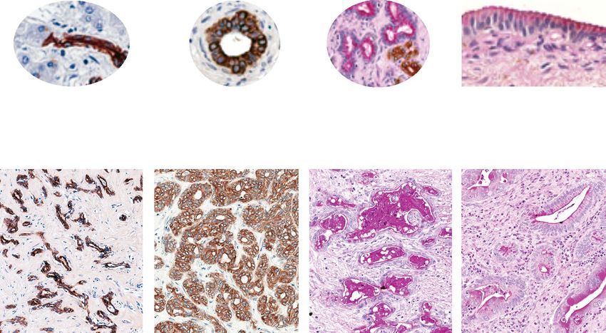
Fig. 3 | Histological classification and putative cells of origin in cholangiocarcinoma. Based on the duct size, the
intrahepatic biliary tree can be further subdivided into small and large intrahepatic bile ducts (iBDs). Small iBDs are lined
by small cuboidal cholangiocytes whereas columnar and mucous cholangiocytes line large iBDs. Typically, large iBDs
contain peribiliary glands within their wall. The extrahepatic biliary tree shares anatomical features with large iBDs.
Histological cholangiocarcinoma (CCA) variants reflect the phenotype of the involved duct and the putative cell of origin.
Conventional intrahepatic CCA (iCCA) has two main variants: small duct-type iCCA arises in small iBDs with cuboidal
cholangiocytes representing the putative cell of origin, and large duct-type iCCA involves large iBDs and is considered to
be derived from columnar cholangiocytes and peribiliary glands (seromucous glands; mucous acini are shown in light pink,
serous acini are shown in green). Cholangiolocarcinoma (CLC) is a frequent histological variant of iCCA and its phenotype
suggests the origin from bile ductules or ductular reaction (DR) that occurs in chronic liver diseases. The vast majority of
perihilar CCA (pCCA) and distal CCA (dCCA) are considered to originate from the lining epithelium and peribiliary glands.
This histological subtyping underlies distinct clinicopathological and molecular features as summarized in Table 2.
eBD, extrahepatic bile duct; HpSC, human pluripotent stem cell.
In PSC, peribiliary gland cell proliferation, muci- senescence in hepatocytes 66,82,83, and that lineage
nous metaplasia, and dysplasia to cancer progression tracing studies must be conducted and interpreted
take place within bile ducts and along the biliary tree, cautiously76,84–87.
mimicking the cancerization field (‘field defect’)49,75.
Controversies exist regarding the cellular origins of Clinical presentation
iCCA based on lineage tracing studies in experimen- Diagnosis
tal carcinogenetic models76. Indeed, there is evidence CCAs are usually asymptomatic during early stages. The
in favour of HpSC, cholangiocyte or hepatocyte origin most frequent symptom of pCCA and dCCA is jaundice
of iCCA from these experimental settings76–81. Thus, due to biliary tract obstruction88. In iCCA, jaundice is
a definitive determination of the origin of iCCA in less frequent and mostly associated with advanced
humans cannot be reached based on current evidence disease. Other symptoms of advanced disease include
and requires further research. Moreover, it should be asthenia, abdominal pain, malaise, nausea, anorexia
underlined that current experimental models of liver and weight loss. iCCA is an incidental finding in around
damage do not fully recapitulate the pathogenesis of 20–25% of cases88. In patients with cirrhosis, ultrasonog-
human chronic liver disease, including proliferative raphy surveillance for HCC enables iCCA diagnosis
562 | September 2020 | volume 17 www.nature.com/nrgastroC o n S e n S u S S tat e m e n t
at an asymptomatic, early stage89. Unfortunately, the with iCCA, washout takes place earlier than 60 s after
majority of iCCA cases occur in the absence of known contrast agent injection; this feature is rarely observed in
risk factors90, when the only chance for early diagnosis is HCC, and the intensity of washout in the portal phase is
by cross-sectional imaging performed for other reasons. more marked in iCCA than in HCC89. These refinements
Imaging techniques, such as ultrasonography, might decrease the risk of misdiagnosis in HCC98, and
contrast-enhanced ultrasonography (CEUS), CT and have been adopted by the Liver Imaging Reporting Data
MRI, play a key part in the management of CCA in System (LI-RADS) for CEUS (LI-RADS-CEUS)99. No
terms of diagnosis, staging, follow-up and assessment evidence supports the use of 18F-FDG PET for comple-
of treatment response. Their diagnostic accuracy is tion of staging, which could be of special value to exclude
influenced by the anatomical location and growth pat- the presence of lymph node or distant metastases100.
terns of CCA, and their use for staging varies accord- As no specific CCA radiology pattern exists, histo
ing to tumour location91. CT is considered the standard pathological or cytological analysis is mandatory to
imaging method for the preoperative assessment of both confirm the diagnosis1,28. This diagnosis is based on the
iCCA and pCCA; it provides a comprehensive evalua- WHO classification of biliary tract cancer showing an
tion of the primary tumour, the relationship with adja- adenocarcinoma or mucinous carcinoma101, with tubu-
cent structures, and potential thoracic and abdominal lar and/or papillary structures and a variable fibrous
spread91. MRI has similar accuracy to CT for diagnosis stroma102.
and staging, but it incorporates specific sequences such
as diffusion-weighted imaging and the potential for per- Staging
forming magnetic resonance cholangiopancreatography There is no widely used staging system for CCA,
(MRCP), which is critical for pCCA staging92. The most although it can be staged according to the American
frequent imaging patterns displayed by iCCA on both Joint Committee on Cancer (AJCC) TNM system103,104.
CT and MRI are arterial peripheral rim enhancement Despite providing a clinically meaningful classifica-
with progressive homogeneous contrast agent uptake tion correlated with prognosis105, the current TNM
until the delayed or stable uptake during late dynamic classification has some limitations. First, it has limited
phases93,94. A targetoid pattern defined as arterial rim discriminatory ability between T2 and T3 tumours in
enhancement, peripheral washout and delayed cen surgically resected iCCAs105,106. T2 tumours include
tral enhancement can also be present in iCCA95. When multifocal disease or disease with intrahepatic vascular
gadoxetic acid is used, the washout should be read in invasion that probably reflect disseminated disease and
the portal phase instead of in delayed phases to prevent the OS in patients with these tumours does not differ
misclassification between HCC and iCCA in a cirrhotic from the OS in patients with extrahepatic metastatic
liver96. More controversial is the use of CEUS in iCCA, disease105. Similarly, there is also evidence supporting
particularly in the setting of underlying chronic liver the negative effect of the presence of multifocal iCCA
disease. iCCA exhibits homogeneous arterial hyper (iCCA with liver metastases; T2) on prognosis (OS)
enhancement followed by venous washout in near 50% when compared with other early stages, which might
of patients, a pattern indistinguishable from that found require consideration in future versions of the AJCC
in HCC94,97. However, in a relevant proportion of patients TNM classifications107. Second, although size has been
Table 2 | Clinicopathological and molecular features of cholangiocarcinoma
CCA type Gross pattern Precancerous Underlying Tissue markersa Frequent mutations
lesion disease
iCCA — CLC Mass-forming None Viral, NCAM IDH1/2, FGFR2 fusions,
cirrhosis BAP1, BRAF, ARID1A,
KRAS, TP53, SMAD4
Increased IDH1 and TP53
iCCA — small Mass-forming None Viral, NCAM, IDH1/2, FGFR2 fusions,
duct type cirrhosis N-cadherin, BAP1, BRAF, ARID1A,
SMAD4, BAP1loss KRAS, TP53, SMAD4
Increased IDH1/2, FGFR2
fusion
iCCA — large Periductal Biliary epithelial Primary Mucinb, IDH1/2, FGFR2 fusions,
duct type infiltrating neoplasia, IPNB, sclerosing MUC5AC, BAP1, BRAF, ARID1A,
(±mass-forming) ITPN, mucinous cholangitis, MUC6, S100P, KRAS, TP53, SMAD4
or intraductal cystic neoplasm liver flukes SMAD4loss, BAP1
Increased KRAS and TP53
growing
pCCA–dCCA Periductal Biliary epithelial Primary Mucinb, KRAS, TP53, SMAD4,
infiltrating or neoplasia, IPNB, sclerosing MUC5AC, ERBB3, PRKACA–PRKACB
intraductal ITPN, mucinous cholangitis, MUC6, S100P, fusions, ELF3
growing cystic neoplasm liver flukes SMAD4loss, BAP1
CCA, cholangiocarcinoma; CLC, cholangiolocarcinoma; dCCA, distal cholangiocarcinoma; iCCA, intrahepatic cholangiocarcinoma;
IPNB, intraductal papillary neoplasm of the bile duct; ITPN, intraductal tubulopapillary neoplasm; pCCA, perihilar cholangiocarcinoma.
a
Markers from single-centre experience; international criteria and consensus on a definite panel of markers are still needed. bMucin
refers to histomorphological stains periodic acid–Schiff (PAS) or Alcian PAS.
NAture RevIewS | GASTROENTEROLOgy & HEPATOLOgy volume 17 | September 2020 | 563C o n S e n S u S S tat e m e n t
included for the first time as a prognostic factor for iCCA and IDH2R172) that cause an accumulation of the onco-
in the eighth edition of the AJCC Cancer Staging Manual, metabolite 2-hydroxyglutarate (2-HG)57, as well as the
the only cut-off size considered is 5 cm in T1 tumours. constitutive active gene fusion event between FGFR2 and
Several authors have shown that a 2 cm cut-off value many different partners, including the most prevalent
might identify very early tumours with very low likeli- (BICC1 (refs50,112–114), PPHLN1 (ref.115), TACC3 (ref.112)
hood of dissemination and potential long-term survival and MGEA5 (ref.112)). These alterations are important as
with low recurrence rates24,108. Finally, the TNM classi- they are driving current marker-based phase III clinical
fication misses relevant prognostic factors such as the trials testing specific agents targeting these alterations in
presence of cancer-related symptoms (such as abdominal FGFR2 fusion-positive CCA (NCT03773302)123,124 and
pain or malaise) or the degree of liver function impair- IDH-mutated CCA (NCT02989857).
ment. As previously shown with HCC, future propos- To date, information on the inherited predisposing
als from society guidelines should focus on stratifying genetic risk factors causing CCA is very limited125. Data
non-surgical patients for clinical studies using clinical mostly stem from GWAS of patient cohorts diagnosed
and imaging data. Notably, Chaiteerakij et al. proposed a with PSC25,126, with increased risk of CCA. However, the
new staging system for pCCA based on tumour size and only detailed genomic association with aetiological risk
number, vascular encasement, lymph node and perito- factors investigated by genome sequencing has been the
neal metastasis, Eastern Cooperative Oncology Group association with liver fluke infection (Opisthorchis viver-
(ECOG) performance status (ECOG-PS), and CA19-9 rini and Clonorchis sinensis), with fluke-positive tumours
level, which has shown a better performance in pre- showing an overall higher mutational rate (median
dicting survival than the TNM staging system109. Also, 4,700 versus 3,143 somatic mutations per tumour)116
important for stratification in clinical trials, radiographic with prevalent mutations in SMAD4 and TP53 as well
staging parameters need to be developed in the absence as ERBB2 amplifications116–118. Furthermore, although
of histological staging, and a radiographic staging system not in a high proportion, KRAS mutations have been
has been proposed for pCCA109. recurrently found in all CCA subtypes56,116,117. A statis-
tically significant association has also been observed
Genetics and epigenetics between TP53 mutation and HBV infection119,120. Few
Genomics studies have investigated the molecular distinction
Initial efforts using integrative genomics approaches to between iCCA, pCCA and dCCA8,56,116,121. Nakamura
stratify CCA based on prognosis have highlighted exten- et al. emphasized the difference in anatomical loca-
sive deregulated transcriptomic landscapes showing aug- tion of the tumour, highlighting IDH, EPHA2 and
mented anti-apoptotic signalling, angiogenesis, signal BAP1 mutations and FGFR2 fusions in iCCA, whereas
transduction and transcriptional control8,110. The main extrahepatic tumours specifically show PRKACA and
oncogenic networks comprised WNT-CTNNB1, MYC, PRKACB fusions as well as mutations in ELF3 (similar to
ERBB, TNF and VEGF signalling, emphasizing cell tumours in the ampulla of Vater)127 and ARID1B56. Based
survival signalling pathways in patients with poor OS8. on these fundamental causal alterations, tumours in dis-
Regarding genomic alterations, CCA falls midway in tinct anatomical sites should probably be treated differ-
the mutational spectrum of cancers111, with an approxi- ently. Besides linking IDH mutations with the response
mately equal content of genomic alterations in iCCA to ivosidenib 128, few studies have related genomic
(median 39 non-synonymous mutations per tumour) alterations to high-throughput drug screening119,129,130.
and eCCA (median 35 non-synonymous mutations Among these, Nepal et al. used an approach of integra-
per tumour)56. Massive sequencing studies56,112–121 have tive genomics in a large cohort of iCCAs to elucidate
improved our understanding of the causal mechanisms unique mutational signatures, structural variants and
in CCA, emphasizing the genomic complexity in prev- epigenomic alterations, emphasizing specific oncoge-
alent oncogenic modules affecting: cell cycle regulation; netic mechanisms in four distinct subsets of patients
DNA damage and genomic instability (TP53, CDKN2A, with potential drug responses and categories: RNA syn-
CCND1, ATM, ROBO2, BRCA1 and BRAC2); MYC thesis inhibition, IDH mutant; microtubule modulator,
amplification; epigenetic regulation including NADPH KRAS mutant; topoisomerase inhibition, TP53 mutant;
metabolism (IDH1 and IDH2), de- u biquitination and mTOR inhibitors119.
(BAP1), SWI–SNF complex (PBRM1, ARID1A, ARID1B,
ARID2, SMARCA2, SMARCA4 and SMARCAD1) and Epigenetics
histone (de-)methylation (MLL2, MML3, KMT2C, Epigenetics was shown to play an important part in the
KDM4A, KDM5D, KDM6A and KDM6B); kinase initiation and progression of CCA, affecting tumour phe-
signalling (KRAS, ERBB1–3, BRAF, PIK3CA, PTEN, notype in the absence of changes in DNA sequences131.
STK11, SMAD4 and FGFR1–3); immune dysregulation Deregulated patterns of methylation, histone modifi-
(JAK–STAT3 signalling); FGFR2 and PRKCA–PRKCB cations and aberrant expression of non-coding RNAs
fusions; the WNT–CTNNB1 pathway (APC); Hippo promote unbalanced transcription and gene expression
signalling (NF2, SAV1 deletion); METLL13 amplifica- that impair cell homeostasis and sustain malignant
tions; and deregulated Notch signalling. Interestingly, transformation. Growing evidence supports deregulated
the predominant genomic alterations in CCA are asso- methylation motifs in CCA cells compared with their
ciated with epigenetic processes122. Indeed, the most normal counterparts, with a prevalent hypermethyla-
clinically significant genomic breakthroughs in iCCA tion of multiple CpG sites occurring in CCA132,133. One
are the discovery of hotspot IDH mutations (IDH1R132 of the largest studies of integrative genetic and epigenetic
564 | September 2020 | volume 17 www.nature.com/nrgastroC o n S e n S u S S tat e m e n t
analyses in CCA, including 489 CCAs from ten coun- epigenetic occurrence seem to have an enrichment
tries/regions, has shown how the molecular make-up of of events within embryonic stem cell-related bivalent
CCA goes beyond the differentiation according to ana- regulation134–136. IDH-mutated tumours instead seem to
tomical site116. Indeed, by combining DNA sequencing resemble the profile of cholangiocellular CCAs that show
with transcriptomic and DNA methylation analyses, gene expression traits of epithelial–mesenchymal tran-
four clusters of CCA with different clinical outcomes sition (EMT)136. Histone modifications have been less
were identified. Two sets of hypermethylated CCAs studied in CCA. Histone deacetylase (HDAC) enzymes
stood out, with an interesting association between CpG are responsible for regulation of histone acetylation that
island hypermethylation and liver fluke-related tumours, ultimately affects chromatin organization. HDAC were
increased mutation rate, downregulation of the DNA found to be upregulated in CCA in vitro137, and are being
demethylation enzyme TET1, upregulation of the his- investigated as targets of treatment. Evidence also sug-
tone methyltransferase EZH2 and an increased level of gests that HDAC inhibitors, as well as dasatinib, might
deamination events. Conversely, the subgroup of iCCAs be particularly active in IDH-mutated tumour cells129,130.
with enrichment in IDH1/2 and BAP1 mutations, as well Non-coding RNAs account for around 98% human RNAs
as FGFR translocations, showed hypermethylation of and include microRNAs (miRNAs) and long non-coding
the CpG shores (the regions immediately flanking CpG RNAs, among others. These non-coding RNAs regulate
islands, up to 2 kb away). This different pattern suggests the expression of a plethora of target genes affecting
how early epigenetic deregulation caused by external all the hallmarks of the cancer phenotype from cell pro-
carcinogenic events (for example, liver flukes) are at the liferation and migration to EMT and the regulation of the
basis of CCA development in the first cluster, whereas in primary cilium in cholangiocytes138–142 (Fig. 4).
the second cluster, epigenetic aberrations probably arise
as a downstream consequence of somatic mutations Signalling and molecular networks
(IDH) that produce oncometabolites responsible for the CCA often arises in the setting of prolonged biliary
DNA hypermethylation. These differences have remark- inflammation and/or cholestasis, which contribute to
able clinical implications, because on the one hand early carcinogenesis. According to transcriptomic profiles,
epigenetic events might be used for early detection of the ‘inflammation’ (38%) and ‘proliferation’ (62%) sub-
tumours in the first cluster (by using quantitative DNA types of iCCA were previously identified and reported
methylation markers in the bile of individuals at risk)80 to be differentially enriched with activation of the pro-
and on the other hand, the tumour clonal mutations inflammatory and oncogenic pathways, respectively110.
might be a marker of effective targeted therapies (such The inflammation subclass of tumours was character-
as IDH inhibitors). ized by induction of immune-related signalling path-
Methylome data can also provide insights into the ways. By contrast, the proliferation subclass was enriched
cells of origin of CCA. Tumours with high genetic and in classic oncogenic pathways, including deregulated
Inflammation
↑ miR-17-92 ↓ miR-125b
↑ miR-200 ↓ miR-605
↓ miR-99a
↓ miR-let7c
EMT or migration or invasion Proliferation and cell cycle
↑ miR-200c ↑ AFAP1 ↓ miR-124 ↑ miR-141 ↑ AFAP1 ↓ miR-101
↑ miR-21 ↑ CCAT2 ↓ miR-138 ↑ miR-181c ↑ CCAT2 ↓ miR-138
↑ miR-221 ↑ CPS1-IT1 ↓ miR-144 ↑ miR-191 ↑ CPS1-IT1 ↓ miR-144
↑ miR-24 ↑ H19 ↓ miR-200b/c ↑ miR-200b ↑ EPIC ↓ miR-148a
↑ miR-421 ↑ MALAT1 ↓ miR-204 ↑ miR-21 ↑ H19 ↓ miR-152
↑ PANDAR ↑ PCAT1 ↓ miR-214 ncRNAs ↑ miR-210 ↑ HOTAIR ↓ miR-34a
↑ TUG1 ↑ UCA1 ↓ miR-376c in ↑ miR-24 ↑ MALAT1 ↓ miR-370
↓ miR-605 cholangiocarcinoma ↑ miR-26a ↑ PANDAR ↓ miR-373
↑ miR-29 ↑ PCAT1 ↓ miR-376c
↑ miR-31 ↑ TUG1 ↓ miR-410
↑ miR-421 ↑ UCA1 ↓ miR-494
Chemoresistance and survival ↑ miR-let7a ↓ miR-605
↑ miR-21 ↑ H19 ↓ miR-204
↑ miR-200b ↓ miR-29b
↑ miR-24 ↓ miR-320
↑ miR-31 Ciliogenesis
↑ miR-let7a Epigenetics
↓ miR-22
↓ miR-148a ↓ miR-433
↓ miR-152
↓ miR-373
Fig. 4 | Non-coding RNAs in cholangiocarcinoma and their relationship with different tumorigenic processes.
Non-coding RNAs (ncRNAs) that have been found to be dysregulated (up or down) in cholangiocarcinoma and that have
key roles in the regulation of cellular processes, such as proliferation, cell cycle, ciliogenesis, epigenetics, inflammation,
chemoresistance, survival, epithelial to mesenchymal transition (EMT), migration and invasion are shown.
NAture RevIewS | GASTROENTEROLOgy & HEPATOLOgy volume 17 | September 2020 | 565C o n S e n S u S S tat e m e n t
JAG1–2
DLL1 LRP5/LRP6 ECM
IL-6 EGF HGF VEGF SDF1 WNT RTKs
stiffness GPCRs
NOTCH1–3
P P P P P P P P P P P P
P P P P P P P P P P CXCR4 AXIN P P
DVL
γ-secretase
JAK JAK EGFR ERBB2 MET VEGFR FGFR2
fusions P
NCID
CK1α MST1/2
APC P
SOCS3 RAS–MAPK PI3K–AKT P
SAV1 YAP/TAZ
AXIN
pathway pathway β-Catenin
GSK3β P P P P P
β-Catenin LATS1/2
KRAS PI3K Ub P
Ub MOB1
P P Ub
NCID
STAT3 BRAF AKT Mitochondria
Citrate
P P
STAT3 Proteosome Citrate
P
MEK1/MEK2 mTOR degradation Isocitrate
P
Isocitrate
P β-Catenin β-Catenin
ERK1/ERK2 α-KG
P β-Catenin
α-KG
IDH1
YAP/TAZ
IDH2
2-HG
Histone and DNA 2-HG
demethylation
inhibition
p300
• Proliferation
ARID1 PBRM1 MAML1 β-Catenin YAP/TAZ Target gene
NCID
• Apoptosis
transcription evasion
SWI/SNF BAP1 CSL TCF/LEF TEAD
• Tumour growth
• Migration
Cytoplasm Nucleus and/or invasion
Fig. 5 | Signalling pathways involved in cholangiocarcinoma development and progression. The process of cholangio-
carcinogenesis, and further tumour evolution and growth, involves complex and heterogeneous processes that include the
interplay of extracellular ligands (such as pro-inflammatory cytokines, growth factors and bile acids, among others), which
are present in the tumour microenvironment, and increased expression and/or aberrant activation of cell surface receptors
and the deregulation of intracellular signalling pathways, finally leading to cell proliferation, survival and migration or
invasion. The most common genes that might be mutated or amplified resulting in the overactivation of some of these
pathways are KRAS, BRAF, ARID1, PBRM1, BAP1, IDH1 and IDH2. The activation of these signalling pathways might also
occur as a result of the interaction between the tumour epithelia and the tumour reactive stroma. 2-HG, 2-hydroxyglutarate;
ECM, extracellular matrix; RTK, receptor tyrosine kinase.
receptor tyrosine kinase (RTK) signalling, RAS–RAF– Chronic inflammation and fibrosis facilitate cholan-
ERK, PI3K–AKT–mTOR, insulin growth factor giocyte transformation in a multistep manner, provid-
receptor 1, MET, polo-like kinase 1, aurora kinase A, ing extracellular ligands that modulate several signalling
KRAS mutations and stem-like genomic traits as well pathways. In particular, sustained IL-6–STAT3 signalling
as a focal deletion in the Hippo pathway (SAV1)8,110,143. was shown to contribute to mitogenesis by upregulat-
Notably, patients with the proliferation subtype of iCCA ing myeloid cell leukaemia 1 (MCL1) or altering EGFR
displayed decreased OS (median 24.3 months versus promoter methylation144,145. Similarly, bile acids are not
47.2 months for those with the inflammation subtype; genotoxic but might also promote cholangiocarcinogen-
P = 0.048). esis through a mechanism involving the activation of
Cholangiocarcinogenesis is orchestrated by a EGFR, induction of COX2, MCL1 and IL-6, and down-
complex interplay of extracellular ligands (such as regulation of farnesoid X receptor (FXR)146,147. Of note,
pro-inflammatory cytokines, growth factors and bile FXR expression was reported to be decreased in human
acids, among others), which are present in the tumour CCA tumours compared with surrounding normal liver
microenvironment (TME), and increased expression tissue, correlating with tumour differentiation140. By con-
and/or aberrant activation of cell surface receptors and trast, the levels of TGR5, another bile acid receptor, were
the deregulation of intracellular signalling pathways, found to be increased in CCA tumours and to be cor-
finally leading to cell proliferation, survival and genetic related with a worse prognosis (perineural invasion)140.
and/or epigenetic alterations (Fig. 5). CCA tumours, and particularly iCCAs and pCCAs, are
566 | September 2020 | volume 17 www.nature.com/nrgastroC o n S e n S u S S tat e m e n t
characterized by a reactive desmoplastic stroma con- which has been shown to be related to well-differentiated
taining cancer-associated fibroblasts (CAFs) that cross- iCCA155, markedly reduced tumour burden in various
talk with CCA cells secreting paracrine factors such as mouse models of liver cancer (including iCCA)81,159,
heparin-binding EGF-like growth factor, stromal-cell whereas overexpression of NOTCH3 was associated with
derived factor 1 (SDF1), platelet-derived growth factor the development and progression of iCCA, promoting
(PDGF)-B and extracellular matrix (ECM) proteins148. cell survival via PI3K–AKT signalling160. Several Notch
Although there are marked differences in the inhibitors are being developed, and their availability
genomic features depending on the anatomical loca- increases interest in this pathway161.
tion and risk factors, activation of the RTK signalling The WNT–β-c atenin signalling pathway is also
pathway is a common event in CCA across subtypes. known to be activated in most CCAs, in part as an effect
In this regard, aberrant EGFR, ERBB2 and MET RTK of the release of Wnt ligands by inflammatory macro
expression has been found in different CCA subclasses phages infiltrating the stroma162,163, but also as a conse-
that are associated with worse prognosis8,110. RTK signal- quence of DNA methylation alterations targeting this
ling mainly triggers the activation of the RAS–MAPK pathway133 and/or mutations encoding key components
and PI3K–AKT–mTOR pathways. Furthermore, RAS– of the canonical WNT–β-catenin signalling pathway164.
MAPK pathway activation due to KRAS-activating Notably, the promoter of the WNT–β-catenin pathway
mutations is found in all CCAs without distinction, inhibitor SOX17 was hypermethylated in CCA tumour
whereas BRAF mutations are more prevalent in iCCA149. tissue compared with healthy tissue, correlating with a
Interestingly, chromosomal oncogenic gene fusion worse prognosis after tumour resection132. Noteworthy,
rearrangements involving FGFR2 RTK occur almost SOX17 was shown to regulate cholangiocyte differentia-
exclusively in iCCA50,52,56,112,113. Besides FGFR2 fusions, tion and to act as a tumour suppressor in CCA in vitro132.
ROS1 kinase protein fusions have also been identified WNT inhibitors successfully inhibit tumour growth in
in iCCA150. Thus, RTK signalling pathways present experimental models163 and clinical trials with agents
actionable molecular alterations that are amenable targeting this pathway are currently being explored164.
for therapeutic targeting at multiple levels. IDH1 and The Hippo–YAP signalling pathway regulates organ size
IDH2 encode metabolic enzymes that interconvert and cell proliferation, among other functions165. YAP is
isocitrate and α-ketoglutarate51,53,113,117,151. Mutations in a transcriptional co-activator that is usually inhibited
IDH1 and IDH2 lead to the production of high levels by Hippo (MST1 or MST2), but can be activated by
of 2-hydroxyglutarate, an oncometabolite that inter- Hippo-independent signals, such as inflammation and
feres with histone and DNA demethylases and inhib- changes in ECM composition and stiffness166. Several
its the mitochondrial electron transport chain. Indeed, groups have reported increased nuclear expression of
IDH-mutant CCAs were shown to exhibit high levels of YAP in CCA specimens and correlation with a worse
mitochondrial and low levels of chromatin modifier gene prognosis167–169. In vitro studies on CCA cell lines have
expression, such as low ARID1A expression due to DNA shown that YAP can be activated by IL-6, PDGF and
hypermethylation121. Besides epigenetic silencing, inac- fibroblast growth factor170,171. PDGF and fibroblast
tivating mutations in multiple chromatin-remodelling growth factor form a feed-forward loop activating
genes (including BAP1, ARID1A and PBRM1) are YAP; YAP transcriptional targets are genes of these sig-
common in iCCA151. nalling pathways, such as FGFR1, FGFR2 and FGFR4
Developmental pathways, including Notch, WNT (refs170–172). Genetic alteration of the YAP pathway seems
and transforming growth factor-β (TGFβ) signalling to be uncommon in CCA, according to an integrative
pathways are prominently active in iCCA compared with genomic analysis of CCA specimens121. However, muta-
HCC, as shown by integrated microarray analysis152. tions in ARID1A have been reported in up to 14% of
During liver repair and in inflammatory conditions CCAs149. ARID1A encodes a subunit of the SWI–SNF
(known risk factors for iCCA), signalling pathways chromatin-remodelling complex that among other
involved in biliary development are activated in ductu- functions reduces YAP transcriptional activity173.
lar reactive cells, including Notch, WNT, Hippo–YAP
and Hedgehog. The Notch pathway is known to be EMT, stemness and plasticity
involved in biliary repair, growth, tubulogenesis, fibro- EMT is a cell plasticity-promoting phenomenon initially
sis and maintenance of the stem cell niche; defective reported to occur during embryogenesis, but that also
Notch function due to JAG1 or NOTCH2 mutations takes place in cancer, enabling epithelial cancer cells to
causes impaired regeneration and Alagille syndrome153, acquire mesenchymal features with invasive proper-
whereas increased Notch activity has been associated ties that lead to metastatic colonization174. The proto-
with primary liver tumours154. Overexpression or aber- type inducer of EMT is the TGFβ-dependent pathway,
rant Notch receptor expression has been reported both whose signature has been identified in iCCA stroma8,175.
in iCCAs and eCCA, including pCCA and dCCA155–157. In CCA, TGFβ induces EMT directly or cooperates with
Activation of Notch signalling was shown to mediate other major EMT inducer pathways such as EGFR176,177.
transdifferentiation of hepatocytes into cholangiocytes During this plastic EMT programme, tumour cells lose
during carcinogenesis79–81,158. In this regard, experimental their epithelial traits and gain mesenchymal features178.
overexpression of the intracellular domain of NOTCH1 Although initially considered as a binary process, it is
receptor (NICD1) in hepatocytes has been associated now well established that epithelial cells undergoing
with the development of iCCA in mouse models79,80,158. EMT become mesenchymal in a gradual manner, known
Similarly, inhibition of NOTCH2, the expression of as partial EMT178,179. Thus, EMT is a dynamic process
NAture RevIewS | GASTROENTEROLOgy & HEPATOLOgy volume 17 | September 2020 | 567C o n S e n S u S S tat e m e n t
that gives rise to intermediate cellular states with both miR-200c with EMT regulators such as ZEB1 and TGFβ
epithelial and mesenchymal traits, contributing to cell has been identified191. Besides EMT, TGFβ is known to
heterogeneity and a broad range of functions from promote stemness in CCA cells in vitro (human CCA
cancer initiation to progression178,179. Notably, EMT is cell line TFK-1)192. A statistically significant correla-
orchestrated by transcription factors (EMT-TFs), com- tion between TGFβ1 and aldehyde dehydrogenase 1
prising SNAIL, ZEB and TWIST family, that regulate the (ALDH1), a functional CSC marker, has been found in
expression of epithelial and mesenchymal genes180. CCAs both iCCA and eCCA192. Furthermore, TGFβ-induced
express EMT-TFs, which are associated with poor prog- EMT resulted in acquisition of mesenchymal traits,
nosis regardless of anatomical localization181. Beyond the ALDH expression and resistance to 5-fluorouracil (5-FU)
EMT programme, EMT-TFs display pleiotropic roles in vitro192. Moreover, new evidence suggests that cell plas-
linking EMT to stemness, metabolic reprogramming, ticity promoted by the EMT programme confers immu-
immune evasion and drug resistance178,182,183. nosuppressive effects on carcinoma cells by mechanisms
Increasing evidence suggests associations between not completely understood178; one mechanism identified
EMT and acquisition of cancer stem cell (CSC) prop- so far is the regulation of the immune checkpoint PD1
erties in different cancer types65,184, and this might also ligand (PDL1) by ZEB1 in breast cancer cells193.
contribute to CCA heterogeneity as well as resistance to
anticancer drugs. Importantly, CSCs represent a pecu- Tumour microenvironment
liar subcompartment of the tumour cell population CCA tumours contain a diverse range of cellular types
crucially involved in recurrence, metastasis and drug (Fig. 6). Although the tumour epithelium is considered as
resistance185–187. A growing body of evidence indicates the coordinator of tumour growth, the importance of the
that CSCs express EMT traits in human CCAs65,187–189. TME cannot be understated. Histopathologically, CCA
Interestingly, CCA emerging in patients with PSC are is typified by an extensive cellular and acellular stroma
characterized by EMT features and high expression of that can comprise the bulk of the tumour194. CCA shares
stem and/or progenitor cell markers in peribiliary glands, many characteristics with scars that form around bile
suggesting a connection between EMT and stemness in ducts in premalignant disease, as usually found in PSC
tumour initiation49. Indeed, EMT-TFs, such as ZEB1, and congenital hepatic fibrosis, suggesting that the ori-
regulate expression of CSC markers by inhibiting miR- gin of the tumour stroma can be found in the regener-
200 family members, well-k nown potent stemness ative microenvironment during bile duct repair195. The
repressors190. In stem-like iCCA, a signature linking CCA stroma consists of cancer-associated endothelial
CD8+ Dendritic
T cells NK cells
cells
CCL2
CCL5
CCL7
IL-1 TGFβ TGFβ TGFβ MMP1
IL-6 PGE2 IDO IDO MMP2
TH2 cells IL-13 PGE2 VEGF MMP3
IL-26
MMP9
ECM
TGFβ
PGE2 CAF
PDGF-D Tenascin
Treg cells FGF Periostin
TGFβ1 Osteopontin
IL-6 IL-1β
SDF1 FGF VEGFA SDF1
M-CSF TGFβ1 VEGFC PDGF-B CCA
HB-EGF
YAP/TAZ
M2 TAM
Nucleus
LEC NOTCH3 Hh
Fig. 6 | Central role of cancer-associated fibroblasts in promoting tumour growth and metastasis of cholangiocarcinoma.
Cancer-associated fibroblasts (CAFs) are recruited and persistently activated by cholangiocarcinoma (CCA) cells, in response
to the effects of PDGF-D, and of FGF and TGFβ1, also released by tumour-associated macrophages (TAMs). In turn, CAFs
enhance cell proliferation and the invasive ability of CCA cells directly, or by influencing the activity of other cells in the tumour
microenvironment. CAFs stimulate tumour-associated lymphangiogenesis (lymphatic endothelial cell (LEC)), support M2
polarization of TAMs and the activation of regulatory T (Treg) cells, while dampening the activity of CD8+ T cells, natural killer (NK)
and dendritic cells. CAFs also induce heavy remodelling of the extracellular matrix (ECM), which becomes stiffer and affects
mechanotransduction of CCA cells, leading to activation of intracellular pathways, including YAP–TAZ. Soluble factors
mediating each cell–cell interplay are shown in boxes of different colours according to their origin (orange from CAFs, green
from CCA cells, light blue from TAMs, red from ECM). Mediators in bold are those with proven effects, the rest are putative
signalling molecules. CAF-derived short-range (Hedgehog (Hh)) and direct (NOTCH3) cell–cell developmental cues also
underlie interactions with CCA cells (lower right corner).TH2 cell, T helper 2 cell.
568 | September 2020 | volume 17 www.nature.com/nrgastroYou can also read