Glial Cells-The Strategic Targets in Amyotrophic Lateral Sclerosis Treatment - MDPI
←
→
Page content transcription
If your browser does not render page correctly, please read the page content below
Journal of
           Clinical Medicine

Review
Glial Cells—The Strategic Targets in Amyotrophic
Lateral Sclerosis Treatment
Tereza Filipi 1,2 , Zuzana Hermanova 1,2 , Jana Tureckova 1 , Ondrej Vanatko 1 and
Miroslava Anderova 1, *
 1    Department of Cellular Neurophysiology, Institute of Experimental Medicine, Academy of Sciences of the
      Czech Republic, 14200 Prague, Czech Republic; tereza.filipi@iem.cas.cz (T.F.);
      zuzana.hermanova@iem.cas.cz (Z.H.); jana.tureckova@iem.cas.cz (J.T.); ondrej.vanatko@iem.cas.cz (O.V.)
 2    2nd Faculty of Medicine, Charles University, 15006 Prague, Czech Republic
 *    Correspondence: miroslava.anderova@iem.cas.cz; Tel.: +420-241-062-230

 Received: 9 December 2019; Accepted: 16 January 2020; Published: 18 January 2020

 Abstract: Amyotrophic lateral sclerosis (ALS) is a fatal neurological disease, which is characterized
 by the degeneration of motor neurons in the motor cortex and the spinal cord and subsequently by
 muscle atrophy. To date, numerous gene mutations have been linked to both sporadic and familial
 ALS, but the effort of many experimental groups to develop a suitable therapy has not, as of yet,
 proven successful. The original focus was on the degenerating motor neurons, when researchers
 tried to understand the pathological mechanisms that cause their slow death. However, it was soon
 discovered that ALS is a complicated and diverse pathology, where not only neurons, but also other
 cell types, play a crucial role via the so-called non-cell autonomous effect, which strongly deteriorates
 neuronal conditions. Subsequently, variable glia-based in vitro and in vivo models of ALS were
 established and used for brand-new experimental and clinical approaches. Such a shift towards glia
 soon bore its fruit in the form of several clinical studies, which more or less successfully tried to ward
 the unfavourable prognosis of ALS progression off. In this review, we aimed to summarize current
 knowledge regarding the involvement of each glial cell type in the progression of ALS, currently
 available treatments, and to provide an overview of diverse clinical trials covering pharmacological
 approaches, gene, and cell therapies.

 Keywords: ALS; astrocytes; microglia; oligodendrocytes; NG2-glia; pericytes; clinical trials

1. Introduction
      Amyotrophic lateral sclerosis (ALS) is a motor neuron disease (MND), resulting in the progressive
degeneration of the upper and lower motor neurons (MNs) in the motor cortex, the brain stem, and the
spinal cord. The paralysis begins focally, depending on the location of primary pathology [1], and
it disseminates in a pattern suggesting that the degeneration is spreading among adjacent pools of
MNs [2]. Survival is highly variable, as ALS manifests in different ways and it has been recognized
for many years that the different clinical presentations correspond with differences in survival [3].
However, the respiratory failure usually leads to death about 3–4 years after onset [4,5]. In spite of
the poor prognosis, there are also studies showing better survival rates [6,7], although there is still no
effective cure that is available to stop or delay the disease from progression. The only therapeutic agents
approved are Riluzole, and, also in some countries, Edaravone (also termed Radicava, previously
MCI-186). They both only slightly prolong the survival of ALS diagnosed patients.
      Traditionally, ALS is classified as either the sporadic (sALS) or familial form (fALS), while the
majority of cases among the population are sporadic. The term sporadic refers to ALS that occurs
without a family history of ALS, but it can sometimes be mistakenly thought to refer to ALS occurring

J. Clin. Med. 2020, 9, 261; doi:10.3390/jcm9010261                                  www.mdpi.com/journal/jcm
J. Clin. Med. 2020, 9, 261                                                                        2 of 47

without a genetic basis. On the contrary, it is now evident that 1–3% of sALS cases reflect mutation in
cytosolic superoxide dismutase 1 (SOD1) [8] and another 5%, or even more, are caused by intronic
expansions in chromosome 9 open reading frame 72 (C9ORF72) [9]. Mutations in other ALS related
genes (e.g., FUS, TARDBP, OPTN) have also been identified in the sALS cases, although they are rare.
The first ALS-related gene, SOD1, was reported in 1993 [10], and, since then, more than 30 genes
were found to cause ALS in the case of their mutation. These findings led to the establishment of
several animal models of the disease, which can improve our understanding of the mechanism of ALS
pathophysiology and possibly lead to the generation of new therapeutic approaches.
     In this study, we discuss certain animal models, individual glial cells, and their participation in
the pathology. We also present an overview of clinical trials that have been made in previous years, in
addition to those that are currently running or recruiting participants.

2. Clinical Phenotype
     A characteristic motor deficit of ALS can affect any voluntary muscle, which results in a
heterogeneous presentation of symptoms. The symptoms usually have focal onset, but the disease
eventually spreads to other regions. According to Gowers’ clinical observation [11], the spreading
seems to be both local (within the same limb) and also between neuro-anatomically linked regions
(rostral-caudal or contra-lateral). The heterogeneous clinical manifestations and variable speed of
progression make the diagnosis difficult. Studies have shown that at least 10% of ALS patients
were misdiagnosed, despite being rather common MND [12], especially in the early stages, due to
nonspecific symptoms that resemble other neurological diseases. The correct identification of specific
ALS phenotype has important implications for patients, particularly with regards to prognosis and
survival, and also for their possible enrolment in clinical trials.
     Amyotrophic lateral sclerosis comprises two main clinical manifestations. Limb-onset ALS is
characterized by a combination of upper and lower MN degeneration signs in limbs, and bulbar-onset
ALS is characterized by dysarthria, dysphagia (which can develop later or simultaneously with
dysarthria), and also with limb features developing later. Less common are primary lateral sclerosis,
which mostly affects corticospinal MNs and is accompanied with symptoms, like hyperreflexia,
spasticity and increased limb tone, but with relatively little muscle atrophy [13,14] and progressive
muscular atrophy, which mainly affects the lower MNs, and patients suffer from weakness, flaccidity,
and atrophy of the limbs [15,16]. ALS also shows clinical overlap with some other adult-onset disorders,
usually frontotemporal dementia (FTD). Atypical presentation of clinical symptoms may further
include weight loss, rapid emotional changes that are associated with frontal lobe-type cognitive
dysfunction, and muscle cramps and fasciculation without muscle weakness [17]. The present clinical
diagnosis mainly relies on the identification of related upper and lower MN dysfunction with clear
disease progression across different body regions, despite the enormous effort to discover reliable
diagnostic markers identifying ALS in its early stages. This current approach is time consuming,
resulting in a definitive diagnosis between eight and fifteen months [18]. Due to the complexity of ALS
symptoms and their variability within the population of ALS patients, misdiagnosing might also occur
as in the case of demyelinating motor neuropathies. Though, recent findings that added new tiles into
mosaic of ALS pathology may point towards other possible therapeutic targets; for example, miR-218
in MNs [19], astrocytic metabolic profile and the membrane transport of mitochondrial specific energy
substrates [20], or astrocytic SERCA pump and store-operated Ca2+ entry [21]. In addition, recent
progress in human induced pluripotent stem cell-based models enables studying the genetic factors
that are related to ALS in patient-derived motor neurons and glial cells [22].

3. Pathophysiology of ALS
     Resolving the relationship between upper and lower motor neuron dysfunction appears to
be crucial in understanding ALS pathogenesis. Three distinct theories of ALS onset have been
proposed [23]. The dying forward hypothesis suggested that ALS originates at a cortical level, with
J. Clin. Med. 2020, 9, 261                                                                          3 of 47

hyperexcitability of cortical MNs, which mediates neuronal degeneration via a trans-synaptic
anterograde mechanism [24]. A contrasting theory, the dying back hypothesis, suggested dysfunction of
lower MNs as a primary event, which begins within muscles or at the neuromuscular junction, and
toxic factors are transported from the periphery to the axon cell body [25]. The third one proposed
an independent and random degeneration of upper and lower MN [26,27]. The development of
new techniques, such as threshold tracking transcranial magnetic stimulation, approved that cortical
hyperexcitability represents an early pathophysiological feature of ALS, which potentially mediates
MN degeneration via a dying-forward, trans-synaptic, glutamatergic mechanism [28].
     The MN degeneration, followed by their death in the anterior horns of the spinal cord, brainstem
motor nuclei, and motor cortex, represents a common denominator within the complexity of ALS. The
degeneration of spinal MNs leads to muscle atrophy and the results in scarring in the lateral tracts of
the spinal cord. Interestingly, in the early phases of the disease ALS spares the MNs, which innervate
extraocular muscles, as well as those that control bowel and bladder function. During the progression,
the affected spinal MNs shrink and accumulate rounded or thread-like deposits of aggregated proteins
that are collectively referred to as inclusions. The inclusions in cytoplasm are often ubiquitinated. The
transactive response DNA binding protein (TDP43) is an early target for ubiquitination, which is the
major component of ubiquitinated inclusions in most ALS cases [29].
     In ALS, the MN degeneration goes together with signs of oxidative stress and mitochondrial
dysfunction, inclusion bodies, impairment of RNA processing, neurofilament aggregation, loss of
axonal transport, disruption of the neuromuscular junction, and axon demyelination [30].
     The development of ALS seems to be caused by numerous interactions of molecular and genetic
pathways. Impaired glutamate uptake from the synaptic cleft leads to glutamate excitotoxicity due to the
dysfunction of the glial excitatory amino acid transporters, which triggers neurodegeneration through
the activation of Ca2+ -dependent enzymatic pathways. In addition, mutations in the SOD1, C9ORF72,
TARDBP, and FUS genes result in dysregulated RNA metabolism, which leads to abnormalities of
translation and formation of intracellular neuronal aggregates. Mutations in the SOD1 gene also
increase oxidative stress and induce mitochondrial dysfunction and defective axonal transportation.
For example, ALS cases that are caused by microsatellite expansions in C9ORF72 show intranuclear
RNA foci [31], distinctive cytoplasmic inclusions that are derived from dipeptide repeat proteins
(DPRs) [32,33], as well as p62-positive; largely TDP-43-negative neuronal cytoplasmic inclusions that
predominantly occur in the cerebellum and hippocampus [34]. Cases of ALS that are caused by
mutations in SOD1 and FUS are pathologically different. They do not exhibit TDP-43 pathology, but
rather inclusions of abnormal SOD1 and FUS proteins. In addition to the pathological findings in MNs,
there is abundant evidence of a significant pathology in non-neural cell types, such as the appearance
of reactive astrocytes and activated microglia, which secrete neurotoxic factors and pro-inflammatory
cytokines [35]. As reviewed below, it is likely that both forms of non-cell autonomous cellular reactivity
adversely influence the ALS progression. Numerous model systems were developed, including in vitro
biochemical systems, cell cultures, invertebrates, non-mammalian vertebrates, rodent models, and also
recently, human patient-derived stem cell models, to study the pathological mechanisms of ALS.

4. ALS Models
      Here, we briefly summarize the basic groups of ALS models and the advantages and disadvantages
of their use, to provide the basic overview, since we mention the majority of these models in the
chapters describing the role of individual glial cells in ALS. We recommend excellent reviews by [36–39]
and guidelines for preclinical animal research in ALS by [40] for readers who are more interested in all
ALS models and their use in various studies.
      Genetic models are based on the known mutations of ALS causative genes. Tables 1 and 2 provide
a list of the most frequently used ALS genetic models, along with the relevant gene mutations and the
affected functions.
J. Clin. Med. 2020, 9, 261 4 of 47
SOD1 transgenic models. The mutation of the gene for SOD1 was the first to be identified as a
cause of ALS [10,41] and Gurney et al. generated the first SOD1 mouse model in 1994 [42]. Since then,
more than ten models that are based on the overexpression of the mutated form of this gene have been
generated [39]. All of the mutant SOD1 (mSOD1) mouse models develop progressive MN degeneration,
similar to the human disease, varying in the onset of symptoms as well as progression rates. Moreover,
astrogliosis, microgliosis, impaired glutamate uptake, mitochondrial vacuolization, and decreased
metabolic support from glia to MNs occur in these models [38]. The absence of degeneration of
MNs in the cerebral cortex, which is one of the main features of human disease and also one of
the diagnostic aspects, is a major disadvantage of models with SOD1 mutation. Moreover, these
mice have a tendency to spontaneously reduce the number of transgene copies, which can affect the
phenotype of this model [37,43]. Corresponding rat models have also been generated [44–46]. In
addition, rodent models in which mSOD1 is selectively expressed in particular types of glial cells or
neurons have been generated, to improve the understanding of the contribution of these cell types to
ALS pathology [47–52], for review see [53]. Aside from rodents, various SOD1 zebrafish models were
also developed [54,55]—see Table 1.
TDP-43 and FUS transgenic models. A common feature of TDP-43 and FUS models is that they
are based on mutations in the genes (TARDBP, FUS) for RNA-binding proteins (TDP-43, FUS/TLS),
both of which are involved in RNA processing and modifications. Similarly to the SOD1 models,
these models exhibit progressive motor dysfunction, MN degeneration, impaired neuromuscular
integrity, astrocytosis, and microglia infiltration. In addition, they are characterized by the presence of
cytoplasmic inclusions positive for TDP-43 and ubiquitin (for review see [56]). A variety of TDP-43
models have been generated, which differ in the type of mutation, the used promoter, as well as the level
of overexpression—see Table 2. Similarly, rat models overexpressing human mutant TDP-43 M337V
were developed [57]. Interestingly, models performing neuron specific mutations of TARDBP were
not shown to develop progressive MN degeneration. On the other hand, using the astrocyte specific
TDP-43 mutation model (M337V) [58] suggested a non-cell autonomous mechanism of neuronal death.
The main limitations of using these models lie in their dependence on the level of overexpression.
Only mild phenotypes are observed in the low overexpression models, and the early mortality of such
animals is not due to progressive muscle atrophy [59]. On the other hand, in high overexpression
models, the symptoms occur, even in the wild type TDP-43 mutants. Zebrafish TDP-43 models (A315T
or Q331K) show defects in MN function, axonopathy, and impaired branching [39,60].
C9ORF72 models. An excessive hexanucleotide (GGGGCC) repeat expansion in the first intron of
C9ORF72 accounts for approximately 40% of fALS [31]. Several recent studies have described different
approaches for developing the C9ORF72 models, but neither the C9ORF72 knockout mice [61] nor
different knockouts [62,63] were successful. Another approach—bacterial artificial chromosomes (BAC)
transgenic mice with human C9orf72 expansion repeat—was described by four groups [64–67], for
review see [68].
Induced pluripotent stem cells (iPSCs) that were derived from ALS patients comprise of a special
group of ALS models [69]. These models have many advantages when compared to animal ALS models.
The main advantage is the natural endogenous mutation, eliminating the need for the overexpression
of genes carrying the pathogenic mutation. This model is one of the few that allows for the study of
the sALS. Furthermore, iPSCs can be differentiated into specialized cell types, such as MNs [70].
Wobbler mice represent an independent group of ALS models, in which the mutation causing
symptoms of the disease is spontaneous. The wobbler mouse spontaneously arose in a C57BL/Fa strain
and the autosomal recessive-mutation is caused by a point mutation in the Vps54 gene. Although
the wobbler mouse is the best-characterized spontaneous mutant with a progressive degeneration of
upper and lower motor neurons and showing striking similarities to ALS patients, the comparable
mutation of VPS54 has not been found in ALS patients yet, for review see [71].J. Clin. Med. 2020, 9, 261 5 of 47
Table 1. Summary of representative SOD1 animal models of ALS.
Protein CNS Overexpression Symptoms Onset Survival
Species Protein/Gene Mutation Promoter Phenotype Ref.
Function (Fold) (Weeks) (Weeks)
D90A human SOD1 20 52 61
G37R human SOD1 4–12 15–17 25–29 [72,73]
G85R human SOD1 0.2–1 35–43 37–45 [74]
ALS-like phenotype,
G86R human SOD1 n/a 13–17 17 [75]
progressive MN deficit,
G93A human SOD1 8 12 40–50 [76]
Cu-Zn ROS axonal degeneration,
Mouse G127X human SOD1 0.5–1 35 36 [77]
SOD/Sod1 detoxification paralysis, gliosis, SOD1
H46 human SOD1 n/a 20 24 [78]
inclusions, mitochondrial
H46R/H48Q human SOD1 n/a 17–26 n/a [79]
vacuolation
T116X human SOD1 n/a 41 43 [80]
L126X human SOD1 0–0.5 28–36 n/a [81]
L126delTT human SOD1 2 17 18 [82]
Cu-Zn ROS H46R human SOD1 6 20 24
Rat ALS-like [83]
SOD/Sod1 detoxification G93A human SOD1
Cu-Zn ROS zebrafish Muscle athrophy, MN loss,
Zebrafish G93R 3 48 72–108 [84]
SOD/Sod1 detoxification SOD1 œsurvival
Cu-Zn ROS A4V 3–5 4 normal
Drosophila glia activation, no MN loss [85]
SOD/Sod1 detoxification G85R 1–2 2 normal
Cu-Zn ROS G85R 4 days decreased SOD1 aggregates, no MN
C. elegans [86]
SOD/Sod1 detoxification H46R/H48Q 4 days loss
6–9 months
DM, axonal lesions,
Cu-Zn ROS from
Dog. E40K/E40K endogenous 1 5 years functional deficit in UMN, [87]
SOD/Sod1 detoxification symptom
LMN
onset
ALS—amyotrophic lateral sclerosis; ROS—reactive oxygen species; SOD—superoxid dismutase; UMN—uper motor neurons; LMN—lower motor neurons; DM—degenerative myelopathy.J. Clin. Med. 2020, 9, 261 6 of 47
Table 2. Summary of representative TDP-43 animal models of amyotrophic lateral sclerosis (ALS).
CNS
Protein Symptoms Survival
Species Protein/Gene Mutation Promoter Overexpression ph-TDP-43 Inclusions Phenotype Ref
Function Onset (Weeks) (Weeks)
(Fold)
Ubiquitous expression
TDP-43+
A315T mPrP 3 13 22 MN degeneration, gliosis [88]
Ub+
TDP-43+
A315T mPrP 4 4 37.5 + gliosis, muscle atrophy [89]
RNA Ub+
Mouse TDP-43/TARDBP
metabolism human
TDP-43+
A315T TDP43 3 7 months >12 months motor and cognitive
Ub+ phenotype, NMJ denervation, [90]
(BAC)
human gliosis
TDP-43+
G348C TDP43 3 7 months >12 months
Ub+
(BAC)
NII/NCI, motor dysfunction,
TDP-43+
M337V mPrP 2.5 3 10 + gliosis, mitochondria [91]
Ub+
aggregation
M337V mPrP 1–1.5 10 months >17 months - - motor dysfunction, ↓ NMJ
[92]
Q331K mPrP 1–1.5 3 months >17 months - - integrity, axonopathy, gliosis
human
RNA MN degeneration, muscle
Rat TDP-43/TARDBP M337V TDP43 high 3 4–8 [57]
metabolism atrophy
(BAC)
RNA motor defects, axonopathy,
Zebrafish TDP-43/TARDBP A315T [60]
metabolism aberrant branching
Q331K lethal
motor deffects [93]
RNA M337V lethal
Drosophila TDP-43/TARDBP
metabolism G348C
abberant branching [94]
A315T
RNA M337V locomotor deffects, paralysis,
C. elegans TDP-43/TARDBP [95]
metabolism A315T MN degeneration
Neuron specific expression
motor and cognitive
RNA ∆NLS CaMK2 7.9 1 + [96]
Mouse TDP-43/TARDBP phenotype, gliosis
metabolism
TDP-43+ motor defects, MN
M337V Thy1.2 1.7 11 days 3 + [97]
Ub+ degeneration, gliosis
RNA Chat TDP-43+ MN degeneration,
Rat TDP-43/TARDBP iTDP43-M337V 2 60 days [98]
metabolism NEFH Ub+ muscle atrophy, gliosis
TDP-43—TAR DNA-binding protein 43; mPrP—murine prion protein; ph-TDP-43—phospho-TDP-43; CaMK2—Ca2+ /calmodulin-dependent protein kinase II; Chat—choline
acetyltransferase; NEFH—neurofilament heavy polypeptide; NMJ—neuromuscular junction.J. Clin. Med. 2020, 9, 261 7 of 47
5. The Role of Glia
The genes, of which mutations cause ALS, are expressed in multiple cell types. This means that the
disease arises from a damage combination within MNs and their glial partners, rather than exclusively
from neuronal damage. For mSOD1, this statement is supported by studies using a mouse model of
ALS, which revealed that ALS does not develop when its expression is restricted to neurons [48,49].
In addition, the ALS progression was slowed down when the mSOD1 was conditionally deleted in
individual glial populations (microglia, astrocytes, or oligodendrocytes) [99–101]. On the other hand,
when the level of mSOD1 in MNs was reduced, no beneficial effect was shown in slowing the rate of
disease progression after onset, not even when its level was reduced pre-symptomatically [99,101,102].
Such findings point towards the fact that glial cells might represent the key determinants of disease
symptoms onset and/or progression. While most of the early findings on glial involvement in ALS
pathogenesis were derived from studies on mutant SOD1, there is also accumulating evidence for glial
contribution in other subtypes of ALS [103]. The development of reactive gliosis—the accumulation
of the morphologically and functionally altered glial cells that are involved in the neuro-immune
response—is a key feature of neuroinflammation. During an acute neuro-immune response, microglia
can adopt an “M1” pro-inflammatory phenotype or “M2” protective/anti-inflammatory function,
depending on the type of insult [104]. Similarly, reactive astrocytes can both contribute to (“A1”) and
prevent (“A2”) damage from inflammation [105,106].
6. Astrocytes
Astrocytes are the largest population of non-neuronal cells in the central nervous system (CNS)
and they possess a wide range of functions in a healthy brain. They provide metabolic support for
neurons, ionic-, and neurotransmitter homeostasis and they are involved in the maintenance of blood
brain barrier (BBB) integrity. Therefore there is increasing evidence that astrocytes strongly contribute
to neurodegeneration, and our understanding of the processes, which occur in the damaged CNS, is
crucial for potential therapy development.
6.1. Downregulation of Glutamate Transporters in Astrocytes
Glutamate buffering is one of the astrocytic physiological features. Clearing glutamate from
excitatory synapses is essential in normal synaptic transmission and its impairment leads to the damage
of neurons. In healthy tissue multiple excitatory amino acid transporters (EAATs) mediate the glutamate
transport. Those mainly expressed on astrocytic membranes are EAAT1 and EAAT2. In normal
tissue, they take up the majority of synaptic glutamate and their transporting ability is driven by the
electrochemical gradient of Na+ (for a review see [107]). During ALS progression, astrocytes may lose
the majority of EAAT2 (in murine models referred to as GLT-1) in the spinal cord of mSOD1G93A murine
models, as well as in animals with transplanted human neural progenitors [45,108,109]. Interestingly,
during development, the level of EAAT2 in glial-restricted progenitors (which differentiate into
astrocytes) is higher in murine mSOD1G93A models, when compared to the controls; moreover, these
cells do not yet show any signs of reactive astrogliosis [108,110].
Caspase-3, which generates two fragments from EAAT2 protein, mediates the glutamate
transport [111]. These are then sumoylated and accumulate within the astrocytic nuclei in the
spinal cord. The accumulation of EAAT2 fragments coincides with the disease progression [112].
This process causes morphological changes within the astrocytes and the EAAT2 protein aggregates
in cellular nuclei dysregulate astrocytic gene expression. The most affected genes are related to
mitochondrial functions and cellular respiration [113]. Damaged mitochondria lack the ability to buffer
intracellular Ca2+ and its cytosolic concentration increases, together with the concentration of reactive
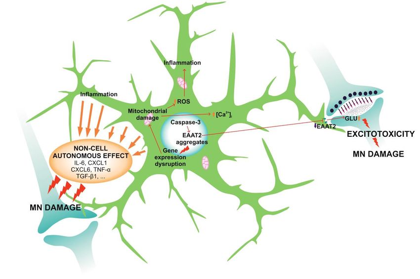
oxygen species (ROS) ([114]; Figure 1).excessive and pathological neuronal stimulation, which disrupts ionic homeostasis in the neurons.
This process is termed glutamate excitotoxicity and contributes to MNs damage and death in ALS
[112,113,115]. In both mice and humans, the loss of EAAT2 proteins does not appear until the
symptomatic stage
J. Clin. Med. 2020, of the disease [116], which means that it is slightly delayed relative to the changes
9, 261 8 of 47
in astrocytes towards reactive phenotype [113].
DuringALS
Figure1.1.During
Figure ALSastrocytes
astrocytesundergo
undergopathological
pathologicalchanges,
changes,which
whichaffect
affecttheir
theirphysiological
physiological
functions and lead to the reduction of motor neuron (MN) survival. ALS astrocytes
functions and lead to the reduction of motor neuron (MN) survival. ALS astrocytes have decreased have decreased
levelsofofEAAT2,
levels EAAT2,which
whicharearecleaved
cleavedbybyCaspase-3
Caspase-3and andcreate
createaggregates
aggregateswithin
withinastrocytic
astrocyticnucleus.
nucleus.
Therefore, the number of EAAT2 receptors on membranes is reduced and the astrocytic ability toto
Therefore, the number of EAAT2 receptors on membranes is reduced and the astrocytic ability
bufferglutamate
buffer glutamate(GLU)
(GLU)from
fromsynapses
synapsesisisimpaired.
impaired.Increased
Increasedlevels
levelsofofGLU
GLUcause
causeexcessive
excessiveneuronal
neuronal
stimulation that damages MNs via the process termed excitotoxicity. The nuclear
stimulation that damages MNs via the process termed excitotoxicity. The nuclear aggregates not only aggregates not
only damage MNs but also astrocytes themselves, as they cause gene
damage MNs but also astrocytes themselves, as they cause gene expression disruption and expression disruption and
subsequently,mitochondrial
subsequently, mitochondrialdamage.
damage.Moreover,
Moreover,astrocytes
astrocytessecrete
secretea arange
rangeofofinflammatory
inflammatorysoluble
soluble
factors in response to the intracellular damage (interleukins, cytokines...), which strongly
factors in response to the intracellular damage (interleukins, cytokines...), which strongly reduce MN reduce MN
survival via the so-called non-cell autonomous
survival via the so-called non-cell autonomous effect. effect.
The decrease in the level of EAAT2 leads to the deterioration of glutamate buffering and its transport
6.2. Reactive Astrogliosis
into the astrocytes. Therefore, glutamate accumulates in the synaptic cleft and it causes excessive and
In neurodegenerative
pathological diseases, such
neuronal stimulation, whichasdisrupts
ALS, astrocytes change their
ionic homeostasis morphology
in the and process
neurons. This functionis
and then glutamate
termed become reactive in response
excitotoxicity and to various stimuli,
contributes to MNs fordamage
example soluble
and deathfactors
in ALSthat are secretedIn
[112,113,115].
by microglia
both mice and[106]. This process
humans, the loss comprises of extensive
of EAAT2 proteins doesmolecular
not appearchanges
until thethat lead to astrocyte
symptomatic stage of
hypertrophy and proliferation and to secretion of pro- and anti-inflammatory
the disease [116], which means that it is slightly delayed relative to the changes in astrocytes towards cytokines,
chemokines, interferons,
reactive phenotype and growth factors, together with components of extracellular matrix [117].
[113].
Such activation has a dual effect. It limits the spread of the lesion and restricts ongoing inflammation
by6.2. Reactive Astrogliosis
preventing infiltration of activated immune cells into the surrounding tissue and, thus, reduces
subsequent neuronal degeneration
In neurodegenerative diseases,[118].
suchOn as the contrary,
ALS, astrocytesthechange
modifications of extracellular
their morphology matrix,
and function
which are an essential part of reactive astrogliosis and glial scar formation, contribute
and then become reactive in response to various stimuli, for example soluble factors that are secreted to the
inhibition of axonal
by microglia [106].regeneration
This process and growth [117].
comprises of extensive molecular changes that lead to astrocyte
hypertrophy and proliferation and to secretionhave
However, the activated astrocytes in ALS slightly
of pro- different properties,
and anti-inflammatory which are
cytokines, common
chemokines,
for astrocytes from human sALS and fALS and murine models [109,119]. Astrocytes that
interferons, and growth factors, together with components of extracellular matrix [117]. Such activation are isolated
from
has amSOD1
dual effect. murine
G93A
It limitsmodels have
the spread of higher proliferative
the lesion and restricts potential
ongoingininflammation
vitro than the by wild-type
preventing
astrocytes [120] and the first reactive cells appear before the disease symptoms
infiltration of activated immune cells into the surrounding tissue and, thus, reduces subsequent manifestation and
neuronal degeneration [118]. On the contrary, the modifications of extracellular matrix, which are an
essential part of reactive astrogliosis and glial scar formation, contribute to the inhibition of axonal
regeneration and growth [117].J. Clin. Med. 2020, 9, 261 9 of 47
However, the activated astrocytes in ALS have slightly different properties, which are common
for astrocytes from human sALS and fALS and murine models [109,119]. Astrocytes that are isolated
from mSOD1G93A murine models have higher proliferative potential in vitro than the wild-type
astrocytes [120] and the first reactive cells appear before the disease symptoms manifestation and
MN degeneration [45]. The mSOD1G93A astrocytes are larger than those in healthy tissue, with
more hypertrophied processes [109]. They express markers that are typical for astrogliosis as well
as markers of immature astrocytes, such as high levels of non-filamentous glial fibrillary acidic
protein (GFAP) [108,109]. In addition, the expression of connexin 43 (Cx43), which is generally
elevated in reactive astrocytes [121], shows a similar increase in mSOD1G93A astrocytes. During the
pre-symptomatic stages of the ALS disease, Cx43 in astrocytes is only slightly increased above the
physiological level. This elevation becomes more substantial during disease progression [116,120,122].
In addition to the above-mentioned proteins and changes, mSOD1G93A astrocytes overexpress the α2
subunit of Na+ /K+ ATPase [123], which directly interacts with astrocytic glutamate transporters and
affects their functioning via an electrochemical gradient [124,125]. How this change in Na+ /K+ ATPase
expression is affected by the reduction of EAAT2 levels (and vice versa) remains to be elucidated.
However, Gallardo and colleagues proposed that the level of α2-Na+ /K+ ATPase is elevated in response
to mitochondrial damage, and it is supposed to anticipate reduced ATP levels within the astrocytes
by the stimulation of mitochondrial respiration. Subsequent to an increase in cellular respiration,
ROS levels increase within the astrocytes and activate an inflammatory response, causing non-cell
autonomous MN degeneration [123].
6.3. Non-Cell Autonomous Effect
Activated astrocytes lack the ability to support the survival and recovery of MNs [106,126]. In fact,
they do the exact opposite. They increase MN damage by the so-called non-cell autonomous effect, which
has been confirmed in several studies using astrocyte-conditioned media for MN cultivation. Results
from both in vitro and in vivo studies with murine (SOD1G93A,G37R,G86R , C9ORF72, and TDP-43A315T
mutants) and human tissue showed significantly declined neuronal survival [109,127–132]. This
process is mediated by astrocyte-specific soluble factors. Several molecules were identified that affect
the physiological functioning of MNs in ALS, such as cytokines or growth factors (IL-6, CXCL1, 10 and
12, tumor necrosis factor-alpha (TNF-α) or transforming growth factor-beta (TGF-β1)). These molecules
are upregulated in ALS astrocytes (together with prostaglandin D2 receptor, Sonic hedgehog (SHH),
SHH-responsive genes, etc.) and they are secreted to the surrounding tissue [74,120,131,133–135], as
in Figure 1. They cause alterations in MN morphology, namely smaller cellular bodies and shorter
axons [74,131]. Apart from morphological changes, these molecules cause axonal swelling and the
accumulation of mSOD1 and ubiquitin-positive aggregates in axons and somata of MNs [74,131]. The
aggregates appear even before the onset of disease symptoms and their level continues to rise during ALS
development [116], which corresponds with the progress of reactive astrogliosis. The mSOD1 protein is
suspected of contributing to MN degeneration via the impairment of mitochondrial functions (hereby
the intracellular levels of ROS and Ca2+ are increased and trigger an MN inflammatory response—for a
review see [136]), together with increasing nitrosative stress [127–129,132,137]. Damaged mitochondria
then become permeable, which leads to the release of ‘pro-cell death factors’, and subsequently to MN
necroptosis [138]. Aside from MNs, astrocytic soluble factors also affect the functioning of other cell
types, such as microglia, and regulate their immunological responses [139].
6.4. Astrocytes in C9ORF72 and TDP-43 Pathology
Similarly to the mSOD1 astrocytes, human and murine-derived astrocytes with C9ORF72 or
TDP-43 pathology also show changed physiological properties and affect functioning of surrounding
cells, especially MNs. The expansion of a non-coding GGGGCC repeats in C9ORF72 in astrocytes
strongly affects RNA metabolism. The transcription of the hexanucleotide repeat leads to the formation
of aggregates of poly-proline-arginine peptides, which bind other mRNAs within astrocytic nucleus.J. Clin. Med. 2020, 9, 261 10 of 47
These aggregates then further affect- and often block-RNA splicing and nuclear export and, subsequently,
protein transcription as well [140–142].
These changes in protein translation lead to a reduction of the metabolic flexibility of C9ORF72
astrocytes, together with an inhibition of proteasome functions and autophagy pathways and eventually
to the activation of heat shock protein (HSP)-response [20,143,144]. Allen and colleagues also found a
reduced ability of the astrocytes to metabolize glycogen and thus to utilize cellular energy sources [145].
Another change that is common for both mSOD1 and C9ORF72 astrocytes is the reduced expression
of glutamate transporters, which causes a reduction in glutamate buffering, leads to glutamate
accumulation in synaptic clefts, and causes excitotoxicity to MNs [146]. Similar changes in glutamate
signalling were described also in C9ORF72 Drosophila model of ALS [147]. C9ORF72 astrocytes
contribute to the MN degeneration in the ALS also via the release of soluble factors (non-cell autonomous
effect), as mentioned above [148,149]. Aside from single soluble molecules, C9ORF72 astrocytes release
extracellular vesicles, containing specific microRNAs, which (after contact with several specific targets,
such as semaphorin proteins) cause axonal retraction and worsen overall MN survival [150].
On the contrary, the information regarding the role of astrocytes with mutated TDP-43 in ALS
is unclear. It was showed that, within astrocytes, intracellular cytoplasmic inclusions, called stress
granules, are formed. Their formation increases with the time and eventually leads to the cell
death [151–154]. These granules consist of insoluble phosphorylated TDP-43 protein together with
ubiquitin and α-synuclein [151,155,156]. Interestingly, the changes in TDP-43 expression and its
solubility were also observed in mSOD1G93A model of ALS, where TDP-43 created similar cytoplasmic
aggregates [157], as well as in the glial cells of Drosophila TDP-43D169G, G298S, A315T, N345K model of
ALS [158]. Similarly to the other ALS models, the TDP-43M337V astrocytes show increased oxidative
damage thanks to the impairment of glutathione antioxidant function [159] and their phenotype
changes towards the reactive one [160,161], which correlates with the global inflammation and
increase in the levels of components of the innate immune complement system in TDP-43Q331K, A315T
pathology [162,163]. The reactive astrocytes then respond with an upregulation of small HSPs [164],
which usually serves as a protective and stress response to cellular damage [165]. In addition, the
number of astrocytes in the motor cortex of TDP-43A315T murine models is higher than in healthy
tissue [163].
TDP-43 cells were shown to contribute to MNs degeneration via the already-discussed non-cell
autonomous effect, which is in agreement with the results from different ALS murine models [58,134],
as well as Drosophila model [166]. However, the role of TDP-43 astrocytes in MN degeneration is not
so clear. Several groups obtained different results. Their results demonstrated that when co-cultured
with MNs TDP-43M337V, A315T astrocytes do not show a non-cell autonomous effect and are not toxic to
MNs [152,153].
Taken together, astrocytes play an important role in the pathology of ALS in human as well as in
murine tissue. They affect numerous cell types, but mainly MNs, whose survival is strongly reduced
due to astrocytic influence. As the role of astrocytes in ALS is not fully understood, these cells represent
an important target for further research towards possible therapy development.
7. Microglia
As the immune-competent cells of the brain and spinal cord, microglia play an important role in
maintaining normal CNS function. They colonize the brain early in development and transform into a
highly ramified phenotype, constantly screening their environment. Microglia are activated by any
kind of pathological event or change in the homeostasis of the CNS. The activation process is diverse
and it depends on the context and type of the stressor/pathology. They influence the pathological
outcome or response, owing to the release of plenty of substances, e.g., cytokines, chemokines, or
growth factors.
Microglia become activated in all instances of ALS and there are numerous studies that have
confirmed the presence of microglial activation at the site of MN damage in both ALS patients [167,168]J. Clin. Med. 2020, 9, 261 11 of 47
and mSOD1 transgenic mice. Furthermore, increased microglial activation in the motor cortex
has been shown to correlate with the severity of upper MN degeneration signs [168]. Studies
using animal models indicate that in vivo resident microglia increase their number with the disease
progression and their activation states represent a continuum between the two classical microglial
phenotypes—neuroprotective M2 and toxic M1 [169,170], see Figure 2. In agreement with the occurrence
J. Clin. Med. 2020, 9, x FOR PEER REVIEW 12 of 49
of two different phenotypes on the basis of their morphology, Ohgomori and his team [171] described
[171]
differentdescribed
types ofdifferent
microgliatypes
in mSOD1 G93A during
of microglia in mSOD1
ALS G93A during ALS
progression. Theprogression.
type “R1” was The typein
found “R1”
the
was found in the early stage and it was poorly branched with short processes. “R2”
early stage and it was poorly branched with short processes. “R2” type was similar to “R1” and onlytype was similar
to “R1” andoccurred
transiently only transiently occurred
in the middle stageinofthe
themiddle
disease.stage of the disease.
The microglia The microglia
appearing in the endappearing in
stage, called
the end
“R3”, stage, called
exhibited short “R3”, exhibited
and thick shortand
processes andlarge
thickcell
processes and large cell bodies [171].
bodies [171].
2. M2
Figure 2.
Figure M2 and
andM1M1microglia
microgliarelease certain
release molecules
certain thatthat
molecules are are
ableable
to influence the survival
to influence of MNs.
the survival of
The molecules and their effect depend on the phenotype of microglia. M1 are toxic, while M2
MNs. The molecules and their effect depend on the phenotype of microglia. M1 are toxic, while M2 microglia
are protective.
microglia are protective.
7.1. Microglia in SOD1 Pathology
7.1. Microglia in SOD1 Pathology
Microglia exhibit an anti-inflammatory profile, an overexpression of IL-10 and, moreover,
Microglia exhibitreceptor
attenuated Toll-like an anti-inflammatory profile, toanaoverexpression
2 (TLR2) responses of IL-10
controlled immune and, moreover,
challenge, in the
pre-symptomatic stage of SOD1-mediated ALS [172]. At the disease onset, the expression in
attenuated Toll-like receptor 2 (TLR2) responses to a controlled immune challenge, the
of M2
pre-symptomatic stage of SOD1-mediated ALS [172]. At the disease onset, the
markers Ym1 and CD206 was upregulated in the lumbar spinal cords of ALS mice, which indicatedexpression of M2
markers Ym1 and
that microglia CD206
at this wasdisplay
stage upregulated
an M2inphenotype.
the lumbar spinal cords of
Eventually, in ALS mice,phase,
the final which microglia
indicated
that microglia at this stage display an M2 phenotype. Eventually, in the final phase,
expressing high levels of NOX2, seem to prevail. NOX2 is the subunit of NADPH oxidase expressed by microglia
expressing high levels of NOX2, seem to prevail. NOX2 is the subunit of NADPH oxidase expressed
macrophages and is considered an M1 prototypic marker [173]. As the disease progresses, mSOD1G93A
by macrophages and is considered an M1 prototypic marker [173]. As the disease progresses,
expressing microglia undergo phenotypic transformation. More specifically, when co-cultured with
mSOD1G93A expressing microglia undergo phenotypic transformation. More specifically, when
MNs early stage M2 microglia enhance MN survival, whereas end-stage M1 microglia show toxic
co-cultured with MNs early stage M2 microglia enhance MN survival, whereas end-stage M1
properties, which increases neuronal death [170]. In addition, mSOD1 microglia exhibit increased
microglia show toxic properties, which increases neuronal death [170]. In addition, mSOD1
expression of molecular players of the endoplasmic reticulum (ER) stress pathway [174], such as C/EBP
microglia exhibit increased expression of molecular players of the endoplasmic reticulum (ER) stress
homologous protein (CHOP), which might be involved in their toxic phenotype.
pathway [174], such as C/EBP homologous protein (CHOP), which might be involved in their toxic
One of the hallmarks of ALS is neuroinflammation, and M1 microglia seem to be hyper-reactive
phenotype.
to inflammatory stimuli [175]. Nuclear factor-kappa β (NF-κβ) protein, which plays a key role in
One of the hallmarks of ALS is neuroinflammation, and M1 microglia seem to be hyper-reactive
regulation of the inflammation, is upregulated in the spinal cords of ALS patients and mSOD1G93A
to inflammatory stimuli [175]. Nuclear factor-kappa β (NF-κβ) protein, which plays a key role in
mice. It was shown that selective NF-κβ inhibition in ALS astrocytes is not sufficient for rescuing
regulation of the inflammation, is upregulated in the spinal cords of ALS patients and mSOD1G93A
MNs from their death. However, the localization of NF-κβ activity and subsequent deletion of NF-κβ
mice. It was shown that selective NF-κβ inhibition in ALS astrocytes is not sufficient for rescuing
signaling in microglia rescued MNs from microglial-mediated death in vitro and extended the survival
MNs from their death. However, the localization of NF-κβ activity and subsequent deletion of
of ALS mice by impairing pro-inflammatory microglial activation. On the contrary, the constitutive
NF-κβ signaling in microglia rescued MNs from microglial-mediated death in vitro and extended
activation of NF-κβ, selectively in wild-type microglia induced gliosis and death of MNs in vitro
the survival of ALS mice by impairing pro-inflammatory microglial activation. On the contrary, the
constitutive activation of NF-κβ, selectively in wild-type microglia induced gliosis and death of MNs
in vitro and in vivo [176]. The stimulation of excessive extracellular production of superoxide is
another mechanism of mSOD1 damage that is produced by microglia. The SOD1 enzyme is not just
catabolic, but it can also regulate NADPH oxidase–dependent superoxide production due to binding
Rac1. This protein is a small GTPase controlling the NADPH oxidase activation. Inhibition ofJ. Clin. Med. 2020, 9, 261 12 of 47
and in vivo [176]. The stimulation of excessive extracellular production of superoxide is another
mechanism of mSOD1 damage that is produced by microglia. The SOD1 enzyme is not just catabolic,
but it can also regulate NADPH oxidase–dependent superoxide production due to binding Rac1. This
protein is a small GTPase controlling the NADPH oxidase activation. Inhibition of GTPase activity due
to mSOD1G93A results in the production of high levels of extracellular superoxide [177].
7.2. Microglia in C9ORF72 Pathology
The microglial function in ALS, which is associated with the mutations in C9ORF72, also undergoes
some disturbances. Once it was known that the C9ORF72 mutation results in decreased expression
levels of this protein in ALS patients [31], it led to speculation that the loss of the C9ORF72 protein
function might contribute to the disease onset/progression. The protein that is encoded by C9ORF72 is
probably a guanine exchange factor for one or more not-yet-identified G proteins. When inactivated
in mice, abnormal microglia and age-related neuroinflammation occurs, which provides evidence
that non-cell-autonomous, microglia-mediated inflammation might contribute to ALS [62,64,178].
Microglia have a proinflammatory phenotype with increased expression of cytokines IL-6 and IL-1β [62].
C9ORF72-knockout mice lacking the expression of C9ORF72 in MNs, however, do not develop MN
degeneration or disease. It seems that the expression of C9ORF72 in innate immune cells, including
macrophages and microglia, is not sufficient to cause MND in a mouse model, unless C9ORF72 is also
expressed in MNs.
Impaired regulation of autophagy and enhanced inflammation can be caused not only by mutations
in C9ORF72, but also in other genes, e.g., OPTN, TBK1, SQSTM, or VCP. The inhibition of autophagy
in mSOD1G93A MNs in transgenic mice accelerated the disease onset, but prolonged lifespan [179].
However, autophagy inhibition in glia might have different effects than in neurons. Additionally, the
responses may vary among cell types. For more details, see the review article [180].
7.3. Microglia in TDP-43 Pathology
In addition to other animal models, transgenic TDP-43Q331K mice also show increased microglial
activation and MN degeneration [162]. In more than 90% of ALS patients, cytoplasmic TDP-43
aggregates that accumulated in spinal cord were observed post mortem [29]. The translocation of
TDP-43 from nucleus to cytoplasm is thought to be a part of the ALS pathogenesis [181], as the
aggregates are strong triggers of microglia immune responses. The NLRP3 inflammasome is an
important part of the innate immune system activated by protein aggregates—an intracellular signaling
complex [182–185]. The activation of this complex requires a priming signal and it results in the
upregulation of NLRP3 and cytokine precursors: pro-IL-1β and pro-IL-18. This process is followed by
an activation step, which involves the recruitment of the inflammasome adapter (apoptosis-associated
protein containing a caspase recruitment domain), activation of the caspase-1 protease, and, finally, the
cleavage and release of IL-1β and IL-18 [186,187]. Deora, V. introduced the first report of increased
inflammasome gene expression in TDP-43Q331K mice [187]. Their study also demonstrates that TDP-43
proteins (as well as SOD1) induce inflammasome activation in primary microglia.
TDP-43 is able to induce microglial activation via interaction with the CD14 receptor on the cell
surface and initiating a proinflammatory cascade and neuronal cytotoxicity. When influenced by
activated TDP-43, microglia start to express NOX2 and produce TNF-α and IL-1. Microglia that are
activated by TDP-25 cause the death of MNs when co-cultured together, but TDP-25 and TDP-43 do
not seem to be toxic to neurons on their own, only when microglia are present [188]. These results
are consistent with the previous work of these authors, where they demonstrated these results in
mSOD1G93A mice [189].
Interestingly, there are also studies showing that microglia in the environment of MNs can even
have a positive impact on their survival [190]. Researchers using the mSOD1 model usually propose the
elimination of microglia as a beneficial strategy [176]. Nevertheless, Spiller and colleagues suggested
that, in TDP-43 pathology (using rNLS8 mutation), it might be more efficient to find ways of how toJ. Clin. Med. 2020, 9, 261 13 of 47
encourage appropriate microglia-mediated inflammation, to clear pathological TDP-43 proteins and
help axonal regeneration during the onset/progress of ALS [190].
In our point of view, the Spiller suggestion for TDP-43 pathology seems to be promising. However,
as they pointed out, their model, unlike mSOD1, has wild-type microglia and intact BBB. Based on this
fact, we hypothesize that the possible modulation of microglia towards their anti-inflammatory state
could be more beneficial in SOD1 and C9ORF72 mutants. In a recent study [191] while using SOD1G93A ,
they ascribe the neuroprotective effect to the shift of microglia from their pro- to anti-inflammatory
state and similarly, study using C9ORF72 mutant carriers [192] showed that immunotherapy can
inhibit the activation of microglia and, thus, prevent the disease onset. Overall, it seems that the ability
to modulate the character of microglia could make an impact on the onset/progress of ALS whether it
is the shift to their neuroprotective state or encouragement of appropriate microgliosis.
8. NG2 Glia
The adult CNS contains a widely distributed population of oligodendrocyte progenitor cells
(OPCs), which are also called NG2 glia. These cells have the capacity to replace injured cells or those
lost due to age-related degeneration. They differentiate not only into myelinating oligodendrocytes,
but also into protoplasmic astrocytes, which are specifically localized in the gray matter [193]. It has
also been suggested that NG2 cells can differentiate into neurons, which might possibly happen in the
different brain regions. Two groups described NG2 cell ability to give rise to pyramidal-like neurons in
dorsal and ventral forebrain cortext [194,195]. Alternatively, Aguirre and colleagues showed that they
express neuronal markers in the subventricular zone or differentiate into GABAergic interneurons in
the hippocampus [196].
Following CNS injury, NG2 glia start to proliferate extensively and their differentiation into
oligodendrocytes is enhanced [100,197]. However, following neurodegeneration, NG2 glia only give
rise to oligodendrocytes [197], while their rate of proliferation remains high during the whole progress
of the disease [100]. The proliferation of NG2 cells in mSOD1 murine models differs among brain
regions and it is more prominent in the ventral gray matter, where the proliferation increases before
the onset of disease symptoms and continues to rise during ALS progression. Interestingly, NG2 glia
of ventral white- and dorsal gray matter display a lower proliferation rate, which occurs in the later
stages of ALS progression [100]. When compared to NG2 cells in healthy white matter, the number of
NG2 cells in TDP-43 mutant mice is markedly increased and they have a different morphology, which
is characterized by enlarged cellular soma and lesser processes [198]. Overall, there is little information
regarding NG2 glial contribution to ALS pathology, therefore more information should be obtained,
and more research undertaken to understand their role in this disease.
9. Oligodendrocytes
Oligodendrocytes in the CNS are responsible for the myelination of axons. Myelin plays an
important role, not only in the electrical insulation of the axons, but also in their provision with
trophic and metabolic support [199]. Oligodendrocytes and myelinated axons are metabolically
coupled. Oligodendrocytes are able to transform glucose to lactate or pyruvate and provide it
to neurons via monocarboxylic acid transporters (MCT1, MCT2) [200,201]. N-methyl-D-aspartate
(NMDA) receptors conduct the regulation of this process, which are present on oligodendrocytes and
are able to recognize neuronal activity, incorporate additional glucose transporters, and thus increase
the import of glucose [202]. Oligodendrocytes and myelin are not only central to the pathological
mechanisms of inflammatory diseases (e.g., multiple sclerosis), but they may also play an important
role in several neurodegenerative diseases, such as ALS The importance of oligodendrocytes in disease
onset/progression was suggested in SOD1-, C9ORF72-, and TDP-43 pathologies.J. Clin. Med. 2020, 9, 261 14 of 47
9.1. Oligodendrocytes in SOD1 Pathology
Oligodendrocytes are strikingly affected during ALS and their degeneration seems to precede
MN death in the mSOD1G93A mouse model [100,203]. Additionally, a rapid proliferation of
oligodendrocyte progenitors has been shown in the spinal cord of SOD1G93A mice; however, newly
derived cells failed to mature and replace degenerated oligodendrocytes. Consequently, axons of MNs
remained demyelinated [100,203]. It was demonstrated that the reduction in mSOD1G93A synthesis in
oligodendrocytes, which occurs during their early maturation, produces a more substantial delay in
the disease onset [100] than that observed in MNs in mSOD1G37A [99,204]. Oligodendrocytes support
MNs by providing the direct supply of the energy metabolite lactate to axons due to the action of
MCT1, as mentioned above. In the ALS mouse model, the mSOD1G93A impairs the expression of MCT1
in oligodendrocytes and a similar reduction in MCT1 accumulation can be found in sALS [200].
In the gray matter of the spinal cord of SOD1G93A ALS mice, mature oligodendrocytes extensively
degenerate, prior to the onset of disease symptoms [100,203]. The selective deletion of mSOD1G93A
from oligodendrocytes markedly delays the onset and prolongs the survival of mice [100]. In vitro
studies showed that oligodendrocytes that are obtained from ALS patients are able to induce MN death
when co-cultured together [205]. These findings suggest that the dysfunction of mature oligodendroglia
that is caused by mSOD1 can have a critical role in the ALS.
One of the studies of mature oligodendrocytes in SOD1 pathology was carried out while using a
zebrafish model [206], in which the mSOD1G93A was only selectively expressed in oligodendrocytes.
They found that mSOD1 directly induces the degeneration of oligodendroglia, via the disruption of
myelin sheaths as well as the downregulation of MCT1, which resulted in the degeneration of the spinal
cord MNs. The dysfunction of oligodendrocytes was also associated with behavioural abnormalities,
learning impairments, and motor defects in the early symptomatic stage of ALS. In addition, treating
the fish with K+ channel inhibitors rescued abnormalities in behaviour, but, unfortunately, without
rescuing the expression of MCT1. These results suggest that myelin disruption induces abnormalities in
behaviour, independently of MCT1. Overall, the dysfunction of mature oligodendrocytes is presumably
sufficient to induce MN degeneration [206].
It was also shown that oligodendrocytes expressing mSOD1G93A are able to induce
electrophysiological changes in wild type MNs, ultimately leading to MN death [205]. These data are
consistent with the findings of Pieri and colleagues [207], who reported that increased persistent Na+
currents are selectively altered and cause hyperexcitability. Data from both mentioned studies [205,207],
while using an in vitro approach or mouse model, confirmed that cortical hyperexcitability happens
to be one of the first neuronal alterations detected in patients with ALS [208] before the symptom
onset [209]. Marcuzzo and colleagues, who worked with cultured neurons from SOD1G93A mice aged
one day, also just recently confirmed this [210].
9.2. Oligodendrocytes in C9ORF72 and TDP-43 Pathology
TDP-43 and FUS-positive inclusions were found in oligodendrocytes of ALS patients in
post mortem analyzed tissue [211], and their presence suggests an impairment of autophagy in
oligodendrocytes. A study comparing both human and mouse ALS oligodendrocytes was performed
since it is not obvious whether observations obtained in mouse models of ALS hold true in a
wide spectrum of ALS patients [205]. The in vitro study showed that oligodendroglia successfully
differentiate from mouse neural progenitor cells (NPCs) and human iPSCs, and neural progenitor cells
iNPCs [130], from both ALS- and non-ALS samples. According to these results, the ability of ALS
oligodendrocytes to pass the toxicity on MNs in vitro does not depend on their origin. The reduction of
mSOD1 in OPC can rescue the toxicity, but the reduction of mSOD1 in differentiated oligodendrocytes
does not have the same effect. The toxicity of cells carrying C9ORF72 repeat expansion shows no
response to the reduction of SOD1 suggesting their SOD1 independence [205]. Moreover, they also did
not display dysfunction in lactate release. These findings suggest that this mutation defines a specificYou can also read


