The Microbiota and the Gut-Brain Axis in Controlling Food Intake and Energy Homeostasis
←
→
Page content transcription
If your browser does not render page correctly, please read the page content below
International Journal of
               Molecular Sciences

Review
The Microbiota and the Gut–Brain Axis in Controlling Food
Intake and Energy Homeostasis
Marina Romaní-Pérez † , Clara Bullich-Vilarrubias † , Inmaculada López-Almela, Rebeca Liébana-García                                            ,
Marta Olivares and Yolanda Sanz *

                                          Microbial Ecology, Nutrition & Health Research Unit, Institute of Agrochemistry and Food Technology,
                                          National Research Council (IATA-CSIC), 46980 Valencia, Spain; marina.romani@iata.csic.es (M.R.-P.);
                                          clarabullich@iata.csic.es (C.B.-V.); inma.lopez@iata.csic.es (I.L.-A.); reliegar@iata.csic.es (R.L.-G.);
                                          m.olivares@iata.csic.es (M.O.)
                                          * Correspondence: yolsanz@iata.csic.es
                                          † These authors contribute equally to this work.

                                          Abstract: Obesity currently represents a major societal and health challenge worldwide. Its preva-
                                          lence has reached epidemic proportions and trends continue to rise, reflecting the need for more
                                          effective preventive measures. Hypothalamic circuits that control energy homeostasis in response
                                          to food intake are interesting targets for body-weight management, for example, through interven-
                                          tions that reinforce the gut-to-brain nutrient signalling, whose malfunction contributes to obesity.
                                          Gut microbiota–diet interactions might interfere in nutrient sensing and signalling from the gut to
                                the brain, where the information is processed to control energy homeostasis. This gut microbiota–

                                          brain crosstalk is mediated by metabolites, mainly short chain fatty acids, secondary bile acids or
Citation: Romaní-Pérez, M.;
                                          amino acids-derived metabolites and subcellular bacterial components. These activate gut–endocrine
Bullich-Vilarrubias, C.; López-Almela,
                                          and/or neural-mediated pathways or pass to systemic circulation and then reach the brain. Feeding
I.; Liébana-García, R.; Olivares, M.;
                                          time and dietary composition are the main drivers of the gut microbiota structure and function.
Sanz, Y. The Microbiota and the
                                          Therefore, aberrant feeding patterns or unhealthy diets might alter gut microbiota–diet interactions
Gut–Brain Axis in Controlling Food
Intake and Energy Homeostasis. Int.
                                          and modify nutrient availability and/or microbial ligands transmitting information from the gut to
J. Mol. Sci. 2021, 22, 5830. https://     the brain in response to food intake, thus impairing energy homeostasis. Herein, we update the scien-
doi.org/10.3390/ijms22115830              tific evidence supporting that gut microbiota is a source of novel dietary and non-dietary biological
                                          products that may beneficially regulate gut-to-brain communication and, thus, improve metabolic
Academic Editors: Rosalía                 health. Additionally, we evaluate how the feeding time and dietary composition modulate the gut
Rodríguez-Rodríguez and                   microbiota and, thereby, the intraluminal availability of these biological products with potential
Cristina Miralpeix                        effects on energy homeostasis. The review also identifies knowledge gaps and the advances required
                                          to clinically apply microbiome-based strategies to improve the gut–brain axis function and, thus,
Received: 6 April 2021
                                          combat obesity.
Accepted: 26 May 2021
Published: 29 May 2021
                                          Keywords: microbiota; gut–brain axis; nutrient sensing; food intake and obesity

Publisher’s Note: MDPI stays neutral
with regard to jurisdictional claims in
published maps and institutional affil-
                                          1. Gut and Brain Control of Energy Homeostasis
iations.
                                                The gastrointestinal tract is a huge sensory organ able to transmit nutrient-related
                                          information to the brain where diverse endocrine and neural inputs converge to ultimately
                                          control feeding behaviour and whole-body energy homeostasis through efferent outputs.
Copyright: © 2021 by the authors.
                                          Nutrient sensors are receptors that bind molecules derived from the macronutrient diges-
Licensee MDPI, Basel, Switzerland.
                                          tion. They are located in enteroendocrine cells (EECs), which are the primary chemosensory
This article is an open access article
                                          cells in the gut, and are in direct contact with the luminal environment [1]. The activation
distributed under the terms and           of nutrient sensors of EECs initiates the secretion of gut hormones that in turn trigger the
conditions of the Creative Commons        downstream processes required to maintain energy homeostasis postprandially. The most
Attribution (CC BY) license (https://     extensively studied gut hormones are cholecystokinin (CCK), gastric inhibitory polypep-
creativecommons.org/licenses/by/          tide (GIP), mainly secreted in the upper part of the intestine, and glucagon-like peptide-1
4.0/).                                    (GLP-1) and peptide tyrosine tyrosine (PYY), mainly secreted in the distal part [2]. EECs

Int. J. Mol. Sci. 2021, 22, 5830. https://doi.org/10.3390/ijms22115830                                            https://www.mdpi.com/journal/ijms
Int. J. Mol. Sci. 2021, 22, 5830 2 of 35
specifically sense carbohydrates, proteins and lipids through a diverse repertoire of nutrient
sensors. Among others, sodium/glucose cotransporter 1 (SGTL1) mediates carbohydrate
sensing, mainly sensed in the form of glucose, and, then, induces GIP and GLP-1 secretion;
calcium-sensing receptor (CaSR) senses dietary amino acids and, then, secretes CCK and
GLP-1; and G-protein coupled receptor (GPR) 120 (FFAR4), GPR40 (FFAR1) and GPR119
sense products from lipid digestion and induce the secretion of CCK, GIP, GLP-1 and
PYY [1,3]. EECs are in close proximity to vagal afferent nerve terminals in the lamina
propria, which express intestinal hormone receptors. Enteroendocrine hormones secreted
after a meal activate nutrient-sensing signalling via endocrine routes, when gut hormones
reach the brain or other organs through systemic circulation, or via paracrine routes, when
hormones stimulate vagal afferents nearby in the intestinal mucosa and, then, the signal
reaches the brain.
The arcuate nucleus (ARC) of the hypothalamus is accessible to humoral signals since
is not fully protected by the blood–brain barrier [4]. The ARC contains two subpopulations
of neurons; those expressing anorexigenic propiomelanocortin (POMC), the precursor
of α-melanocyte-stimulating hormone (αMSH), and the cocaine and amphetamine reg-
ulated transcript (CART); and those neurons expressing the agouti gene-related peptide
(AgRP) and neuropeptide Y (NPY) [5]. The activation of the POMC/CART neurons after
feeding induces the release of α-melanocyte stimulating hormone (α-MSH) that binds
to melanocortin 4 receptor (MC4R) in the paraventricular nucleus (PVN) where the in-
formation is integrated to suppress food intake and regulate body weight [6]. Thus, the
increased levels of gut hormones such as CCK, GLP1 or PYY after a meal reach, via en-
docrine routes, the ARC to suppress food intake [7]. Via paracrine routes, gut hormones
can stimulate vagal afferents [8], which are bipolar neurons whose somas converge in the
nodose ganglion and their proximal extensions terminate in the nucleus of the solitary tract
(NTS) in the brainstem [9]. This, together with the hypothalamus, represents an important
integrative gut–brain hub. The NTS also contains POMC neurons and, through monoamine
neurotransmission, transmits sensory information to upstream brain areas including the
PVN and the dorsal vagal complex (DVC) that send efferent outputs involved in the vago–
vagal reflex [5]. In summary, there are different paths for transmitting nutrient signals
to the brain. The hypothalamus senses nutrients through the action of enteroendocrine
hormones released in the intestine, which reach the brain by humoral pathways [10–12],
that is through the circulatory system, or by paracrine pathways, that is by activating the
nerve terminals of intestinal vagal afferents, which are the main focus of the present review.
The increased intake of energy-dense and palatable foods impairs the brain circuits
controlling energy homeostasis, whose deficient response to nutrient signals alters feeding
behaviour, which contributes to obesity. Accordingly, the restoration of nutrient signalling
via the gut–brain axis represents a promising strategy to improve the central control of
energy homeostasis in response to meals and, thus, help combat obesity [13–15]. The gut
microbiota is a biological factor that might directly or indirectly influence nutrient-sensing
and, theoretically, its modulation could aid in the restoration of gut-to-brain communication
and maintaining energy homeostasis. The role of gut microbiota in obesity has been proven
through faecal transplantation experiments, which produce the metabolic phenotype of
the donor in the recipient organism [16]; however, the mechanisms by which microbiota
influence energy homeostasis and body-weight regulation are not yet fully understood.
Western diets, rich in saturated fat and simple sugars, alter the gut ecosystem reducing
bacterial diversity and increasing the abundance of potential pathogens [17]. This, in turn,
could alter the metabolism of macronutrients and, thereby, the ligands available for nutrient
sensors in the intestinal lumen as well as the presence of structural microbial components
that might also act as ligands of sensors that mediate the gut-to-brain communication.
Sensors of microbially produced metabolites and bacterial components are located in EECs,
vagal afferents and, occasionally, in the hypothalamus and can be activated by ligands
reaching the brain through the systemic circulation. These receptors sense microbial-
derived metabolites such as short chain fatty acids (SCFAs), secondary bile acids (BAs) andInt. J. Mol. Sci. 2021, 22, 5830 3 of 35
amino acid-derived metabolites and subcellular bacterial components such as caseinolytic
peptidase B (ClpB), lipopolysaccharide (LPS) or muramyldipeptide (MDP).
Here, we review the role of dietary factors, including feeding patterns and diet
composition, in modulating gut microbiota structure and function and potentially affecting
gut-to-brain nutrient sensing, mainly via endocrine and neural routes. We also compile
evidence of microbial molecules able to modulate specifically hypothalamic-mediated
control of the energy homeostasis, placing special emphasis on their role in regulating food
intake and, thereby, obesity.
2. Circadian Rhythms, Eating Patterns and Gut Microbiota in Energy
Homeostasis Control
Most of the physiological functions display circadian rhythms. At cellular level,
these are governed by clock proteins that generate self-sustained daily oscillations of the
biological processes that allow cells to anticipate and thus to optimally respond and adapt
to environmental variations [18,19]. In mammals, most cells throughout the body express
clock genes that elicit autonomous circadian oscillations [20]. The hypothalamic clock
located in the suprachiasmatic nucleus acts as a master pacemaker that synchronizes the
secondary clocks [21,22]. Eating behaviour and energy metabolism show a well-defined
circadian pattern through the day. In humans and rodents, the timed feeding patterns are
governed by metabolic hormones and nutrients [23] and shift certain secondary clocks
without affecting the suprachiasmatic nucleus, whose circadian oscillations are mainly
governed by light–dark cycles [24,25]. Circadian rhythmicity, specially that affecting eating
behaviour, is required to maintain energy homeostasis. Indeed, humans with disrupted
circadian rhythms have higher risk of developing obesity and type 2 diabetes (T2D) [26,27]
and mistimed eating increases adiposity in rodents [28] and also favours obesity and
increases postprandial glycaemia in humans [29,30].
Circadian rhythmicity is involved in gut microbiota–host interactions. Gut microbiota
play a role in maintaining the circadian rhythms of the host, including those related to
eating patterns and energy homeostasis, and vice versa. The abundance of bacterial species,
as well as of their derived metabolites, vary throughout the day, suggesting associations
between gut microbes and eating behaviours in humans [31]. A recent study by Reitmeier
et al. [32] supports the link between diurnal fluctuations of the gut microbiota and metabolic
health in humans. The authors identified these diurnal oscillations in faecal samples of
three independent large-scale population studies and detected aberrant oscillations in
subjects with metabolic disorders such as obesity or T2D [32].
2.1. Gut Microbiota Influences Circadian Rhythms Affecting Metabolism
The influence of gut microbiota on the host circadian rhythms and on energy metabolism
has been demonstrated in peripheral tissues such as the gut, liver or white adipose tissue. In
mice, gut microbiota is required to maintain the circadian rhythmicity of the expression of
the Toll-like receptors (TLRs), through which the microbiota-associated molecular patterns
(MAMPs) communicate with the intestinal epithelial cells (IECs) of the host [33]. The
absence of gut microbiota impairs the circadian clock of the IEC leading to an increased
synthesis of ileal corticosterone, which impairs systemic metabolic homeostasis. This
is evidence for the role of the microbiota–IEC dialogue oscillation during the day in
metabolic health maintenance [33]. Similarly, germ free mice show altered expression
of circadian-related genes in liver and white adipose tissue [34]. Microbiota-derived
molecules, especially those that activate aryl hydrocarbon receptor (AHR) and pregnane
X receptor (PXR), are crucial to maintain the rhythmicity and sexual dimorphism of the
growth hormone secretion, which maintains rhythmic gene expression and metabolome in
liver in a sex-dependent manner [34].
2.2. Eating Patterns, Gut Microbial Diurnal Oscillations and Energy Homeostasis
The eating rhythms of the host cause microbiota oscillations, which seem flexible
and able to adapt to the nutritional environment. Mice deficient in the clock gene Per1/2Int. J. Mol. Sci. 2021, 22, 5830 4 of 35
or mice submitted to an experimental “jet-lag” show altered eating rhythms throughout
the day linked to aberrant microbiota fluctuations [35]. Time-restricted feeding of Per1/2
knock-out mice partially restores the microbiota oscillations, proving that eating behaviour
is the main driver of microbiota diurnal oscillations [35]. A fat-rich diet also blunts the
cyclical changes of the gut microbiota [36,37] and, consequently, their derived-metabolites,
thus affecting host metabolism. For instance, the fluctuations of species belonging to the
family Lachnospiraceae and their derived metabolite butyrate disappear under a high
fat diet (HFD), which affects hepatic clock genes and metabolism [36]. Additionally, the
spatial distribution of microbes in the gut seems to follow daily fluctuations. In rodents,
bacterial adherence to the intestinal epithelium and their proximity to the mucosal surface
are higher in the dark than in the light phase and, over the course of the day, some bacterial
species show oscillations of epithelial adherence, findings that are dependent on feeding
time [38]. Importantly, the nutrient-induced microbiota oscillations throughout the day
lead to fluctuations of luminal and serum metabolites, such as amino acids and polyamines,
responsible for the circadian hepatic transcriptome required for physiological processes
such as detoxification functions [38]. Eating rhythmicity persists in antibiotic-induced
microbiota depletion [38] but obese-associated hyperphagia might be transmissible through
gut microbiota transplants to germ-free mice; this occurs in hyperphagic TLR5 knock-out
mice [39] although not in ob/ob mice. This suggests that eating rhythmicity is driven mainly
by the host circadian clock machinery driving endocrine hormone secretion, but nutrient
signalling could also be modulated by gut microbiota activated pathways.
Interestingly, the diurnal bacterial growth dynamics, which are controlled by bacterial
quorum sensing, availability of nutrients, gut motility and immunity, apparently overlap
with host-feeding cycles [40]. In this regard, a bacterial growth-based model of appetite
control has been proposed, in which the exponential and stationary growth phases of
the bacteria are associated with satiety-induced signals of the prandial and postprandial
phase, respectively, while the decline growth phase is coupled with hunger-related signals,
defining intermeal intervals [40]. Therefore, to understand how the gut microbiota shapes
eating rhythms it is necessary to investigate the bacterial growth fluctuations parallel
to the generation of dietary and bacterially derived ligands of nutrient sensors able to
centrally control energy homeostasis (such as SCFAs, secondary BAs or amino acid-derived
metabolites—see Section 4 for details). For instance, ClpB, an antigen mimetic of αMSH
produced by Escherichia coli [41], activates host satiety pathways following nutrient-induced
bacterial growth [42]. Similarly, nutrient-induced bacterial growth might enhance the gen-
eration and release of bacterial ligands, such as cell wall and membrane components (e.g.,
lipopolysaccharide, LPS, or muramyldipeptide, MDP) or quorum sensing molecules, which
in turn activate nutrient sensing pathways in the gut. Although the causal link between
the diurnal oscillations of these molecules and feeding rhythms has yet to be proven, LPS
and MDP are known to suppress food intake through immune sensing pathways linked
to satiety and sickness behaviour [43–45]. Through an immune-related cascade in which
LPS and MDP bind to TLR4 and NOD2, respectively, both molecules seem to modulate
GLP-1-mediated signalling [46–48]. Nevertheless, the extent to which nutrient-induced
bacterial growth and the consequent release of LPS or MDP synchronize meal-related host
rhythms under physiological conditions is still unknown. Indeed, the effects of LPS and
MDP on feeding behaviour have mainly been described in the context of bacterial infection,
where these bacterial molecules reach the systemic circulation. However, their role as
modulators of nutrient signalling in the gut, for instance as GLP-1 secretagogues, remains
mostly unexplored. In this regard, LPS could act as an inducer of GLP-1 secretion when
administered orally only if the gut barrier integrity is impaired [46]
3. Diet Composition Influences Gut Microbiota and Gut-to-Brain Nutrient-Sensing
Adherence to a particular dietary pattern, such as a Western, vegetarian or Mediter-
ranean diet, or dietary interventions characterized by large variations in the macronutrients
proportions, such as high protein or high fat (ketogenic) diets, impacts gut microbiotaInt. J. Mol. Sci. 2021, 22, 5830 5 of 35
composition and function [49–51]. The composition in macronutrients of these diets ex-
erts an important effect on the availability of microbially derived ligands of dietary and
non-dietary nature in the luminal content able trigger gut-to-brain sensing routes and
controlling food intake and energy metabolism (summarized in Table 1). The availability of
these ligands depends on multiple biological processes, including the microbiota-mediated
catabolism of ingested nutrients and their absorption by enterocytes.
Most of the simple carbohydrates, proteins and fats are absorbed in the proximal
regions of the small intestine while indigestible complex carbohydrates, the preferred
carbon source of gut microbiota, reach the colon, favouring the growth of anaerobic
bacteria and species diversity. The gut microbiota structure and composition are flexible,
showing rapid adaptations to macronutrient shifts (24–48 h) that remain for short periods
while more permanent changes might require longer adherence to a dietary pattern [52,53].
When the dietary macronutrient composition is high in non- or low-fermentable nutrients
(e.g., lipids or proteins) these macronutrients overwhelm their intestinal absorption and
reach the colon, where they are used by the best adapted microbes with the subsequent
shift in their ability to activate gut nutrient sensing routes.
Although the diet per se modulates the energy metabolism, here we focus on effects
possibly mediated by the interactions between the gut microbiota and the main macronutri-
ents of different diets for which there is substantial scientific evidence. A special emphasis
is placed on those interactions that potentially affect gut-to-brain nutrient sensing and,
thereby, energy homeostasis.
3.1. Western Diets
Over the last two decades, the dietary patterns of modern societies have exhibited
a shift towards a diet low in fibre and high in saturated fats, simple sugars and refined
foods, termed the “Western diet” [54]. The Western diet is frequently associated with
altered eating patterns, leading to hyperphagia and obesity onset. Specifically, the over-
consumption of saturated fats and simple sugars in Western diets contributes to impair
eating behaviour as a consequence of an overstimulation of the nutrient sensing routes that
disturbs how brain senses these nutrient-related signals to control food intake patterns.
Particularly, the intake of dietary fats amplifies meal sizes in humans; this phenomenon is
exacerbated in obese subjects [55] that also showed reduced PYY secretion following a lipid
load [56,57]. Studies in obese individuals reported increased postprandial levels of GLP-1
and CCK and occasional reductions of GLP-1 secretion [58,59] while investigations in
rodents demonstrated that HFD diminished the sensitivity of GLP-1 and CCK contributing
to alter eating behaviour and energy homeostasis [60,61]. In addition to the effects on
eating behaviour, mouse studies also demonstrate that diets rich in saturated fats impair
the enteric detection of glucose required to induce glycogen depots in muscle through a
GLP-1 receptor mechanism in the arcuate NPY-expressing neurons [62].
Sugar-enriched foods also contribute to increasing weight gain as demonstrated by the
meta-analyses of randomised controlled intervention trials and observational studies [63].
Excessive intake of sugar is associated with overeating due to impaired hedonic and
homeostatic brain circuits [64–66] related to defective gut-to brain glucose sensing. In
lean humans, calories from dietary sugars negatively correlate with glucose-induced GLP-
1 secretion and positively with dorsal striatal food cue reactivity to palatable food [67].
Obesity in combination with increased dietary sugars has an additive effect reducing
circulating levels of GLP-1 after a glucose load [68]. Functional magnetic resonance imaging
also reveals that, compared with lean individuals, obese subjects show altered sucrose-
related functional connectivity of lateral hypothalamus and NTS with reward-related brain
areas resulting in reduced sucrose-associated hedonic responses [69] while studies in mice
indicate that the Western diet impairs glucose sensing in POMC-expression neurons [70].
Together with the altered feeding behaviour and energy homeostasis, the Western diet
also decreases the bacterial diversity in the human gut, partly due to its reduced contentInt. J. Mol. Sci. 2021, 22, 5830 6 of 35
of complex carbohydrates [71] and especially if diets are rich in high saturated fatty acids
(SFAs) [72].
Compared with children from rural areas of Africa, European children eating a typical
Western diet showed a higher abundance of Firmicutes with an overrepresentation of
Faecalibacterium, while genera from Bacteroidetes, such as Alistipes and Prevotella, are
increased and decreased, respectively [50]. These associations can reflect a metabolic
adaptation of the intestinal bacteria to a new nutritional environment. How gut microbiota,
through their interactions with Western diet-derived nutrients (i.e., saturated fats and
simple sugars), influences the pool of bacterial-derived metabolites potentially activating
gut nutrient sensing routes to control eating patterns and energy homeostasis is in an early
stage of investigation. Herein, we compile associative studies in humans and mechanistic
studies in rodents which provide evidence of these interactions affecting the central control
of energy homeostasis.
In humans, short-term adaptations of the gut microbiota to an animal-based diet
consist of a predominance of bile-tolerant microorganisms such as Alistipes or Bilophila and
a decrease in bacteria adapted to metabolize dietary plant polysaccharide such as Roseburia,
Eubacterium rectale and Ruminococcus bromii [53]. In vitro human gut simulator assays have
demonstrated the predominance of lipid degradation-related genes and the downregulation
of carbohydrate degradation genes, along with an increase in Gammaproteobacteria and the
genera Alistipes and Bilophila in bacterial communities exposed to a fat-based medium [73].
In mice, a SFAs-enriched diet, especially high in palmitic acid, boosts overgrowth of
Bilophila wadsworthia, which aggravates the HFD-induced metabolic disturbances [74]. The
increased abundance of B. wadsworthia seems to be a secondary consequence of the SFAs
on BAs metabolism [75]. Specifically, the SFAs-enriched diet increases the host production
of BAs conjugated with taurine, which increase the fitness and growth of B. wadsworthia,
while inhibiting other bacteria. B. wadsworthia also exacerbates the HFD-induced decrease
in deoxycholic acid (DCA) and hyodeoxycholic acid (HDCA), secondary BAs that mediate
gut nutrient sensing routes [75] (see Section 4 for details).
Studies in mice fed saturated fat-enriched diets combined with microbiota transplants
or antibiotic treatments have provided insights into the causal relationship between specific
bacterial genera or species and nutrient sensing routes involved in the central control of
glucose homeostasis. For instance, the abundance of Lactobacillus gasseri (which is reduced
by Western diets) helps to sense oleic and linoleic acid in the small intestine to ultimately
reduce hepatic glucose production through downregulation of the bile acids receptor
farnesoid acid receptor (FXR) and upregulation of the long-chain acyl-CoA synthetase-
3 (ACSL3) [76]. In addition, the Western diet-induced lactobacilli decrease seems to be
involved in the defective GLP-1 receptor/nitric oxide synthase (nNOS)-mediated signalling
in response to oral glucose load to centrally control insulin secretion and gastric emptying,
a process initiated by LPS and MDP [47].
Western diet also provides an excess of non-absorbable glucose and fructose to the
small intestine and subsequently reach the colon. Gavage of [13C]fructose in mice reveals
that high doses of fructose saturate the fructose-to-glucose conversion in the small intestine,
enhancing fructose utilization by the microbiota in the colon via the hexokinase pathway
to further generate tricarboxylic acid cycle (TCA) intermediates, such as essential amino
acids (valine and leucine) and SCFAs (succinate, butyrate and acetate) [77,78]. In addition,
sucrose-derived glucose and fructose in the colon downregulate a gene involved in gut
bacteria colonization, reducing the fitness of bacterial species such as Bacteroides thetaio-
taomicron associated with a lean phenotype by some studies [79]. The Western diet-induced
decrease in the abundance of Lactobacillus spp. in the small intestine also impairs the
glucose transporter SGLT1 and the GLP-1 receptor-mediated glucose sensing in the gut
required to centrally control energy homeostasis in rats [80].Int. J. Mol. Sci. 2021, 22, 5830 7 of 35
3.2. Vegetarian Diets
Vegetarian diets largely vary in composition according to the interindividual choice
of foods. Commonly, they are devoid of meat and can include eggs and dairy products
(lacto-ovo-vegetarian, lacto-vegetarian or ovo-vegetarian); exclude eggs and dairy products
(vegan diets) or only include vegetables, fruit, nuts, seeds, legumes and sprouted grains
(raw vegan diet) [81].
Compared with omnivorous diets, individuals adhering to vegan or vegetarian diets
show less uncontrolled eating and emotional eating [82]. In the short-term, compared
with a processed meat meal, a plant-based tofu meal enhances the secretion of GLP-1 in
T2D individuals with a concomitant increase in satiety and a reduction of triglycerides in
plasma [83,84].
Deciphering which are the vegetarian/vegan diet-associated nutrients and molecules
that facilitate the gut-to brain nutrient sensing would provide valuable knowledge for
designing effective anti-obesity dietary interventions for subjects who chose this dietary
pattern. Overall, well-implemented vegetarian diets meet or exceed the recommended
fibre intake [81,85]. A comparative study demonstrated that while the daily intake of
sugars does not differ between vegan, vegetarian and omnivorous diets and the intake
of proteins shows slight differences, subjects who adhered to a vegetarian/vegan diet
consumed fewer calories and saturated and monounsaturated fats and higher amounts
of dietary fibre [86], pointing out a main role of fibres in the ability of plant-based diets to
modulate the gut–brain axis.
Dietary fibres, complex carbohydrates unable to be digested and absorbed in the upper
part of the human intestine and which pass to the distal part, serve as substrate for intestinal
microbes [87]. These complex polysaccharides, highly abundant in plant-based diets, per se
display protective effects against the progression of obesity; moreover, new investigations
suggest that the gut microbiota might be an intermediate player of the fibre benefits. Fibre
fermentation generates diverse molecules including lactate, pyruvate and succinate as
well as the SCFAs including acetate, propionate and butyrate in a molar ratio of 60:20:20,
approximately [88]. Specifically, acetate is produced by phosphate acetyltransferase and
acetate kinase; propionate is catalysed via the succinate, acrylate and propanediol pathways;
and butyrate is produced by phosphate butyryltransferase and butyrate kinase [89].
Overall, an overrepresentation of the genera Prevotella and Ruminococcus is present in
faeces of humans who adhere to plant-based diets [90]. Interventional studies in humans
also show that different types of fibres favour the growth of bacteria with the enzymatic
machinery directly involved in their fermentation. For instance, the abundance of Bifi-
dobacterium spp., whose genome encodes transporters and enzymes involved in complex
carbohydrate catabolism [91], are increased in the human colon as a result of dietary supple-
mentation with inulin-type fructans [92], resistant starch [93], galactoologosacharides [94]
or arabyno-oligosaccharides, which also increase Prevotella [95,96]. Some studies also asso-
ciate the bifidogenic effect of the inulin-type fructans supplementation with improvements
in oral glucose tolerance in obese women and reductions in body weight z-score in obese
children [92]. Nonetheless, most of the human studies only show associations between
specific bacterial taxa and metabolic benefits, and further studies are needed to demon-
strate causality. Thus, it is also possible that the microbiota-mediated metabolic benefits of
prebiotic fibres could depend on the initial microbiota configuration of the human subject
and its capability to enhance the production of enough fermented products (e.g., SCFAs or
others) and that this variation explains the inconsistency of the results across intervention
trials [96,97].
Nevertheless, some studies in germ-free mice intentionally colonized with specific mi-
crobiotas have already demonstrated that, for example, plant-based diets induce microbiota-
dependent benefits. In particular, Prevotella copri seems to mediate the fibre-induced glucose
tolerance improvement as revealed by a study conducted in germ-free mice colonized
with the gut microbiota from individuals that favourably respond to barley kernel-based
bread consumption [98]. An important route of communication between the gut and theInt. J. Mol. Sci. 2021, 22, 5830 8 of 35
brain is the intestinal gluconeogenesis that, via portal glucose sensing, is essential for
maintaining the postprandial regulation of the hypothalamic energy homeostasis control,
including food intake and endogenous production of glucose [99]. It seems that P. copri
could modulate the gut–brain axis to centrally control glucose homeostasis antagonizing
the effects of other intestinal bacteria. Particularly, P. copri seems to limit the effects of Bac-
teroides thetaiotaomicron on energy metabolism impairment, possibly due to the ability of the
latter bacterium to reduce colonic gluconeogenesis [98]. This idea was later demonstrated
by de Vadder et al. 2016 [100] who evidenced the capacity of the P. copri colonization in
mice to increase the intraluminal content of succinate, which could be used as substrate
for the intestinal gluconeogenesis to then reduce hepatic glucose production via portal
glucose sensing.
3.3. Mediterranean Diet
The Mediterranean diet is characterized by a high-level consumption of fruits, vegeta-
bles, grains, and fish and seafood as the main animal protein [101,102]. Scientific evidence
supports that most of the benefits, mainly cardiovascular, of the Mediterranean diet are
attributed to the high content of monounsaturated fatty acids (MUFAs), mainly oleic acid;
ω-3 polyunsaturated fatty acids (PUFAs), mainly α-linolenic acid, eicosapentaenoic acid
and docosahexaenoic acid; polyphenols, flavonoids and non-flavonoids, and fibre [103,104].
Contrary to SFAs, unsaturated fats are associated with metabolic benefits [105,106]
and, accordingly, the Mediterranean diet is considered adequate to promote metabolic
health without restricting total fat intake. Oleic and α-linolenic acid are GLP-1 secret-
agogues [107–109] and initiate lipid sensing routes to centrally control the endogenous
production of glucose [110].
Dietary polyphenols present in vegetables and fruits might also display protective
effects against obesity by the modulation of the hypothalamic function. Polyphenols can
reach the brain and also initiate gut nutrient sensing routes. In the gut, polyphenols are
able to induce the secretion of GLP-1, and PYY [111] in postprandial periods favouring
the insulin-mediated glucose-lowering effects [112,113]. Whether or not these effects are
mediated by the central nervous system should be further ascertain.
The Mediterranean diet also shapes the gut microbiota and the associated metabolome.
Obese individuals adhere to a Mediterranean diet for 2 years show increases in the gen-
era Bacteroides, Prevotella, and Faecalibacterium, and most importantly of Roseburia and
Ruminococcus and the species Parabacteroides distasonis and Faecalibacterium prausnitzii [114].
Compared with Western diet non-human primate’s consumers, Mediterranean diet con-
sumers also show higher levels of genera Lactobacillus, Clostridium and Oscillospira [115].
The overrepresentation of these gut bacteria species could represent an adaptation of the
microbial ecosystem to the higher preabsorptive abundance of PUFAs, polyphenols and
complex carbohydrates, not absorbed in the upper gut, and thus able to interact with intesti-
nal bacteria to modulate the pool of nutrient and microbe-derived metabolites activating
the gut–brain axis.
Studies in mice reveal that, compared with saturated fats, intake of polyunsaturated
fats limit the progression of obesity and induce different changes to the gut microbiota
composition [116,117]. Overall lipids are not primarily digested by intestinal microbes but
lipid-induced gut microbiota changes can equally influence how the gut senses nutrients.
Lactobacillus spp. might be overrepresented under a diet rich in linoleic acid since they
specifically develop resistance to its toxicity [118] and also use this ω6 fatty acid to pro-
duce PUFAs-derived metabolites such as 10-hydroxy-cis-12-octadecenoic acid (HYA) [119].
HYA improves metabolic health, suppressing food intake through lipid sensing-mediated
signalling in L cells, in which GPR40 and/or GPR120 activation induces GLP-1 secretion in
mice [119]. In addition, gut bacteria might influence lipid sensing routes and the expor-
tation of fatty acids to extra-intestinal tissues by influencing the fatty acid storage in the
enterocyte. For instance, Lactobacillus paracasei and Escherichia coli decrease the intestinal
secretion of the absorbed oleic acid but through different metabolites derived from theInt. J. Mol. Sci. 2021, 22, 5830 9 of 35
fermentation of complex carbohydrates [120]. Specifically, intestinal storage of oleic acid is
promoted by L. paracasei-produced L-lactate by inhibiting malonyl-CoA-induced fatty-acid
beta oxidation, a route activated by E.coli-produced acetate, which increases oleic acid
degradation [120].
Additionally, gut microbiota favours the bioavailability of polyphenols from food
in the intestinal lumen through complex multienzymatic reactions [121] modulating the
polyphenol-associated gut nutrient sensing. To date, the identification of bacteria species
specifically modulating polyphenol metabolic routes is under an early stage of investigation.
Nevertheless, some evidence is available indicating, for instance, that Bacillus subtilis,
which protects against obesity in mice [122], may produce protocatechuic acid from dietary
quercetin in the human gut, which can virtually bind to the GLP-1 receptor as predicted by
molecular docking simulations [123].
3.4. Diets Based on Macronutrients Ratio Variations
Herein we review the effects of diets used for weight loss, which are based on varia-
tions of the macronutrients proportions, such as high protein diets and low carbohydrate-
high fat diets. We also focus on the potential role of these diets in the postprandial modula-
tion of the gut–brain axis by changes in the pool of microbial and nutritional ligands in the
intestinal lumen derived from the dietary macronutrients.
High protein diets are characterized by an increased intake of food rich in proteins
(25–35% of energy compared with 12–18% of the standard protein diets) [124] and fre-
quently associated with reduced carbohydrate consumption. These type of diets seem to
be appropriate to rapidly induce weight loss [125]. Indeed, high protein diets positively
regulate energy metabolism since, compared with other macronutrients, proteins strongly
induce satiety and stimulate intestinal gluconeogenesis and thermogenesis [99,126]. Never-
theless, in the long-term, diets with different macronutrients ratios but the same energy-
restricted content have similar effects on body weight maintenance, which could also be
due to a poor long-term adherence to all diets [127–129].
In normal weight or obese human subjects, high protein meals induce the greatest sati-
ety compared with isocaloric diets with high content of carbohydrates or fats, an effect that
is dependent on PYY, the secretion of which is preferentially enhanced by proteins [130].
In addition to gut hormones, high protein diets modulate the gut–brain axis to control
food intake and energy metabolism by stimulating the intestinal gluconeogenesis from
postprandial to postabsorptive periods [99]. Digested peptides in the upper gut antagonize
the µ-opioid receptors in the spinal and vagal afferents of the portal vein, a signalling that
centrally activates the intestinal gluconeogenesis involved in the glucose sensing-induced
food intake suppression [131,132]. Additionally, enriched-protein meals are a source of glu-
coneogenesis substrates for the gut such as glutamine and glutamate [99]. Compared with
carbohydrates and fats, proteins also have the highest effect inducing thermogenesis [126].
Although the underlying mechanisms needs to be clarified specifically for proteins, some
studies have identified thermogenesis-dependent gut–brain axis mechanisms mediated
by gut hormones. For instance, GLP-1 centrally enhances thermogenesis through sym-
pathetic efferents [133] and the duodenal hormone secretin postprandially activates the
thermogenesis to induce satiety [134].
High protein diets also increase the amount of amino acids that can be fermented
by gut microbiota in the colon to obtain energy and to produce nutrient sensing ligands.
These include amino acid-derived SCFAs, branched chain fatty acids (BCFAs: isobutyrate,
2-methylbutyrate and isovalerate) and other molecules derived from tryptophan or gluta-
mate, among others.
Compared with carbohydrates, the fermentation of proteins produces fewer SCFAs,
although it still contributes substantially to microbial organic acid production. Amino
acid-derived acetate is produced from glycine, alanine, threonine, glutamate, lysine and
aspartate; butyrate is produced from glutamate and lysine and propionate from threo-
nine [135].Int. J. Mol. Sci. 2021, 22, 5830 10 of 35
Tryptophan is also biochemically transformed by gut microbiota, leading to the pro-
duction of either serotonin (5-HT), indole, kynurenine or other derivative compounds [136].
Intestinal bacteria modulate the production of 5-HT in the gut directly or indirectly through
microbe–host interactions. Members of the gut microbiota also possess tryptophanase
activity, mediating the conversion of tryptophan into indole, which serve as interspecies
signalling molecule that control bacterial physiology [137]. Indole-producing bacteria
include, among others, species belonging to Bacillus, Clostridium, Enterococcus, Bacteroides,
Enterobacter, E. coli, Prevotella, Shigella and Vibrio [138]. In addition, the gut microbiota
modulates the expression of the rate-limiting host enzyme involved in the conversion of
tryptophan to kynurenine, indoleamine 2,3-dioxygenase (IDO1), thus influencing the levels
of kynurenine and its derivatives such as kynurenic acid [139]. Although some of these
compounds might be toxic, such as indoxyl sulphate and quinolinic acid [140,141], others
can potentially activate 5-HT receptors, aryl hydrocarbon receptor (AhR), and GPR35,
which affect energy homeostasis (see Section 4 for details).
Intestinal bacteria can also decarboxylase glutamate, producing γ-aminobutyric acid
(GABA) via the enzymatic activity of the glutamate decarboxylase, which helps to maintain
the intracellular pH of the bacteria [142]. Strains belonging to the genera Lactobacillus,
Bifidobacterium, Lactococcus, Streptococcus, Escherichia, Listeria, and Aspergillus have been
reported to produce GABA [143–145], which also modulate nutrient sensing in the gut (see
Section 4 for details).
Nevertheless, few studies address associative or causative links between the gut
microbiota and high-protein diets on the control of food intake and energy homeostasis via
the gut–brain axis.
Overall, diets high in proteins and low in carbohydrates reduce faecal abundance
of SCFAs; i.e. butyrate [146], acetate and propionate [147,148], while increasing branch
chain fatty acids; i.e., 2-methylbutyrate [146], isobutyrate and isovalerate [53,148,149].
Additionally, high intake of dietary proteins often increases the levels of kynurenic acid
and indoxyl sulphate in plasma [150].
Concerning the effects on gut microbiota composition, interventional studies in hu-
mans with high protein diets initially suggest that variations (such as reductions of Bifi-
dobacterium, Roseburia and Eubacterium rectale), were a consequence of the reduced intake
of dietary fibres or the caloric restriction associated with these interventions [151]. More
recent studies comparing the effects of different protein sources suggest that proteins also
play a direct role in driving at least microbial-mediated metabolite changes with potential
health impacts [146]. This study shows associations between bacterial abundance and fae-
cal metabolites in overweight subjects conducting a 3-week high-protein diet intervention
(either soy- or casein-based diet). For example, the study identified a positive correla-
tion between Oscillospira and Odoribacter and amino acid-derived bacterial metabolites
measured by targeted metabolomics and 1H nuclear magnetic resonance [146].
In humans, protein intake is positively associated with ClpB-like gene function [152]
and in vitro studies demonstrate that, compared with other macronutrients (D-fructose
and oleic acid), E.coli requires protein supplementation (bovine serum albumin) to increase
the mRNA and protein levels of ClpB, which induces a dose-dependent stimulating effect
on PYY secretion [153]. However, future investigations are needed to assess whether
postprandial increases in ClpB coupled with bacterial growth account for the PYY-mediated
food intake suppression in high protein diets in humans [130].
Larger-scale human interventions are needed to elucidate to what extent changes in
microbiota-derived metabolites from dietary proteins affect nutrient sensing and, ultimately,
control energy homeostasis, with an especial emphasis on intestinal gluconeogenesis and
gut-mediated enhancement of thermogenesis. This will shed light on the link between
protein-derived microbial metabolites and high protein-associated weight loss. Further-
more, additional studies are needed to assess the risk and benefits of high-protein dietary
interventions in improving metabolic health, since these reduce butyrate production (theInt. J. Mol. Sci. 2021, 22, 5830 11 of 35
main energy source for colonic enterocytes) and increase levels of mucosal and renal toxic
compounds (e.g., tyrosine-derivate p-cresol and indoxyl sulphate) [151,154].
Low carbohydrate diets are frequently defined as diets with less than 20% of calories
from carbohydrates with high content of fats (55–65%) and occasionally high in proteins
(25–30%) [155]. These types of diets include ketogenic diets, frequently characterized by
high content of fats, which were originally used to treat epilepsy due to their associated
anticonvulsant effect driven by the increased production of ketone bodies and the modula-
tion of GABA neurotransmission and mitochondrial metabolism [156]. Currently, these
diets are also conceived for weight loss purposes. The metabolic benefits of ketogenic
diets are based on their reduced capacity to postprandially increase glycemia and insuline-
mia and the concomitant enhancement of cellular catabolic routes using fats as the main
source of energy. Accordingly, fat depots are reduced and gluconeogenesis, followed by
ketogenesis, are also enhanced to supply glucose to cells [157]. These diets are effective to
rapidly lose weight although long-term adherence poses challenges due to their associated
contraindications [158]. Some studies reveal that ketogenic diets also induce less hunger
and reduce the desire to eat in humans when comparing the appetite assessments before
and during adherence to the diet [159]. Low carbohydrate diets also induce satiety in T2D
patients [160] and, compared with diets with high content in carbohydrates, have lower
capacity to stimulate food intake-related brain areas [161]. Nevertheless, the exact underly-
ing mechanisms of the satiety effects caused by these diets remained elusive, especially
those possibly dependent on gut microbiota.
Some studies indicate that diet-induced ketogenesis seems to mediate the reduced
circulating levels of ghrelin associated with lower appetite in overweigh/obese individuals
following a low energy diet [162] although ketone bodies inhibit the GLP-1 release by
the EECs [163] and directly activate orexigenic hypothalamic routes in the brain [164].
Similar to SCFAs, ketone bodies also initiate GPR41 and GPR43 signalling to control
energy metabolism. In particular, the acetate binds to GPR43 to induce lipid utilization
in plasma [119] and β-hydroxybutyrate antagonizes GPR41 in sympathetic neurons to
attenuate the sympathetic mediated metabolism [165]. To date, how these ketone bodies
impact on the hypothalamus via the GPR41/43 has not been explored.
Ketogenic diets also change the gut microbiota structure and function in obese in-
dividuals with an overall decrease in butyrate and butyrate-producing bacteria, such as
Roseburia spp. and Eubacterium rectale and Bifidobacterium spp. These changes are mainly
attributed to the reduced content of the diet in complex carbohydrates and the concomitant
reduction of SCFAs production [148].
Since ketogenic diets are also diets rich in fats, the identification of the main mech-
anisms through which these diets impact on the gut–brain axis through gut microbiota-
dependent mechanisms is complex. These mechanisms could be related to the reduction
of SCFAs generated in the gut by the intestinal microbiota. Nevertheless, the role of other
specific gut microbiota adaptations to these diets in shaping the specific pool of bioactive
ligands and the existence of potential interactions with the levels of ketone bodies and their
effects remain to be investigated.
In this regard, it is unknown whether the ketogenic diet-associated gut microbiota play
a causative role in the postprandial control of the gut–brain axis. A recent study conducted
in humans and mice identified that, compared with a conventional HFD, a ketogenic diet,
high in fats and low in proteins, specifically reduces species from the genus Bifidobacterium
by a direct effect of intestinal ketone bodies inhibiting their growth [166]. Additionally, the
ketogenic diet displays metabolic improvements in mice, although these were attributed
to a protective effect on the intestinal immunity rather than to the modulation of the
gut–brain axis [166]. A direct role of ketogenic diets modulating the gut–brain axis was
evidenced in another study in mice demonstrating that a carbohydrate-restricted diet with
high content of fats favours cross-feeding between Akkermansia muciniphila and Parabac-
teroides, resulting in a reduced gamma-glutamyltranspeptidase activity in faeces driving
an increased GABA/glutamate ratio in the hippocampus that in turn confers protectionInt. J. Mol. Sci. 2021, 22, 5830 12 of 36
Int. J. Mol. Sci. 2021, 22, 5830
axis was evidenced in another study in mice demonstrating that a carbohydrate-restricted 12 of 35
diet with high content of fats favours cross-feeding between Akkermansia muciniphila and
Parabacteroides, resulting in a reduced gamma-glutamyltranspeptidase activity in faeces
driving an increased GABA/glutamate ratio in the hippocampus that in turn confers pro-
against
tection refractory epilepsyepilepsy
against refractory [167]. Although how ketogenic
[167]. Although diets centrally
how ketogenic control energy
diets centrally control
homeostasis through gut
energy homeostasis microbiota-mediated
through mechanisms
gut microbiota-mediated remains uncertain,
mechanisms remains given the
uncertain,
role
givenof the
GABAergic neurotransmission
role of GABAergic on the central
neurotransmission control
on the of control
central energy of
homeostasis and
energy homeo-
food
stasisintake (seeintake
and food Section(see
4 for details)
Section it seems
4 for details)plausible
it seems that ketogenic
plausible diet modulates
that ketogenic the
diet mod-
gut–brain axis in postprandial periods by a similar gut microbiota-dependent
ulates the gut–brain axis in postprandial periods by a similar gut microbiota-dependent mechanism
operating
mechanism in the hypothalamus.
operating On the other hand,
in the hypothalamus. On the theother
gut microbiota,
hand, the by gutmodulating
microbiota,theby
lipid metabolism,
modulating seems
the lipid to influence
metabolism, the circulating
seems to influencelevels of β-hydroxybutyrate,
the circulating which
levels of β-hydroxy-
exerts neuroprotective
butyrate, which exertseffects on the brain,
neuroprotective although
effects on thethis effectalthough
brain, has not been explored
this effect has for
not
its relationship with metabolic health [168].
been explored for its relationship with metabolic health [168].
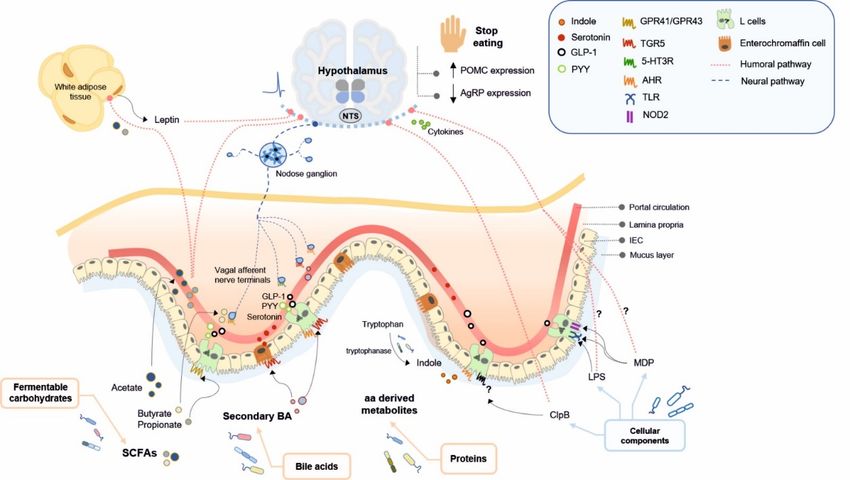
4. Microbial Ligands Mediating Gut–Brain Communication and Energy Homeostasis
4. Microbial Ligands Mediating Gut–Brain Communication and Energy Homeostasis
Here, we review the microbial products, including bacterial metabolites and bacterial
Here, we review the microbial products, including bacterial metabolites and bacterial
cell components, that might impact on brain functions by modulating nutrient sensing
cell components, that might impact on brain functions by modulating nutrient sensing
signalling through enteroendocrine humoral and neural pathways, and could contribute
signalling through enteroendocrine humoral and neural pathways, and could contribute
to controlling energy homeostasis (Figure 1, Table 1). Beyond the control of food intake,
to controlling energy homeostasis (Figure 1, Table 1). Beyond the control of food intake,
gut microbiota might influence the whole-body energy metabolism by modulating the
gut microbiota might influence the whole-body energy metabolism by modulating the para-
parasympathetic and sympathetic efferent tone [47,169], although this mechanism has been
sympathetic and sympathetic efferent tone [47,169], although this mechanism has been the
the subject of far fewer studies. Thus, this section focuses on the role of gut microbiota in
subject of far fewer studies. Thus, this section focuses on the role of gut microbiota in control-
controlling food intake and energy homeostasis, mainly through effects on the hypothalamus-
ling food intake and energy homeostasis, mainly through effects on the hypothalamus-medi-
mediated food-intake suppression and, specially, in the postprandial periods.
ated food-intake suppression and, specially, in the postprandial periods.
Figure 1. Bacterially produced metabolites from dietary nutrients and structural components of non-dietary origin mod-
Figure 1. Bacterially produced metabolites from dietary nutrients and structural components of non-dietary origin modulate
ulate food intake in the brain (hypothalamus) through humoral and/or enterodocrine and neural signalling pathways.
food intake in the brain (hypothalamus) through humoral and/or enterodocrine and neural signalling pathways. Here, we
Here, we represent the pathways by which bacterial metabolites and non-dietary bacterial components (LPS, MDP and
represent the pathways
ClpB) induce by which
an anorexigenic bacterialinmetabolites
response postprandialand non-dietary
periods and a bacterial
long-term components
food intake(LPS, MDP
control. and ClpB)
5-HT3R, induce
5-hydroxy-
an anorexigenic response in postprandial periods and a long-term food intake control. 5-HT3R, 5-hydroxytryptamine
tryptamine type 3 receptor; aa, amino acid; AgRP, agouti gene-related peptide; AHR, aryl hydrocarbon receptor; BA, bile
type 3 receptor;
acids; aa, amino acid;
ClpB, caseinolytic AgRP, agouti
peptidase gene-related
B; GLP-1, peptide;
glucagonlike AHR, arylGPR41/FFAR3,
peptide-1; hydrocarbon receptor; BA, acid
free fatty bile acids; ClpB,3;
receptor
caseinolytic
GPR43/FFAR2, peptidase B; GLP-1,
free fatty glucagonlike
acid receptor peptide-1;
2; IEC, intestinal GPR41/FFAR3,
epithelial cells; LPS,free fatty acid receptor
lipopolysaccharide; 3; GPR43/FFAR2,
MDP, free
muramyldipeptide;
NOD2,
fatty acidNucleotide-binding oligomerization
receptor 2; IEC, intestinal domain
epithelial cells; 2; NTS, nucleus tractus
LPS, lipopolysaccharide; solitarius; PYY, peptide
MDP, muramyldipeptide; NOD2, YY; POMC,
Nucleotide-
proopiomelanocortin;
binding oligomerizationSCFA,
domainshort-chain fatty acids;
2; NTS, nucleus TGR5,
tractus takedaPYY,
solitarius; G protein-coupled
peptide YY; POMC,receptor 5; TLR, Toll-like receptor.
proopiomelanocortin; SCFA,
short-chain fatty acids; TGR5, takeda G protein-coupled receptor 5; TLR, Toll-like receptor.Int. J. Mol. Sci. 2021, 22, 5830 13 of 35
Table 1. Main microbially derived ligands of dietary and non-dietary nature involved in gut-to brain nutrient sensing and control energy homeostasis.
Dietary Gut Bacterial-Derived Bacterial-Producing
Bacterial Producers Receptor Pathway Function References
Nutrients Ligand Enzyme
Food intake suppression, ARC
neuronal activation, increase in
Phosphate
acetyl-CoA carboxylase activity
acetyltransferase and
FFAR2/GPR43 (L cells) Humoral pathway and AMPK inducing an [89,170–173]
acetate kinase for
increase in POMC and
acetate
reduction in AgRP expression,
Prevotella [90], leptin release from adipocytes
Fermentable SCFAs (acetate, Ruminococcus [90], Enzymes involved in Food intake suppression, leptin
FFAR3/GPR41 (L cells, Humoral pathway, gut
carbohydrates propionate, butyrate) Bifidobacterium sp. [91], succinate, acrylate and release from adipocytes, control
enteric neurons, nodose nutrient sensing [89,132,173–177]
Prevotella [95,96] propanediol pathways of postprandial glucose, control
ganglion neurons) pathways (GLP-1, PYY)
for propionate of intestinal gluconeogenesis
Food intake suppression,
Phosphate
FFAR3/GPR41 (L cells, Gut nutrient sensing stimulation of POMC
butyryltransferase and
enteric neurons, nodose pathways (GLP-1, GIP, expression, suppression of [89,178–181]
butyrate kinase for
ganglion neurons) vagal afferents) AgRP expression, suppression
butyrate
of orexigenic neurons activity
Food intake suppression in
Members of the genera: synergy with CCK1R
Lactobacillus [182–184], Bacterial bile salt activation, activation of
TGR5 (L cells, vagal Humoral pathway, gut
Bile acids (BAs) Bifidobacterium [182,185], hydrolases (BSH) POMC/CART-expressing
afferents, nodose nutrient sensing
(involved in Secondary BAs Enterococcus [186,187], (deconjugation of hypothalamic neurons, glucose [190–200]
ganglion neurons, pathways (GLP-1, PYY,
lipid digestion) Clostridium [182,188], primary BA to homeostasis, 5-HT3R activation
hypothalamic neurons) 5-HT, vagal afferents)
Listeria [182,189], secondary BA) in intestinal vagal afferent
Bacteroides [182] terminals (probably
modulating food intake)
Members of the genera:
Bacillus, Clostridium,
Enterococcus, Bacteroides, Tryptophanase Gut nutrient sensing Contribution to eating patterns
Indole AHR (L cells) [136,201–203]
Enterobacter, Escherichia, (tryptophan to indole) pathways (GLP-1) unknown
Prevotella, Shigella and
Vibrio [138]
Proteins Members of the genera:
Lactobacillus,
Bifidobacterium, Glutamate Gut nutrient sensing
GABAA , GABAB (L Contribution to nutrient
GABA Lactococcus, decarboxylase pathways (potentially [142,204–207]
cells, vagal afferents) sensing in the brain unknown
Streptococcus, Escherichia, (glutamate to GABA) through vagal afferents)
Listeria, and Aspergillus
[143–145]You can also read