COVID-19 2nd Edition PULMONOLOGIST'S CONSENSUS ON - INTERNATIONAL - II
←
→
Page content transcription
If your browser does not render page correctly, please read the page content below

INTERNATIONAL
                  PULMONOLOGIST’S CONSENSUS ON

                               COVID-19   2nd Edition

         Chief Editor
         Dr.rr. Tinku Joseph (India)

Published on 22nd April 2020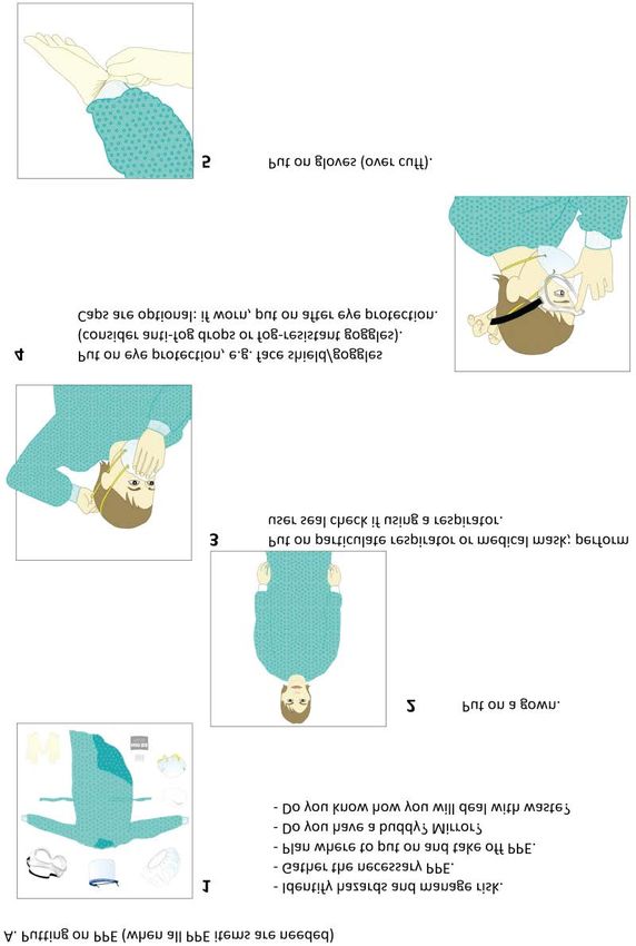
Editors Note
The novel Corona virus disease (COVID-19) has been spreading at a rapid rate across
the world, which made World health organization (WHO) to declare it as a pandemic
disease. A lot is still unknown about this virus. In view of this Pulmonologist’s from
different nations affected by this disease joined hands to frame an E-book titled
“International Pulmonologist’s consensus on COVID-19, which focused on various
preventive and treatment strategies. The first edition of E-book was officially
published on March 14th 2020 and it was accepted by health care professionals
across the world. Inspite of the collective efforts from all human beings we are still
finding it difficult to contain the spread of this disease.
Over the past one month a lot has changed in the way we approach COVID-19. In
view of this my team of Pulmonologist’s regrouped to create a second edition of the
E-book and I would like to thank all of them who have contributed immensely in
between there busy working hours. Like the first edition, the second one is also made
available to everyone at free of cost. Also on behalf of all contributors I would like to
dedicate this E-book to every health care worker who has been contributing
immensely in the fight against this deadly disease. Let us all join and fight against
COVID-19

Dr.Tinku Joseph
Editor in chief
International pulmonologist’s consensus on COVID-19
Associate Prof. & Interventional Pulmonologist
Amrita Institute of Medical Sciences, Kochi, Kerala, India
Dedicating our E-book to all health care workers
 who are fighting against COVID-19 pandemic

           WHAT MAKES A GREAT TEAM?

   Nursing staff                               Housekeeping staff
   Medical/Nursing students                    Ward clerks
   (Interns/Residents)                         Bronchoscopy & operation theatre staff
   Phlebotomist’s                              Infection & quality control staff
   Respiratory Therapist’s                     Outpatient services
   Radiology & Imaging Technicians             Dieticians
   Pharmacists                                 Hospital administration staff
   Admission staff                             Finance & Accounting team
   Physical/occupational/speech therapists     Central supply
   Coders & Transcriptionists                  Public relations & Marketing team
   Social workers                              Doctors
   Ambulance drivers

  Together we can. “Never stop. Do your best. Today you are someone’s hope and
                            one day someone’s hero”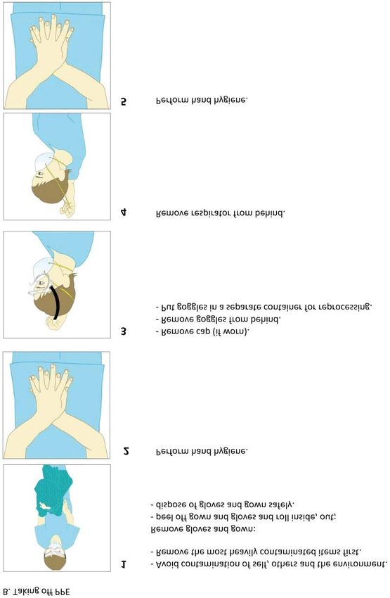
Chief Editor
                        Dr. Tinku Joseph (India)
                        Associate Prof & Interventional Pulmonologist
                        Bronchoscopy course director
                        Amrita Institute of Medical Sciences & Research center, Kochi, Kerala.
                        Corresponding Author
                           +917034564871
                           tinkujoseph2010@gmail.com
                           www.drtinkujoseph.com

Contributors
      Dr.Mohammed Ashkan Moslehi                                         Dr. Anantham Devanand
      (Iran)                                                             (Singapore)
      Head of Pediatric Pulmonary Division
                                                                         Head of Lung Centre
      Shiraz University Of Medical Sciences
                                                                         Respiratory & Critical care Medicine
       hair o e iatri se tion in or sso iation
                                                                         Singapore General Hospital, Singapore
      for Bronchology and Interventional pulmonology (WABIP)

      Dr. Kyle Hogarth                                                   Dr. Lucia Viola
      (USA)                                                              (Columbia)
      Prof. of Medicine                                                  Faculty of Medicine
      Director of Bronchoscopy services                                  Fundacion Neumologica
      University of Chicago, Chicago, IL                                 Colombiana, Bogota

      Dr. Michela Bezzi                                                  Dr. Jasleen Pannu
      (Italy)                                                            (USA)
      Director of Interventional Pulmonology                             Director of Interventional Pulmonary
      University Hospital Asst Spedali Civili Brescia                    Translational Research
      Florence                                                           Ohio State University Werner Medical Center

      Dr. Aji Kavidasan                                                  Dr. Nitesh Gupta
      (UK)                                                               (India)
                                                                           ssistant ro     o a o fi er
      Consultant Chest Physician &
                                                                         (COVID-19 outbreak)
      Interventional Pulmonologist
                                                                         Dept. of Pulmonary Medicine
      at Croydon University Hospital, London
                                                                         VMMC & Safdarjung Hospital, New Delhi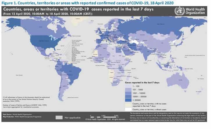
Contributors

      Dr.Mayank Vats                                Dr. Tushar Sahasrabudhe
      (UAE)                                         (India)
      Senior Pulmonologist                          Professor
      Rashid Hospital, Dubai health authority       Dept. of Pulmonary Medicine
      Dubai                                         D.Y. Patil Medical college, Pimpri, Pune

                                                    Dr. Nader Faseeh
      Dr.Elena Mitrofan                             (Egypy)
      (Romania)                                     Prof. of Pediatrics, Head of
      Consultant Pulmonologist                      Respiratory and Allergy Unit,
      Hospital of Pulmonary Diseases, Lasi county   Faculty of Medicine. Alexandria University.

                                                    Dr. Otis Rickman
      Dr.Calvin N G                                 (USA)
      (China)                                       Associate Professor of Medicine &
      Division of Cardiothoracic Surgery,           Thoracic Surgery
      The Chinese University of Hong Kong.          Division of Pulmonary & Critical care
      Prince of Wales Hospital. Hong Kong           Vanderbilt University Medical Center
                                                    Nashville, TN

      Dr. Arvind Perathur                           Dr. Basil Elnazir
      (India)                                       (IreLand)
      Clinical Head & Associate prof.               Senior Pediatric Respiratory consultant
      Dept. of Pulmonary Medicine                   Tallaght university hospital
      Amrita Hospital, Kochi                        Trinity college, Dublin, Ireland

                                                    Dr. Henri Colt
      Dr. Lili Zhong                                (USA)
      (China)                                       Founder chairman of WABIP
      Director of Pediatric Respiratory division    Professor Emeritus
      Hunan provincial peoples hospital, Hunan.     Pulmonary & Critical care Medicine
                                                    University of California

      Dr. Jamalul Azizi                             Dr. Omer Elgaili
      (Malaysia)                                    (Sudan)
      Head of Respiratory Medicine &                Associate professor
      Chief Interventional Pulmonologist            Faculty of Medicine
      Hospital Serdang, Kualalumpur                 Alneelain university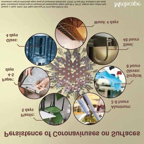
Contributors

      Dr. Antonio Gonzalo                                    Dr. Sumita Agrawal
      (Bolivia)                                              (India)
                                                             Consultant & Head
      Consultant Pulmonologist
                                                             Dept. of Pulmonary & Critical care Medicine
      Hospital Elizabeth seton
                                                             MediPulse Hospital, Jodhpur, Rajasthan

      Dr. Ali Sadoughi                                       Dr. Samaher Hashim
      (USA)                                                  (Saudi Arabia)
      Director, Interventional Pulmonology & Bronchoscopy.
                                                             Consultant Pulmonologist
      Montefiore Medical Center
                                                             University of Tabuk
      Divisions of Pulmonary and Critical Care
                                                             Tabuk city, KSA
      Albert Einstein College of Medicine

      Dr.Roseleen Bali
      (India)
                                                             Dr. Adnan Majid
                                                             (USA)
      Consultant                                             Chief of Interventional pulmonary
      Dept. of Pulmonary & Critical care Medicine            Boston,MA
      Apollo Hospital, New Delhi

      Dr. Renato Cutrera                                     Dr. Kostas Priftis
      (Italy)                                                (Greece)
      Clinical Head & Associate prof.
                                                             Associate Prof. in pediatric Pulmonology
      Dept. of Pulmonary Medicine
                                                             National & Kapodistrian university of Athens
      Amrita Hospital, Kochi

      Dr. Dr.Chen Meng
      (China)
      Director of pediatric Respiratory division
      Qilu children’s Hospital
      Shandong university, Jinan.
Contents

   01   Introduction                      01

   02   Mode of transmission              02

   03   Epidemiology                      04

   04   Pathophysiology                   07

   05   Clinical features                 09

   06   COVID-19 in Pediatric age group   11

   07   Diagnosis                         13

   08   Initial management                29

   09   Treatment options                 33

   10   Critical care management          43

   11   Prognostic Factors                52

   12   Prophylaxis                       53

   13   Prevention                        56

   14   Conclusion                        72
International Pulmonologist’s Consensus On Covid-19 2nd Edition
                                                                                      Page 01

1    Introduction
    Corona virus comprises of a large family of viruses that are common in human
    beings as well animals (camels, cattle, cats, and bats). There are seven different
    strains of corona virus. [1].
        229E (alpha coronavirus)
        NL63 (alpha coronavirus)
        OC43 (beta coronavirus)
        HKU1 (beta coronavirus)
        MERS-CoV (the beta coronavirus that causes Middle East Respiratory
        Syndrome, or MERS)
        SARS-CoV (the beta coronavirus that causes severe acute respiratory
        syndrome, or SARS)
        SARS-CoV-2 (the novel coronavirus that causes coronavirus disease 2019,
        or COVID-19)

    Sometimes corona virus from animals infect people and spread further via human to
    h an trans ission s h as with           o          o an now with this
    (Corona disease 2019). The virus that causes COVID-19 is designated as severe
    acute respiratory syndrome corona virus 2 (SARS-CoV-2); previously, referred to as
    2019-nCoV.

      owar s e e er            this no e orona ir s was i entifie as a a se o             er
    and lower respiratory tract infections in Wuhan, a city in the Hubei Province of China.
    It rapidly spread, resulting in an epidemic throughout China and then gradually
    spreading to other parts of the world in pandemic proportions. It has affected almost
    every continent in this world, except Antarctica. In February 2020, the World Health
    Organization designated the disease COVID-19, which stands for corona virus
    disease 2019 [2].
International Pulmonologist’s Consensus On Covid-19 2nd Edition
Page 02

 2         Mode of Transmission
          Our understanding of the mode of transmission is currently incomplete. Epidemi-
          o ogi in estigation in han at the eginning o the o t rea i entifie an initia
          association with a seafood market where most patients had worked or visited [2]. The
          seafood market also sold live rabbits, snakes and other animals. The initial concept
          was that the virus originated from snakes, however later studies proved that it had
          more similarity with bats. However, as the outbreak progressed, person-to-person
          transmission through droplets and fomites became the primary mode of transmis-
          sion.

          Viability of SARS-CoV-2 in aerosols is about 3 hours [5], on plastic and stainless
          steel for up to 72 hours, on copper up to 4 hours and on cardboard up to 24 hours [6].
          The survival for SARS-CoV has been shown to be affected by temperature; a lower
          environmental temperature favours persistence of virus on surfaces [7]. In a analysis
          of spread of infection across the globe and its correlation with local environmental
          conditions it was contemplated that absolute humidity (AH) above 10g/m2 could
          possible slowdown the transmission of 2019-nCoV [9,10].

          Effect of epidemic would depend upon the transmission and the severity of infec-
          tion. The basic reproduction number (R0 – R naught) is 3 for SARS-CoV-2; which
          means that 3 new cases are being infected with the virus by the index case [10]. The
          R0 indicates whether the transmission would be sustained or not; a R0 of less than
          1 means that the transmission would die out and a R0 of more than 1 means that it
          would be sustained.

 2A How does Person-to-person transmission occur?

          Droplet transmission
          The virus is released in the respiratory secretions when an infected person coughs,
          sneezes or talks. These droplets can infect others if they make direct contact with
          the mucous membranes. Infection can also occur by touching an infected surface
          and followed by eyes, nose or mouth.

          Droplets typically do not travel more than six feet (about two meters) and do not
          linger in the air. However, given the current uncertainty regarding transmission
          mechanisms, airborne precautions are recommended routinely in some countries
          an in the setting o s e ifi high ris ro e res Patients are thought to be most
          contagious when they are symptomatic [7]. Some spread might be possible before
          symptoms appear, but this is not thought to be a common occurrence [4-6].
International Pulmonologist’s Consensus On Covid-19 2nd Edition
                                                                                        Page 03

Extensive environmental contamination from infected patients has been document-
ed in terms of contamination of air vents, door handles, toilet bowl, sink and on
personnel protective equipment [4]. Asymptomatic carriers (super spreaders) have
been shown to spread the COVID-19. These are cases that have no symptoms and
radiological manifestations, but can transmit the virus to others [14].

Close contact is defined as
 a     t east        in tes within eet     eters o a atient with onfir e
      COVID-19 , or individuals who was exposed to the COVID-19 patient
                [16]

      within 2 meters for more than 1 hour within 2 days before the symptom
      onset of the patient,
 b) Cohabiting family members of the COVID-19 patient or suspected patient
    (17). Also it is essential to assess the duration of exposure and the clinical
    symptoms of the patient with COVID-19 [18].

The high viral load during the initial days of the illness suggests that patients could
be most infectious during the first week period, and it might account for the high
transmissibility of SARS-CoV-2 and also high viral load at presentation. Older age
groups are associated with high viral loads and thereby severe infection [19].

The mean viral load of severe cases is around 60 times higher than mild case,
suggesting that poor clinical outcomes might be associated with viral load [20]. This
can probably explain the severe infection in healthcare workers.

Other possible modes of transmission
Formite Spread: It may be possible that a person can get COVID-19 by touching a
surface or object that has the virus on it and then touching their own mouth, nose,
or possibly their eyes, but this is not thought to be the main way the virus spreads.

SARS-CoV-2 RNA has been detected in stools and persists for long duration in high
concentration even if the patient is asymptomatic, this indicates active replication in
the gastrointestinal tract, but till now there are no reports of faecal transmission. [14, 21]
Further studies are thus, required to document this mode of transmission.

SARS-CoV has already been demonstrated to survive in infected water and sewage
for weeks [22]. Thus, this mode of transmission should be actively looked into. Since
the demonstration of virus in conjunctival secretions has been noted, the possibility
of transmission through ocular surfaces has also been contemplated [23]. Adsorption
of virus on airborne dust and particulate matter also needs to be investigated for the
possible transmission of virus [24].
International Pulmonologist’s Consensus On Covid-19 2nd Edition
Page 04
Though, anecdotal reports of COVID positivity has been reported in newborns, a case
series of 9 pregnant patients from Wuhan, China who delivered in January 2020, did
not note a vertical transmission to their newborns [25, 26, and 27].
Nosocomial transmission is an important source for infection in healthcare profes-
sionals including physicians, nursing and paramedical staff during examination,
transport, non-invasive ventilation, intubation and bronchoscopy of COVID patients
[28]
.
The policies that have been used in many countries to break transmission are
Lockdown of infected areas
Travel restrictions
Continuous surveillance and active contact tracing [29].
Quarantine of close contacts is essential, as they may not have symptoms
initially but may develop symptoms later on as the incubation period varies
from 5-14 days [30].
To wear a face mask in public on a mandatory basis
3 Epidemiology
in e the first re orts o ases ro han at the en o ore than
ases ha e een re orte in hina in ing a a orator onfir e
cases as well as clinically diagnosed cases in the Hubei Province. Increasing num-
bers of cases have also been reported in other countries across all continents except
Antarctica.
From the discovery of the disease, up to now, more than 509 154 COVID-19 cases
have been reported globally, number of cases that tripled compared to last week,
and a total of 23,335 deaths have been reported by WHO. In this moment European
egion ta ain ran e er an wit er an ontain ore tota onfir e
ases o tsi e o hina where the ir s first s rea estern a ifi region hina
Republic of Korea, Japan, Malaysia, Australia), Eastern Mediterranean Region (with
Iran), region of the Americas have also reported a high number of cases (Figure 1) [31].International Pulmonologist’s Consensus On Covid-19 2nd Edition
Page 05
Figure 1: Countries, territories or areas with reported confirmed cases of COVID-19, 18th April 2020
Around 800 cases and 10 deaths have also been reported in passengers travelling
on cruise ships [32]. Subsequently the world health organization (WHO) to declare
COVID-19 as a pandemic on 12th March 2020. Table 1 gives an estimate of the
number of COVID-19 cases; which may be lesser than the actual number as number
of asymptomatic individuals is unknown and the testing facilities may vary from
region to region [33]. Residents of nursing homes, hospices and elderly homes are
more susceptible to the viral infection and are in vulnerable group for severe infec-
tion considering age and presence of comorbidities [34].
Health care workers are another susceptible group as they are taking care of severe
cases of the infection (likely with high viral loads), close proximity to the case during
procedures such as examination, transport, blood sampling, intubation and bron-
choscopy. Ancillary reports from China claim 3300 health care professional have
been infected and similarly 20% of health care workers from Italy have contracted
the infection [35]. With the paucity of personal protective equipment across the globe
and long hours of work to deal with extra load on the infrastructure; this risk further
increases.
The case fatality rate (CFR) has shown been shown to have a broad range from
0.25-7% may also vary as the denominator comprising of asymptomatic cases is not
fully known [10,36]. The case fatality rate is 2-7% depending on age and presence of
co-morbid conditions [10,37], especially in the elderly. Preliminary data from United
states of America suggest also that younger adult (20-44 years) are 20% hospital-
ized and 12 % are admitted in ICU [37]. In Italy, the country with a higher mortality rate,
60% of coronavirus cases and 70% of deaths in the country so far have been in men.
Similarly 64% mortality in China has also been in men [38].International Pulmonologist’s Consensus On Covid-19 2nd Edition
Page 06
Further the WHO has shown that the mortality occurring due to COVID-19 was
between 2-8 weeks [33]. Thus, the number of deaths may be underestimated. The
presentation of the disease is heterogenous with mild disease in 80-95% cases;
severe disease is associated with high viral loads (up to 60 times higher in severe
versus mild cases) and prolonged viral shedding [20]. The CFR also depends on the
age of the patient; early data from United States of America suggest that elderly age
group >85 years have 10-27% mortality, 3-11% amongst 65-84 years, 1-3% in 55-64
years andInternational Pulmonologist’s Consensus On Covid-19 2nd Edition
Page 07
Case Comparison
WHO Regions
Europe
Americas 821, 860
confirmed cases
Western Pacific 130, 696
confirmed cases
Eastern Mediterranean- 124, 691
confirmed cases
South-East Asia 27, 319
confirmed cases
Africa 13, 892
confirmed cases
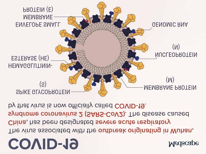
4 Pathophysiology of COVID-19
he orona ir ses are a arge a i o sing e stran e ir ses ss that
an e iso ate in i erent ani a s e ies he ha e a rown i e a earan e n er
an electron microscope (coronam is the Latin term for crown) due to the presence of
s i e g o roteins on the en e o e hese ir ses an a so in e t h ans an
cause illness ranging from the common cold to more severe diseases such as MERS,
an now o i o ate se en h an o s o s a a e o in e ting
h ans ha e een i entifie
SARS-CoV-2 belongs to the betaCoVs. It has round or elliptic and often pleomorphic
form, and a diameter of approximately 60–140 nm (Figure 2) or ing to re ent
research, a spike mutation, which probably occurred in late November 2019,
triggered jumping to humans. In particular, Angeletti et al. compared the SARS-Cov-2
gene se en e with that o o he ana e the trans e rane he i a
segments in the ORF1ab encoded 2 (nsp2) and nsp3 and found that position 723
presents a serine instead of a glycine residue, while the position 1010 is occupied by
ro ine instea o iso e ine [155]International Pulmonologist’s Consensus On Covid-19 2nd Edition
Page 08
Figure 2: Ultrastructure of SARS CoV-2
On getting deposited in the nasal and pharyngeal mucus membrane, the virus starts
proliferating rapidly and causes Covid-19. The lymphocytes are highly vulnerable to
this virus and hence lymphocytopenia is a common feature.
The disease progression can be divided into three distinct phases:
Early infection phase
Pulmonary phase
Severe hyperinflammatory phase
During the early infection phase, the initial inflammatory response may cause local
symptoms like throat irritation & dry cough and constitutional symptoms like fever,
myalgia and headaches. Many patients may be asymptomatic. During this phase,
the patient is infective and can transmit the disease. Large number of patients may
not progress beyond this phase and recover slowly over a period of 2-6 weeks.
During the pulmonary phase the ir s infi trates the ng aren h a an egins to
proliferate. This stage is characterized by injury to lung parenchyma leading to vaso-
dilation, increased endothelial permeability and leukocyte recruitment leading to
further pulmonary damage, hypoxemia and cardiovascular stress.International Pulmonologist’s Consensus On Covid-19 2nd Edition
Page 09
In a subset of patients, the host inflammatory response continues to amplify and
results in systemic inflammation. This, often labelled as cytokine storm, can injure
istant organs he rotagonist o this stor is inter e in is ro e
by activated leukocytes and acts on a large number of cells and tissues. It is able to
promote the differentiation of B lymphocytes, promotes the growth of some catego-
ries of cells, and inhibits the growth of others. This hyperinflammatory response can
e onfir e in rease erritin e e s inter e ins an rea ti e rotein in the
serum.
Interestingly two distinct types of respiratory failure are being recognized. One is
ARDS or a type H patient that is characterized by high elastance, high right-to-left
shunt, high lung weight, and high recruitability. Few case reports of biopsy of
in o e ngs showe i atera i se a eo ar a age with e ar fi ro oi
exudates, desquamation of pneumocytes and hyaline membrane formation, typical
of ARDS. [156] hese atients a nee int ation an enefit ro e hani a enti-
lation with high PEEP.
Another subgroup of patients have been labelled as type L phenotypes with lungs
having low elastance, low ventilation-perfusion ratio, low lung weight, and low
re r ita i it hese atients are o ten se ere h o i witho t signifi ant s -
noea. Some have compared this to high altitude mountain sickness kind of presenta-
tion hese atients a res on to o gen thera a one an a not enefit ro
high pressure ventilation. [157]
Although the prominent site of infection and hence inflammation is lungs, the ampli-
fie in a ator res onse an ha e e eterio s e e ts on other organs in ing
the heart.
Consistent with this notion, biomarkers of cardiac injury (raised Trop-I and BNP) and
electrocardiographic abnormalities correlate with elevated inflammatory markers.
SARS-CoV infection appears to down regulate ACE2 receptors, which may contribute
to left ventricular dysfunction. There is some evidence of direct myocardial injury as
we to sies ha e on or e onon ear infi trates with ne rosis th s satis -
ing criteria for viral myocarditis. [158] Heart may also get stressed secondary to
respiratory failure, especially in patients with pre-existing underlying heart disease.
The kidneys often get affected in serious illness, especially ARDS and Covid-19 is no
e e tion retros e ti e st o atients with onfir e ne o-
nia in China showed that 4.5% developed acute kidney injury (AKI). [159]
The cytokine
storm alone cannot explain AKI and only a small percentage of ARDS patients devel-
oped AKI. Fluid dysregulation, cardiac failure, rhabdomyolysis and sepsis can all
contribute to AKI.International Pulmonologist’s Consensus On Covid-19 2nd Edition
Page 10
5 Clinical Features
5A Incubation period
The incubation period for COVID-19 is thought to be approximately4 to 14 days
following exposure [43,44] he a erage onset ro first s to s to hos ita i ation is
7 days. [46, 50]
5B Spectrum of severity
In a report from the Chinese Center for Disease Control and Prevention that included
a ro i ate onfir e in e tions with an esti ation o isease se erit [45]
Mild (no or mild pneumonia) 81%.
Severe disease 14%.
Critical disease 5 %.
In another multi-centre study of 1099 patients, 15.7% of patients met ATS criteria for
severe pneumonia [44].The overall case fatality rate was 2.3 percent; no deaths were
reported among non-critical cases. This rate rose to 8% in those aged 70-79, 14.8%
in those >80 and 49% in those requiring critical care [45].
Co morbidities such as cardiovascular disease, diabetes mellitus, chronic lung
disease, hypertension and malignancies are believed to increase the risk of mortality
[45]
.
5C Age
COVID-19 can affect all age groups and asymptomatic infection has been well
described. In the large Chinese report, 2 % of infections were in individuals younger
than 20 years old. Similarly, in South Korea, 6.3 % of nearly 8000 infections were in
those younger than 20 years old [47].
5D Clinical manifestations
Initial presentation
Pneumonia is the most frequent serious manifestation of infection, characterized
primarily by fever, cough, dyspnea [49] here are no s e ifi ini a eat res that an
yet reliably distinguish COVID-19 from other viral respiratory infections.International Pulmonologist’s Consensus On Covid-19 2nd Edition
Page 11
Common clinical features at the onset of illness were [44,50]
Fever in 88-99 %
Fatigue in 38-70 %
Dry cough in 59-68%
Anorexia in 40 %
Myalgias in 15-35 %
Dyspnea in 19-31 %
Sputum production in 27-34 %
Other, less common symptoms have included
Headache.
Sore throat.
Rhinorrhea.
Gastrointestinal symptoms (eg, nausea and diarrhea) .
[46]
Acute respiratory distress syndrome (ARDS) is a major complication in patients with
severe disease17-29% [46]
5E Complications in ICU patients [46, 50,51]
Arrhythmias 44%
Acute cardiac injury 22-31%
Shock.23-20%
Acute Kidney Injury8-23%
Secondary infection 31%
Cardiomyopathy 33%.International Pulmonologist’s Consensus On Covid-19 2nd Edition
Page 12
6 COVID-19 in pediatric population
In this outbreak, compared with adult cases, there are relatively fewer cases of
children, milder symptoms and better prognosis. Also, children are less frequently
exposed to the main sources of transmission. Most infected children recover one to
two weeks after the onset of symptoms, and no deaths had been reported by April
2020. According to the recent report of the China-WHO Joint Mission Expert Group,
the current domestic case data show that children under 18 years of age account for
2.4% of all reported cases, and no deaths have been reported. [52]
6A Probable reasons why children are less affected by COVID-19
1. The time period of the outbreak, is the winter vacation time of the university,
middle school and kindergarten. It is a good time for everyone to stay in their
own families, which is equivalent to active home isolation. It is a good time to
avoid the collective cluster disease by chance.
2. Secondly, humoral and cellular immune development in children is not fully
developed. This may be one of the mechanisms that lead to the absence of
severe immune responses after viral infection.
3. As COVID-19 virus exploits the ACE2 receptors to gain entry inside the cells,
under expression, immaturity of ACE2 receptors in children is another hypothe
sis in this regard.
4. Moreover, recurrent exposure to viruses like respiratory syncytial virus in
winters can induce more immunoglobulins levels against the new virus infec
tion compare to adults. There is no direct evidence of vertical mother-to-child
transmission, but newborns can be infected through close contact.
6B Clinical features
n re ent st ies in hina there was no signifi ant gen er i eren e in hi ren an
it was suggested that alleges ranged from 1 day to 18 years were prone to infected
by the COVID-19 [53]. The symptoms of COVID-19 are similar in children and adults.
owe er hi ren with onfir e ha e genera resente with i
symptoms and usually recover within 1 to 2 weeks. Reported symptoms in children
may include cold-like symptoms, such as fever, dry cough, sore throat, runny nose,
and sneezing. Gastrointestinal manifestations including vomiting and diarrhea have
also been reported.
n the e iatri atients that ha an i entifie histor o ose
contact with COVID-19 diagnosed family members. Fever (12/20, 60%) and cough
(13/20, 65%) were the most common symptoms. [52] Children with underlying medi medi-
cal conditions and special healthcare needs may be at higher risk for severe illness.
There is much more to be learned about how the disease impacts children.International Pulmonologist’s Consensus On Covid-19 2nd Edition
Page 13
6C Laboratory findings
In the early stage of the disease, the total number of peripheral white blood cells is
normal or decreased, the lymphocyte count is reduced, and some children have
increased liver enzymes, lactate dehydrogenase (LDH), muscle enzymes, and myo-
globin; some critically ill patients have increased troponin, D-dimer and ferritin and
the number of peripheral blood lymphocytes have progressively reduced. Like adults,
the children with severe and critical illness may be accompanied by elevated levels
of inflammatory factors such as interleukin (IL)-6, IL-4, IL-10, and tumor necrosis
a tor [52]
6D Radiology
here are no a nor a fin ings in the ear stages o the isease in the hi ren s
plain X-rays with COVID-19 thus plain X-rays it is not recommended especially in the
early stages and in whom without symptoms or any positive risk factors. Suspected
cases should undergo chest CT examination as soon as possible. The most impor-
tant fin ing in ear stages is a sing e or ti e i ite gro n g ass o a it whi h
mostly located under the pleura or near the bronchial blood vessel bundle especially
in the lower lobes. Severe period is very rare, manifested by diffuse unilateral or bilat-
eral consolidation of lungs and a mixed presence of ground glass opacities [54]. Also
compared to adults, consolidation with surrounding halo signs is more common in
pediatric patients and was suggested as a typical sign in pediatric patients. [52]
6E Treatment
ain s orti e no s e ifi anti ira e i ations are a ai a e or hi ren or
more details please refer to treatment section [page no: ]International Pulmonologist’s Consensus On Covid-19 2nd Edition
Page 14
7 Diagnosis
7A Case Definition [55]
Suspected case
Based on the epidemiologic characteristics observed so far in China, everyone is
assumed to be susceptible, although there may be risk factors increasing suscepti-
bility to infection.
1) A patient with acute respiratory tract infection (sudden onset of at least one
of the following: cough, fever, shortness of breath) AND with no other aetiology
that fully explains the clinical presentation AND with a history of travel or
residence in a country/area reporting local or community transmission
during the 14 days prior to symptom onset;
OR
2) A patient with any acute respiratory illness AND having been in close contact
with a onfir e or ro a e ase in the ast a s rior to onset
of symptoms;
OR
3) A patient with severe acute respiratory infection (fever and at least one
sign/symptom of respiratory disease (e.g., cough, fever, shortness breath))
AND requiring hospitalisation (SARI) AND with no other aetiology that fully
explains the clinical presentation.
Probable case
A suspected case for whom testing for virus causing COVID-19 is inconclusive
(according to the test results reported by the laboratory) or for whom testing was
ositi e on a an orona ir s assa
Confirmed case
erson with a orator onfir ation o ir s a sing in e tion irres e -
tive of clinical signs and symptomsInternational Pulmonologist’s Consensus On Covid-19 2nd Edition
Page 15
Close contacts
Close contact of a probable or confirmed case is defined as
A person living in the same household as a COVID-19 case;
A person having had direct physical contact with a COVID-19 case (e.g. shaking
hands);
A person having unprotected direct contact with infectious secretions of a COV
ID-19 case (e.g. being coughed on, touching used paper tissues with a bare
hand);
A person having had face-to-face contact with a COVID-19 case within 2 metres
and > 15 minutes;
A person who was in a closed environment (e.g. classroom, meeting room,
hospital waiting room, etc.) with a COVID-19 case for 15 minutes or more and at
a distance of less than 2 metres;
A healthcare worker (HCW) or other person providing direct care for a COVID-19
case, or laboratory workers handling specimens from a COVID-19 case without
recommended personal protective equipment (PPE) or with a possible breach of
PPE;
A contact in an aircraft sitting within two seats (in any direction) of the COVID-19
case, travel companions or persons providing care, and crew members serving
in the section of the aircraft where the index case was seated (if severity of
symptoms or movement of the case indicate more extensive exposure, passen
gers seated in the entire section or all passengers on the aircraft may be consid
ered close contacts). [56]
7B Laboratory Findings
White blood cell count (WBC)
White blood cell count can vary. It does not provide accurate information about
COVID-19. [57]
Leukopenia, leukocytosis, and lymphopenia have been reported.
Lymphopenia is more common, seen in more than 80% of patients [57]
Mild thrombocytopenia is commonly seen. However thrombocytopenia is
considered as a poor prognostic sign. [57, 58]International Pulmonologist’s Consensus On Covid-19 2nd Edition
Page 16
Inflammatory markers
Serum Procalcitonin
Serum procalcitonin is often normal at the time of admission; however it
increases in patients who require ICU care. In one study high D-Dimer and
lymphopenia are associated with poor prognosis. [57, 58]
C - reactive protein (CRP)
COVID-19 increases CRP. This seems to track with disease severity and progno
sis. In patients suffering from severe respiratory failure with a normal CRP level
an alternative diagnosis should always be sought. [57, 58]
7C Types of diagnostic tests for COVID-19
Tests to detect the virus
Tests to detect antibodies to the virus
Patients, who meet the criteria for suspect cases, as discussed above, should under-
go testing for SARS-CoV-2 and also other respiratory pathogens.
Table 2: o r e fi e [59]
COVID-19 TESTING POSITIVITY RATES
Sl No. Type of Specimen Positive
1 Bronchoalveolar lavage fluid 93%
2 Bronchoscopic brush biopsy 46%
3 Sputum 72%
4 Nasopharyngeal swab 63%
5 Oropharyngeal swab 32%
6 Feces 29%
7 Blood 1%
8 Urine 0%International Pulmonologist’s Consensus On Covid-19 2nd Edition
Page 17
Note
Nasal swab will detect only 2/3rd of cases and pharyngeal swabs will detect
only 1/3rd of cases and Nasal swab testing is better of two for unadmitted
patients. (Table 2)
Preferably avoid performing bronchoscopy for diagnosing COVID-19 (Aerosol
generating procedure)
Respiratory specimen collection from the upper and in particular lower respiratory
tract should be performed under strict airborne infection control precautions [60].
Preferably these samples should be obtained as early as symptom onset, since it
yields higher virus concentrations.
Recommendations for collection of samples for diagnosis of COVID-19
Collection of specimens to test for SARS-CoV-2 from the upper respiratory tract
(nasopharyngeal and oropharyngeal swab) is the preferred method for diagnosis
Induction of sputum collection is not recommended
Bronchoscopy being an aerosol generating procedure has got the potential to
transmit infection to others. In view of this preferably avoid performing it and
limit its usage clearing secretions/mucous plugs in intubated patients [61]
All respiratory specimen collection procedures should be done in negative pres
sure rooms
Additional specimens (eg: Blood, stool, urine) can also be collected to rule out
alternative/supportive diagnosis.
Specimen collection is very important and any mistake in that will result in
false negatives.
Tests to detect the virus
To date, the diagnostic test of choice during the SARS–CoV-2 outbreak has been
polymerase chain reaction (PCR) / Reverse transcriptase polymerase chain reaction
(RT-PCR) testing (sequencing of the viral genome). For the past 20 years, PCR has
been the gold standard for diagnosing viral infectious agents. Using this technique
ir s was i entifie as a no e an ni e entit the hinese in han
a i a ifi ation ase artri ge at or s se wi e or t er osis is
now available for various viral diseases including SARS-CoV2. This unique diagnos-
tic tool has the potential of being used as a point of care test.International Pulmonologist’s Consensus On Covid-19 2nd Edition
Page 18
RT-PCR tests takes about 4 hours to perform and with a single real time PCR
machine about 100-120 tests can be done in a day. The cartridge based test can be
done in 1-2 hours, but the turnaround is much smaller as many samples cannot be
done simultaneously.
Advantages of PCR tests
Primers needed can be produced on needed basis as soon as the viral sequence
is known
igh s e ifi s e ifi it
Tests becomes positive in the early phase of the disease
Disadvantages of PCR tests
RT–PCR is complicated, expensive, and is thus mainly suited to centralized refer
ence laboratories. The actual test only takes 4-6 hours to complete (given
enough supplies and reagents); however, the turnaround time is typically 12-24
hours due to logistic hurdles related to the collection, shipping, and batching
of the samples
Sensitivity may be as low as 50-70%. Reasons: Number of viral particles may
not be large in some infected patients. The best results are obtained using BAL,
how ever being an aerosol generating procedure with a potential for transmitting
infection to others this procedure is not been preferred much as a diagnostic
modality. Currently more emphasis is given for nasopharyngeal swabs and
sputum sample.
PCR can become negative in the later phases of disease as the patients immunity
builds up.
Tests to detect antibodies to the virus
Antibody based tests for SARS-CoV2 has been developed. These are mainly of two
types. The standard test is ELISA (Enzyme linked immunosorbent azzay). Rapid
tests can be done at the point of care without highly trained personal. Two types of
anti o ies are teste g anti o whi h rises first a ter in e tion an it is an
indicator for an active infection. B) IgG type of antibody rises later and is an indica-
tive of past infection. [59,60] (Table 3)International Pulmonologist’s Consensus On Covid-19 2nd Edition
Page 19
Advantages of antibody based tests
It can be used for the rapid screening of SARS-CoV-2 carriers, symptomatic or
asymptomatic, in hospitals, clinics, and test laboratories.
COVID-19 Rapid Test qualitatively detects IgG and IgM antibodies to
in human whole blood, serum and plasma samples
The IgM-IgG combined assay has better utility and sensitivity compared with a
single IgM or IgG test
Antibody based tests are cheaper and the results are faster
e ifi it is a so oo or a s reening test
Disadvantages of antibody based tests
Negative in the early phase of the disease. IgM titers starts to rise only 3-7 days
after the onset of symptoms
he s e ifi it o the test an a so e a on ern when it is se ri ari as a
standard diagnostic test.
Figure 3: COVID-19 Rapid Test kitInternational Pulmonologist’s Consensus On Covid-19 2nd Edition
Page 20
Table 3: Rapid point of care testing for SARS CoV-2
Rapid point of care testing for SARS CoV-2
Biomerica California, USA Rapid POC lateral flow immunoassay Commenced shipping
IgM/IgG antibody samples; seeking FDA
test EUA approval
Caspr Biotech California, USA Ultrasensitive, rapid, Based on CRISPR-Cas12 Proof of principle
and portable evaluation
coronavirus
SARS-CoV-2
sequence detection
Cepheid California, USA Xpert Xpress Rapid PCR test that runs Received FDA
SARS-CoV-2 on GenXpert benchtop emergency use
system – delivers result in authorization
two hours from sample
collection to delivery of
result
Guangzhou Guangzhou, China Wondfo SARS-CoV-2 Lateral flow 15-minute National Medical
Wondfo antibody test immunoassay that detects Products Administration
Biotech IgM and IgG antibodies EUA in China; CE mark
directed against in Europe
SARS-CoV-2
Innovita Hubei, China SARS-CoV-2 Lateral flow 15-minute National Medical
Biological antibody assay immunoassay that detects Products Administration
Technology IgM and IgG antibodies EUA in China
directed against
SARS-CoV-2
Jiangsu Nanjing, China SARS-CoV-2 rapid Lateral flow 15-minute Shipping
Medomics combined IgM/IgG immunoassay that detects
Medical antibody test kit IgM and IgG antibodies
Technologies directed against
SARS-CoV-2
Mammoth Massachusetts, SARS-CoV-2 30-minute lateral flow In validation studies
Biosciences USA DETECTR assay
Pharmact Berlin, Germany SARS-COV-2 POC 20-minute test for CE-marked and shipping
Rapid Test detecting SARS-CoV-2
exposure through
i entifi ation o g an
IgM antibodies
Sherlock California, USA Rapid CRISPR-based Combines SHERLOCK Intended as proof of
Biosciences, tests for SARS-CoV-2 Cas12 and Cas13 enzymes concept for a broad
Cepheid and other pathogens for nucleic acid detection product development
witCepheid's’s GeneXpert alliance in infectious
test-processing instruments diseaseInternational Pulmonologist’s Consensus On Covid-19 2nd Edition
Page 21
Snibe Shenzhen, China MAGLUMI 2019- Automated central CE mark received
Diagnostic nCoV IgM/IgG kit laboratory rapid test that 19 February 2020
runs on MAGLUMI
chemiluminescence
immunoassay system
Sona Nanotech Halifax, Nova Rapid SARS-CoV-2 Lateral flow screening test Assay development and
Scotia antigen detection for S1 domain of testing with GE
test SARS-CoV-2 S1 protein Healthcare Life
Sciences underway
Sugentech Daejeon, SGTi-flex Ten-minute lateral flow CE Mark
South Korea COVID-19 IgM/IgG immunoassay that detects
IgM and IgG antibodies
directed against
SARS-CoV-2
Xiamen Fujian, China), COVID-19 IgM/IgG Ten-minute lateral flow CE Mark
AmonMed test kit immunoassay that detects
Biotechnology (Colloidal gold) IgM and IgG antibodies
directed against
SARS-CoV-2
Zhejiang Zhejiang, China) COVID-19 IgG/IgM Solid-phase Aytu Bioscience has
Orient Gene Rapid Test immunochromatographic sublicensed US
Biotech assay distribution rights from
L.B. Resources
(Hong Kong) and plans
to obtain EUA; already
has CE mark
Voxtur Bio Ltd India COVID-19 IgG/IgM Ten-minute lateral flow Indian Council of
Rapid Test immunoassay that detects Medical Research
IgM and IgG antibodies (ICMR) approved
directed against
SARS-CoV-2
HLL life care India Makesure COVID-19 POC 20-minute test for ICMR approved
limited rapid test kit detecting SARS-CoV-2
exposure through
i entifi ation o g
and IgM antibodies
Vanguard India COVID-19 IgM/IgG Ten-minute lateral flow ICMR approved
diagnostics Antibody Detection immunoassay that detects
Card Test IgM and IgG antibodies
directed against
SARS-CoV-2
Accucare India IgM/IgG Lateral Lateral flow screening ICMR approved
Lab-care Flow Assay kit test for S1 domain of
diagnostics SARS-CoV-2 S1 proteinInternational Pulmonologist’s Consensus On Covid-19 2nd Edition
Page 22
Current Recommended Diagnostic Modality For Covid 19
Use RT-PCR as the primary diagnostic modality for detecting SARS-CoV-2 RNA
sing e ositi e test sho e onfir e a se on assa targeting a
different SARS-CoV-2 gene
If initial testing is negative but the suspicion for COVID-19 remains, the WHO
recommends re-sampling and testing from multiple respiratory tract sites
Point of care cartridge PCR may be used in centers where it is available.
Antibody based tests need not be used as a primary diagnostic test. The main
use of antibody tests would be to study the incidence and prevalence of the
disease and local outbreaks
For safety reasons, specimens from a patient with suspected or documented
COVID-19 should not be submitted for viral culture.
Respiratory & serum samples should also be tested for other viral/bacterial
pathogens.
7D Bronchoscopy
Deciding the need for bronchoscopy during COVID-19 pandemic is tricky. Bronchos-
o ist s sho e wise eno gh in hoosing an ro e re is ers s enefit ratio
should be considered). Bronchoscopy being an aerosol generating procedure has
the potential to transmit infection to others. Need for all procedures should be
reviewed case by case basis and if not an urgent one it should be rescheduled based
on clinical priorities (Table 4-7)
Broad screening protocol should be followed prior to scheduling any Bronchoscopic
procedure. Patients should be asked about symptoms, contacts and history of travel
to COVID-19 zones. If any of these criteria is met, then the procedure needs to be
rescheduled or a nasopharyngeal swab test should be done.
Table 4: Indications for Emergency Bronchoscopic procedure
Indications for Emergency Bronchoscopic procedure (To be performed on same day)
Symptomatic central airway obstruction:
1 Mass
Foreign body
Mucous plug
2 Massive Haemoptysis
3 Symptomatic Tracheal stenosis
4 Migrated stent (Silicon/metallic)International Pulmonologist’s Consensus On Covid-19 2nd Edition
Page 23
Table 5: Indications for semi-urgent Bronchoscopic procedure
Indications for Semi urgent Bronchoscopic procedure
(can wait for 2 to 4 days, preferably send a swab to rule out COVID-19 infection)
1 Evaluation of lung mass or nodule (Diagnosis/staging)
2 Evaluation of Mediastinal lymphadenopathy
3 Whole lung lavage
4 Suspected pulmonary infection in immunocompromised patients
5 Post lung transplant recipients – Evaluation of Bronchiolitis obliterans syndrome
6 Suspected pulmonary infection in bone marrow/solid organ transplant
7 Evaluation of lobar atlectasis
Table 6: Indications for elective Bronchoscopic procedure
Indication for Elective Bronchoscopy (Re-schedule till your locality is free of COVID-19)
1 Bronchial Thermoplasty
2 Tracheobronchomalacia evaluation
3 r o io s or histo atho ogi a onfir ation o etio og o
4 Bronchoscopic lung volume reduction procedures
RIGID BRONCHOSCOPIC PROCEDURES DURING COVID-19
Performing any rigid Bronchoscopic procedure carries a high risk of transmission of
infection (Open circuit, excessive air leak through the side of rigid tube due to
absence of cuff) compared to endotracheal tube. Also most of the rigid Broncho
scopic procedures are prolonged ones.
Until COVID-19 pandemic ends all rigid Bronchoscopic procedures should be
performed by the highly skilled team member as it saves time and there by reduces
the chance of transmission of infection.
Powered Air purifying Respirators (PAPR) kit is the ideal PPE while performing any
rigid Bronchoscopic procedure (prolonged) as it avoids breathing resistance/suffo
cation and moisture build up associated with using N95 mask along with goggles/
face shield.International Pulmonologist’s Consensus On Covid-19 2nd Edition
Page 24
Table 7: n i ations or ron hos o in s s e te onfir e ases
Indications for Bronchoscopy in COVID-19 suspected/confirmed cases
1 Relatively contraindicated
2 For swab and sputum negative patients who are strong clinical suspects
3 For patients who are on mechanical ventilator: Mucous plug clearance
4 Evaluation for alternative infection
5 To rule out differentials of non –resolving pneumonia
6 Massive haemoptysis – airway interventions
Essential steps to be followed if bronchoscopy is needed in COVID-19 suspect/confirmed case
Avoid performing bronchoscopic procedures under conscious sedation( high chance for dissem
ination of aerosols when patient coughs)
Preferably do all bronchoscopic procedures under general anesthesia (patient should be sedated
& paralysed) since it avoids dissemination of aerosols to a certain extend.
Consider using a disposable bronchoscope if available, especially in an ICU care setting.
All Bronchoscopic procedures (COVID-19) should be performed in negative pressure isolation
rooms
Minimize the staff for all bronchoscopic procedures and avoid training your fellows, since it
increases the procedure duration.
All personnel should use standard PPE kits during bronchoscopy and Powered Air purifying Respi
rators (PAPR) are preferred for prolonged rigid Bronchoscopic procedures.
Use of safety/ aerosol box have been used at many centers in the world to add extra protection
from COVID-19, while performing aerosol generating procedures
onning o fing roto o s o sho e stri t o owe
bronchoscopes,
Standard disinfection protocols should be followed for cleaning your flexible/rigid bronchoscopes
electrosurgical equipments and video monitorsInternational Pulmonologist’s Consensus On Covid-19 2nd Edition
Page 25
Recommendations for performing bronchoscopic procedures during COVID-19
[61, 160,161]
Bronchoscopy is not considered as a diagnostic modality for COVID1-9. Being an
aerosol generating procedure performing bronchoscopy has got a high potential for
transmission of infection
Primary/Preferred method for diagnosing COVID-19 is evaluation of nasopharynge
al/oropharyngeal swab and sputum analysis
wa s e a ate the nee or ron hos o gra e the a or ing to ris enefit
ratio (Emergency/semi-urgent & elective)
Avoid performing bronchoscopic procedures under conscious sedation( high chance
for dissemination of aerosols when patient coughs)
Preferably do all bronchoscopic procedures under general anesthesia (patient
should be sedated & paralysed) since it avoids dissemination of aerosols to a certain
extend.
Consider using a disposable bronchoscope if available, especially in an ICU care
setting.
All Bronchoscopic procedures (COVID-19) should be performed in negative pressure
isolation rooms
Minimize the staff for all bronchoscopic procedures and avoid training your fellows,
since it increases the procedure duration.
All essential personal protective equipments (PPE) should be used by health care
professionals performing any bronchoscopic procedure.
onning o fing roto o s o sho e stri t o owe
Standard disinfection protocols should be followed for cleaning your flexible/rigid
bronchoscopes, electrosurgical equipments and video monitors.
Rigid Bronchoscopic procedures carries maximum risk of transmission of infection
and hence it should be performed only by a highly skilled individual (shortens the
procedure duration)
Powered Air purifying Respirators (PAPR) kit is the ideal PPE while performing any
rigid Bronchoscopic procedure (prolonged) as it avoids breathing resistance/suffo
cation and moisture build up associated with using N95 mask along with goggles/
face shield.
Be wise in choosing any bronchoscopic procedureInternational Pulmonologist’s Consensus On Covid-19 2nd Edition
Page 26
7E Radiology In Covid-19 Infection
he fin ings on hest i aging are not s e ifi o the in e tion an o o er a
with other entities. There are also recommendations about the performance of the
chest radiography, including the fact that it is better to avoid the movement of the
patient within the hospital.
Chest Radiography (CXR).
he fin ings on are not s e ifi an in the initia hases o the isease the st -
ies could be normal. The most common features include lobar/ multi-lobar / bilater-
al lung consolidation. [62]
Computed Tomography (CT Chest).
Recent studies have reported the features on CT imaging. Pan et al [63] described the
tomographic changes of 21 patients with mild to moderate disease who recovered
from the disease, and they describedfour stages
Early stage (0-4 days after the onset of the symptoms), in which ground glass
opacities (GGO) are frequent, with sub-pleural distribution and involving predom
inantly the lower lobes. Some patients in this stage could have a normal CT.
Progressive stage a s a ter the onset o the s to s the fin ings
usually evolved to rapidly involvement of the two lungs or multi-lobe distribution
with GGO, crazy-paving and consolidation of airspaces.
Peak stage (9-13 days after the onset of the symptoms), the consolidation
e o es enser an it was resent in a ost a o the ases ther fin ing was
residual parenchymal bands.
Absorption stage (>14 days after the onset of the symptoms), no crazy paving
pattern was observed, the GGO could remain.
hi et a a so es ri e the fin ings in atients in han hina o the
patients had an abnormal CT, and the features include: GGO, smooth and irregular
interlobular septal thickening, crazy paving pattern, air bronchogram and irregular
pleural thickening. Usually affecting the subpleural regions and the lower lobes.
The Radiological Society of North America (RSNA) Expert Consensus Statement
on reporting chest CT Findings related to COVID-19 was released on March 25th
2020, in order to standardize the reports. [65] They proposed four COVID-19 imaging
assifi ations (Table 8)International Pulmonologist’s Consensus On Covid-19 2nd Edition
Page 27
Typical appearance
Indeterminate appearance
Atypical appearance
Negative for pneumonia.
Table 8: The Radiological Society of North America (RSNA) Expert Consensus
Statement on reporting chest CT Findings related to COVID-19
COVID-19 penumonia Suggested Reporting
CT Findings Language
i aging assifi ation
Typical Appearance Peripheral, bilateral, GGO with or “Commonly reported imaging
without consolidation or visible features of (COVID-19) pneumonia
intralobular lines (“crazy paving”) are present. Other processes such
Multifocal GGO of rounded as influenza pneumonia and
morphology with or without organizing pneumonia, as can be
consolidation or visible intralobular seen with drug toxicity and
lines (“crazy paving”) connective tissue disease, can
e erse ha o sign or other fin ings cause a similar imaging pattern.”
of organizing pneumonia
Indeterminate Appearance Absence of typical features AND “Imaging features can be seen with
Presence of: Multifocal, diffuse, (COVID-19) pneumonia, though are
perihiliar, or unilateral GGO with or nons e ifi an an o r with a
without consolidation lacking a variety of infectious processes”.
s e ifi istri tion an are
non-rounded or non-peripheral.
Few very small GGO with a
non-rounded and non-peripheral
distribution
Atypical Appearance Absence of typical or indetermi- “Imaging features are atypical or
nate features AND Presence of: uncommonly reported for (COV-
Isolated lobar or segmental ID-19) pneumonia. Alternative
consolidation without GGO diagnoses should be considered.”
Discrete small nodules (centrilobu-
lar, “tree in bud”) Lung cavitation
Smooth interlobular septal
thickening with pleural effusion
Negative for Pneumonia No CT features to suggest o fin ings resent to in i ate
pneumonia pneumonia.” (Note: CT may be
negative in the early stages of
COVID-19).International Pulmonologist’s Consensus On Covid-19 2nd Edition
Page 28
Recommendations
CT chest is not a substitute for RT-PCR, consider testing according to local
recommendations and procedures for and availability of RT-PCR
Routine screening CT for diagnosis or exclusion of COVID-19 is currently not
recommended by most professional organizations.
Lung ultrasound. (USG)
he fin ings are a so not s e ifi or in e tion itt e in or ation is
a ai a e to ate on this atter he fin ings in e rreg ar e ra ines s e -
ral areas of consolidation, areas of White lung and thick B lines . It is a tool that
[66]
could be used at bed side avoiding the need for shifting infected patients to a Radiol-
ogy suite [67].
7F Pulmonary Function Tests (PFT)
Sources of cross infection in pulmonary function lab can occur due to close contact,
direct contact and through aerosolized particles. Among these Droplets/aerosolized
particles is the most common mode of transmission of infection. Numerous factors
play a role in the virulence of an organism: source & strain of pathogen, route of
infectivity, particle size, room temperature and infective dose of pathogen. [68,69]
Recommendations
All kinds of pulmonary function tests should be avoided among patients with
a strong suspicion of upper or lower Respiratory tract infection.
In COVID 19 endemic zones it would be wise to avoid PFTs for a major propor
tion of patient to avoid spread of infection and usage of PFT should be limited
for time being for only pre-operative fitness assessment.
All patients who are enrolled to perform a PFT should be segregated, since this
helps in preventing the spread of infection. Performing a chest x-ray prior to PFT
would help to rule out Respiratory infections to certain extent. [68]
Contact in waiting room with potentially infectious patients should be minimized.
Surgical facemasks, tissues, and waste container, alcohol-based sanitizers
-should be made easily available for infectious patients.
All connections between the patient and the PFT machine (tubing’s & valves)
should be cleaned and disinfected before re-use.
Disposable items in PFT lab like mouth pieces can be a reservoir of microorganisms
and hence should be disposed carefully.
Usage of personal protective equipments helps in reducing the risk of cross
contamination.International Pulmonologist’s Consensus On Covid-19 2nd Edition
Page 29
Table 9 : n estigations to e one or a hos ita i e atients onfir e s e te
Investigations to be done for all hospitalized COVID-19 patients ( Confirmed/Suspected)
Lab parameters to be assessed CBC, RFT, LFT, metabolic panel, CPK
Viral serologies HIV,HBV, HCV panel
For risk stratification (may be repeated every D-dimer
2-3 days if abnormal or in case of clinical Ferritin
deterioration) Procalcitonin
CRP/ESR
LDH
ECG and Cardiac enzymes
Blood cultures -2 sets (Aerobic& Anaerobic To be done if clinically indicated
bacterial)
SARS CoV-2 test sing e ositi e test sho e onfir e
(Nasopharyngeal swab/sputum) by a second RT-PCR assay targeting a
different SARS-CoV-2 gene
Sputum bacterial culture (aerobic) To be send at the time of admission
Induced sputum (to be avoided) Anaerobic culture: Only if indicated
Respiratory viral panel Send if available
AFB smear/Gene Xpert/Fungal culture In case of clinical suspicion
Atypical pneumonia panel In case of clinical suspicion
Radiology Portable chest x-ray to be done at the
time of admission and can be repeated every
3 day/ in case of clinical deterioration
hest as got i ite ro e o s e ifi
pattern is seen in COVID-19. Indicated in
case of non-resolving pneumonia to rule
out alternative pathologies
Bronchoscopy Avoid using for diagnosing COVID-19
( aerosol generating procedure)
Indications:
Mucous plug clearance in ventilated patients
Lung malignancies (diagnose & stage)
Haemoptysis
Foreign body aspiration
IL-6 In case of clinical deterioration/features suggestive of
ARDS. IL-6 levels needs to be assessed to rule out
cytokine release syndrome
Immunocompromised patients Rule out Pneumocystis infection Sputum: Quantitative
PCR Avoid induced sputum. If patient cannot
e e torate s t sen oo or g anYou can also read