Circumferential Negative Pressure Wound Therapy with Instillation and Dwell Prior to Delayed Flap Coverage for a Type IIIB Open Tibia Fracture ...
←
→
Page content transcription
If your browser does not render page correctly, please read the page content below
Open Access Case
Report DOI: 10.7759/cureus.4511
Circumferential Negative Pressure Wound
Therapy with Instillation and Dwell Prior to
Delayed Flap Coverage for a Type IIIB Open
Tibia Fracture
Ian G. Hasegawa 1 , Patrick C. Murray 2
1. Orthopedic Surgery, John A. Burns School of Medicine, University of Hawaii, Honolulu, USA 2.
Orthopedics, University of Hawaii, Honolulu, USA
Corresponding author: Ian G. Hasegawa, iangh@hawaii.edu
Disclosures can be found in Additional Information at the end of the article
Abstract
Gustilo and Anderson type IIIB open tibia fractures are associated with high rates of surgical
site infection, wound complications, and flap failure. Controversy surrounds the optimal timing
and method of wound management prior to flap coverage. No studies to date have investigated
the use of negative pressure wound therapy with instillation and dwell for open type IIIB tibia
fractures. We present a single case of an open type IIIB tibia fracture that was managed with 21
days of circumferentially applied negative pressure wound therapy with instillation and dwell
prior to flap coverage. Our results suggest that negative pressure wound therapy with
instillation and dwell may minimize infection risk, decrease wound size, and allow for delayed
soft tissue coverage.
Categories: Plastic Surgery, Orthopedics, Trauma
Keywords: tibia fracture, open fracture, 3b, gustilo anderson, free flap, infection
Introduction
A systematic approach is required to avoid the high rates of wound infection and subsequent
flap failure that have been reported following Gustilo and Anderson open type IIIB tibia
fractures [1,2]. Typically this consists of early antibiotic administration, thorough wound
debridement, postoperative antibiotics, and timely flap coverage. Often, multiple wound
debridements are required prior to flap placement. Appropriate wound coverage during this
transitory period is critical to reduce the risk of hospital contamination and subsequent wound
infection [3].
Received 03/01/2019
Review began 03/02/2019
Temporary management of type IIIB open tibial fractures with negative pressure wound therapy
Review ended 04/16/2019
Published 04/20/2019
(NPWT) has shown some success in the past [2,4]. More recently, NPWT combined with saline
or antiseptic instillation and dwell (NPWT-id) has emerged as a treatment method for acute and
© Copyright 2019
chronic wounds [5,6]. In a recent review, NPWT-id was shown to be superior to standard wound
Hasegawa et al. This is an open
access article distributed under the
care therapy, including NPWT alone [5]. Surprisingly, no studies to date have reported on the
terms of the Creative Commons use of NPWT-id for type IIIB open tibia fractures. We present a case report of an open type IIIB
Attribution License CC-BY 3.0., which tibial shaft fracture that was managed with NPWT-id for 21 days prior to free flap coverage.
permits unrestricted use, distribution,
and reproduction in any medium,
provided the original author and Case Presentation
source are credited.
History
How to cite this article
Hasegawa I G, Murray P C (April 20, 2019) Circumferential Negative Pressure Wound Therapy with
Instillation and Dwell Prior to Delayed Flap Coverage for a Type IIIB Open Tibia Fracture. Cureus 11(4):
e4511. DOI 10.7759/cureus.4511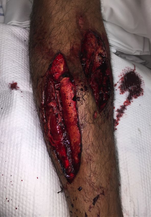
A 27-year-old Caucasian male presented to the emergency department with an open right tibia
                                  and fibula shaft fractures following a high-speed motorcycle crash. The patient was helmeted at
                                  the time of the crash and there was no reported loss of consciousness. He reported isolated
                                  right lower extremity pain without neurologic complaints.

                                  Exam and diagnostics
                                  This was an isolated injury with no clinical or radiographic evidence of intracranial, -thoracic, -
                                  abdominal, or -pelvic injury. A FAST exam (Focused Assessment with Sonography in Trauma)
                                  was performed prior to our orthopaedic exam, which demonstrated no signs of hemorrhage.

                                  Inspection of the right lower extremity revealed two large wounds to the anterolateral and
                                  anteromedial tibial diaphysis. The anterolateral and anteromedial wounds measured
                                  approximately 20 cm and 12 cm in length, respectively. Both wounds exhibited gross
                                  contamination with road debris as well as exposed muscle and fracture fragments (Figure 1).
                                  There was no clinical evidence of compartment syndrome. There were no sensory or motor
                                  deficits involving the superficial peroneal, deep peroneal, or tibial nerves. A strong dorsalis
                                  pedis pulse was palpable, however the posterior tibial pulse was unidentifiable on palpation or
                                  Doppler ultrasound. A computed tomography (CT) angiogram was obtained which
                                  demonstrated vascular stenosis of the posterior tibial artery at the level of the fracture. All
                                  hematologic and metabolic labs were within normal ranges.

2019 Hasegawa et al. Cureus 11(4): e4511. DOI 10.7759/cureus.4511                                                               2 of 10
FIGURE 1: Injury photograph
Right lower extremity wounds over the anterolateral and anteromedial tibia with exposed bone and
gross contamination.
Time from the emergency department to the initial operative
encounter
Dual antibiotic prophylaxis, consisting of cefazolin and gentamycin, was administered
2019 Hasegawa et al. Cureus 11(4): e4511. DOI 10.7759/cureus.4511 3 of 10promptly upon arrival to the emergency department. Antibiotic administration was estimated
to be within three hours from the time of injury. A brief bedside irrigation with 3 L of sterile
saline was performed and the wounds were dressed with moist gauze. The patient was then
provisionally stabilized with a moldable long leg fiberglass splint and sent for
additional preoperative imaging. Preoperative radiographs are provided in Figure 2A-2C. After
imaging was completed, the patient was brought to the operating room for urgent wound
debridement and open reduction internal fixation of the right lower extremity.
FIGURE 2: Injury radiographs
(A) Anteroposterior radiograph of the right tibia and fibula, (B) lateral radiograph of the right ankle,
(C) lateral radiograph of the right tibia and fibula.
Initial operative encounter
The patient was placed under general anesthesia. A thigh high tourniquet was placed prior to
the sterile preparation and draping of the surgical field. Gross wound contaminants were
removed sharply including all devitalized bone. The superficial and deep peroneal nerves were
identified and found to be grossly uninjured. Nine liters of sterile saline was then used to
irrigate the wound. Next, the segmental tibia fracture was provisionally stabilized with multiple
one-third tubular plates and unicortical 3.5 mm screws. A reamed intramedullary tibia nail was
then introduced via a suprapatellar approach. Once in place, multiple cerclage wires were
applied for further stabilization of large fracture fragments (Figure 3). An additional 3 L of
sterile saline was used to irrigate the wounds. Once complete, all wounds were left open and a
circumferential VeraFlo (Acelity, San Antonio, TX, USA) wound vac was applied (Figure 4A-4B).
See technique described below. Postoperative radiographs are provided in Figure 5A.
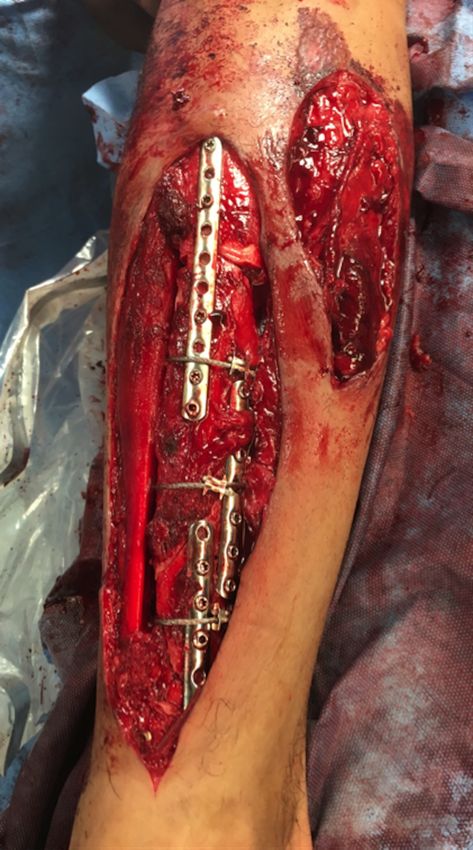
2019 Hasegawa et al. Cureus 11(4): e4511. DOI 10.7759/cureus.4511 4 of 10FIGURE 3: Initial wound debridement and fracture stabilization 2019 Hasegawa et al. Cureus 11(4): e4511. DOI 10.7759/cureus.4511 5 of 10
Intraoperative photograph of the right lower extremity wounds following initial wound debridement
                                     and fracture stabilization. The skin bridge separating the anterolateral and anteromedial wounds
                                     was later excised during the second wound debridement.

                                     FIGURE 4: Circumferential application of negative pressure
                                     wound therapy with instillation and dwell
                                     (A) Lateral view, (B) medial view.

                                     FIGURE 5: Postoperative radiographs

2019 Hasegawa et al. Cureus 11(4): e4511. DOI 10.7759/cureus.4511                                                                        6 of 10
(A) Immediate postoperative anteroposterior radiograph, (B) anteroposterior radiograph at one year
follow-up.
Circumferential wound vac technique
The anterolateral and medial wounds were lined with one inch adhesive draping strips. The
wounds were then lightly packed with Cleans Choice sponge (Acelity, San Antonio, TX,
USA) and sealed with adhesive drapes. Cleans choice sponge was placed directly over exposed
bone and hardware. When sealing the sponge, care was taken to lay drapes over the skin with
minimal skin tension. Once sealed, instillation was set at a volume of 50 mL, at 10-minute soak
times, cycled every three hours. Suction was set at -125 mmHg. Instillation consisted of
Prontosan Irrigation Solution (B. Braun Medical Inc., Bethlehem, PA, USA) for three days
followed by sterile saline for an additional four days. This allowed for a total of seven days of
NPWT-id between wound vac exchanges. Next, one-inch adhesive strips were used to line the
proximal and distal tibia circumferentially. Negative pressure sponge dressing was then applied
circumferentially within this region and sealed with adhesive drapes. Again, care was taken to
create as little tension on the skin when sealing the wound vac sponge. Once sealed, suction
was set at continuous -125 mmHg.
Hospital course
Postoperatively the patient received 72 hours of intravenous cefazolin. Prophylactic
anticoagulation was started on postoperative day one and continued until 24 hours prior to
each subsequent return to the operating room. The patient performed daily physical and
occupational therapy. The right lower extremity was restricted to partial weight bearing.
However, no range of motion restrictions was implemented.
The patient returned to the operating room two additional times for a total of three wound
debridements and two wound vac exchanges prior to flap placement. Each wound vac exchange
consisted of the same circumferential application and instillation and dwell settings as
previously described. Time between each return to the operating room was on average seven
days (i.e., hospital day one, eight, fifteen). During the first repeat wound debridement (hospital
day eight), we excised a necrotic skin bridge separating the anterolateral and medial
wounds. The single wound consequently measured roughly 20 cm x 20 cm at its longest and
widest points. Assessment of the wound each week demonstrated progressive wound bed
granulation and a decrease in overall wound size. More importantly at no time point was there
concern for superficial or deep wound infection. This included physical (e.g., erythema,
malodor, gross purulence) and hematologic (e.g., rising white blood cell count, sedimentation
rate, C-reactive protein) signs. At three weeks from the initial injury (i.e., hospital day 22), the
patient returned to the operating room for placement of an anterolateral thigh (ALT) free flap.
The size of the flap measured approximately 15 cm x 10 cm. Postoperatively the patient
remained in the hospital for seven days for flap monitoring. Outpatient follow-up ensued at six
weeks, three and six months, and one year. At the one-year follow-up there was complete
healing of the fracture site and ALT flap (Figure 5B and Figure 6A-6B).
2019 Hasegawa et al. Cureus 11(4): e4511. DOI 10.7759/cureus.4511 7 of 10FIGURE 6: Anterolateral thigh free flap at one-year follow-up
(A) Anterior view, (B) medial view.
Discussion
To date, this is the first study to report on the wound management of an open type IIIB tibia
fracture exclusively with NPWT-id prior to flap coverage. Our major findings were the absence
of infection at any time point, a dramatic decrease in wound size, and an uncomplicated delay
in flap coverage despite a highly contaminated wound, Tscherne grade III.
Infection and flap failure are well known complications following open type IIIB tibia fractures.
As pointed out by Patzakis et al. wound contamination following open fractures likely occurs
during the acute hospitalization period as opposed to the time of injury [3]. NPWT applied
under sterile conditions should theoretically prevent subsequent infection by isolating the
2019 Hasegawa et al. Cureus 11(4): e4511. DOI 10.7759/cureus.4511 8 of 10wound from hospital environmental exposure. Surprisingly, however, infection and flap failure
rates following type IIIB open tibia fractures have ranged between 34-46% and 9-14%,
respectively, despite management with NPWT [2,7].
The safety of prolonged NPWT has been questioned. Several studies have linked NPWT greater
than seven days prior to flap coverage for type IIIB tibia fractures with higher rates of infection
[7,8]. On the other hand, prolonged NPWT may be associated with a reduction in wound size
and fewer flap procedures being performed [2,9,10]. We utilized a total of 21 days of NPWT
prior to flap coverage without any infectious complications. This is an important finding as
delayed flap coverage is an often unavoidable consequence following high-energy mechanism
injuries due to time needed for resuscitation and medical optimization. We also demonstrated a
dramatic decrease in wound size. We placed an ALT free flap that measured nearly half the size
of the wound after the second debridement.
The optimal timing between wound vac exhanges has not been established. However, standard
NPWT protocols without instillation and dwell typically implement a two to three day interval
between scheduled exchanges [2,7-10]. In contrast, we implemented a weekly wound vac
exchange frequency. This was done to minimize return trips to the operating room as well as to
reduce the risk of hospital contamination that may occur during bedside wound vac exchanges.
It is unclear how our instillation and dwell protocol impacted our outcomes, as this was not
directly tested. It is possible that our instillation and dwell protocol facilitated bacteria removal
from the wound bed. Previous studies have demonstrated reduced bacterial bioburden in acute
and chronic wounds following NPWT with antiseptic instillation and dwell [6,11]. Future
studies would benefit from a comparison between NPWT-id and NPWT alone to assess the true
efficacy of instillation and dwell.
We are also the first to report on the circumferential application of any form of vac therapy.
NPWT is thought to act by improving local tissue perfusion, decreasing wound edema, and
stimulating tissue granulation [12]. It is possible that our circumferential application may have
enhanced these responses, in addition to the removal of bacteria as previously mentioned,
given the larger regional surface area covered.
We recognize there are limitations to our study. Primarily, our study design consisting of a
single case report limits the generalizability of this study. It is also questionable whether our
instillation and dwell protocol consisting of three days of an antiseptic solution followed by
four days of saline is more or less effective than either alone. As such, in addition to those
already stated, future studies should include larger populations, and a comparison between
saline and antiseptic solutions.
Conclusions
This is the first case to report on the wound management of a type IIIB open tibia fracture
exclusively with NPWT-id. While further studies are required to assess the true efficacy of our
methods, our results suggest that circumferentially applied NPWT-id is not only safe, but may
be associated with a reduced risk of infection, smaller wound size needing coverage, and allow
for delayed soft tissue coverage.
Additional Information
Disclosures
Human subjects: Consent was obtained by all participants in this study. Conflicts of interest:
In compliance with the ICMJE uniform disclosure form, all authors declare the following:
2019 Hasegawa et al. Cureus 11(4): e4511. DOI 10.7759/cureus.4511 9 of 10Payment/services info: All authors have declared that no financial support was received from
any organization for the submitted work. Financial relationships: All authors have declared
that they have no financial relationships at present or within the previous three years with any
organizations that might have an interest in the submitted work. Other relationships: All
authors have declared that there are no other relationships or activities that could appear to
have influenced the submitted work.
References
1. Gopal S, Majumder S, Batchelor AG, Knight SL, De Boer P, Smith RM: Fix and flap: the radical
orthopaedic and plastic treatment of severe open fractures of the tibia. J Bone Joint Surg Br.
2000, 82:959-966.
2. Dedmond BT, Kortesis B, Punger K, et al.: The use of negative-pressure wound therapy
(NPWT) in the temporary treatment of soft-tissue injuries associated with high-energy open
tibial shaft fractures. J Orthop Trauma. 2007, 21:11-17. 10.1097/BOT.0b013e31802cbc54
3. Patzakis MJ, Bains RS, Lee J, et al.: Prospective, randomized, double-blind study comparing
single-agent antibiotic therapy, ciprofloxacin, to combination antibiotic therapy in open
fracture wounds. J Orthop Trauma. 2000, 14:529-533. 10.1097/00005131-200011000-00002
4. Joethy J, Sebastin SJ, Chong AK, Peng YP, Puhaindran ME: Effect of negative-pressure wound
therapy on open fractures of the lower limb. Singapore Med J. 2013, 54:620-623.
10.11622/smedj.2013221
5. Kim PJ, Attinger CE, Crist BD, et al.: Negative pressure wound therapy with instillation: review
of evidence and recommendations. Wounds. 2015, 27:2-19.
6. Goss SJ, Schwartz JA, Facchin F, Avdagic E, Gendics C, Lantiz JC 2nd: Negative pressure
wound therapy with instillation (NPWTi) better reduces post-debridement bioburden in
chronically infected lower extremity wounds than NPWT alone. J Am Coll Clin Wound Spec.
2012, 4:74-80. 10.1016/j.jccw.2014.02.001
7. Hou Z, Irgit K, Strohecker KA, Matzko KE, Wingert NC, DeSantis JG, Smith WR: Delayed flap
reconstruction with vacuum-assisted closure management of the open IIIB tibial fracture. J
Trauma. 2011, 71:1705-1708. 10.1097/TA.0b013e31822e2823
8. Bhattacharyya T, Mehta P, Smith M, Pomahac B: Routine use of wound vacuum-assisted
closure does not allow coverage delay for open tibia fractures. Plast Reconstr Surg. 2008,
121:1263-1266. 10.1097/01.prs.0000305536.09242.a6
9. Schlatterer DR, Hirschfeld AG, Webb LX: Negative pressure wound therapy in grade IIIB tibial
fractures: fewer infections and fewer flap procedures. Clin Orthop Relat Res. 2015, 473:1802-
1811. 10.1007/s11999-015-4140-1
10. Karanas YL, Nigriny J, Chang J: The timing of microsurgical reconstruction in lower extremity
trauma. Microsurgery. 2008, 28:632-634. 10.1002/micr.20551
11. Ludolph I, Fried FW, Kneppe K, Arkudas A, Schmitz M, Horch RE: Negative pressure wound
treatment with computer-controlled irrigation/instillation decreases bacterial load in
contaminated wounds and facilitates wound closure. Int Wound J. 2018, 15:978-984.
10.1111/iwj.12958
12. Streubel PN, Stinner DJ, Obremskey WT: Use of negative-pressure wound therapy in
orthopaedic trauma. J Am Acad Orthop Surg. 2012, 20:564-574. 10.5435/JAAOS-20-09-564
2019 Hasegawa et al. Cureus 11(4): e4511. DOI 10.7759/cureus.4511 10 of 10You can also read