Classical presentation of a newborn infant with renal vein thrombosis - SWISS SOCIETY OF NEONATOLOGY
←
→
Page content transcription
If your browser does not render page correctly, please read the page content below
SWISS SOCIETY OF NEONATOLOGY

Classical presentation of a
newborn infant with renal
vein thrombosis

                               December 2017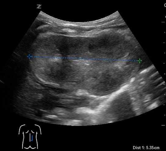
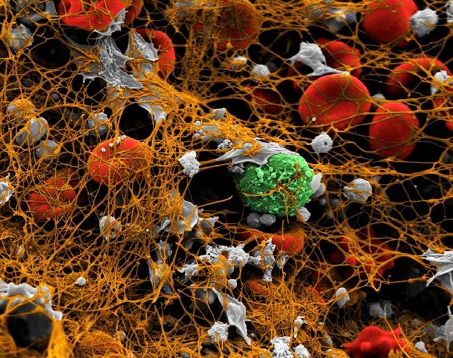
Wendelspiess M, Schifferli A, Rudin C, Kaempfen S,
Department of Neonatology (WM, KS), Department of
Pediatric Nephrology (RC), Department of Oncology /
Hematology (SA), University of Basel Children’s Hospital
(UKBB), Basel, Switzerland

Title figure:
Blood clot (source: www.biocurious.com)

© Swiss Society of Neonatology, Thomas M Berger, Webmaster
3
Thrombosis in neonates is a rare event, but associated INTRODUCTION
with significant morbidity and even mortality. Renal
vein thrombosis (RVT) accounts for approximately
10 % of venous thromboses in neonates. Compa
red to other regions of thrombosis, RVT is less often
assoc iated with central venous catheters (1). Various
pathomechanisms, which may lead to a RVT, reduce
blood flow to the kidneys or provoke hyperos
m o
larity, hypercoagulability or increased blood viscosity
(2, 3). Fetal/neonatal as well as maternal risk factors
are summarized in Fig. 1. RVT can also occur as an
extension of inferior vena cava thrombosis.
Two thirds of neonatal RVTs are diagnosed during the
first three days of life. RVTs are associated with male
sex and preterm birth. Clinical presentation includes
macrohematuria, thrombocytopenia and a palpable
abdominal mass in approximately half of the cases
(4, 5).4
CASE REPORT This male infant was born to a 33-year-old G2/P2 at
38 0/7 weeks of gestation after an uncomplicated
pregnancy. The mother developed fever during labor
and received amoxicillin clavulanate prior to delivery
for suspected chorioamnionitis. The infant was deli
vered by secondary Caesarean section and adapted
well with Apgar scores of 8, 9 and 10 at 1, 5 and
10 minutes, respectively. Arterial and venous umbilical
cord pH values were 7.28 and 7.42, respectively. Birth
weight was 3160 g (P50), length 49 cm (P50) and head
circumference 33 cm (P30).
At 34 hours of age, the infant developed macrohema
turia. Apart from a palpable mass in the right flank,
physical examination was normal. On admission to
the NICU, vital signs including blood pressure (80/40
(56) mmHg) were normal. Laboratory investigations
showed thrombocytopenia (61 G/l) and renal failure
(serum creatinine 124 µmol/l with a maternal serum
creatinine prior to delivery of 49 µmol/l). Hemoglobin
concentration was 135 g/l, and clotting studies (INR,
aPTT, fibrinogen) were within the normal range. Mar
kers of inflammation were not elevated. Urine analysis
confirmed macrohematuria, and the urine output was
normal (3.1 ml/kg/hour). An abdominal ultrasound
examination showed an enlarged right kidney with
abolished corticomedullary differentiation and echo
genic streaks in the lower pole, compatible with partial
renal venous thrombosis (Fig. 2, 3). Apart from a per
sistent foramen ovale, echocardiography was normal.5
Fetal/neonatal risk factors Maternal risk factors
Catheters T Infertility
H Oligohydramnion
CHD
R
RDS O Thrombotic states
Sepsis M Chorioamnionitis
Dehydration B Preeclampsia
O Diabetes mellitus
Birth asphyxia
S
Polycythemia I Antiphospholipid
Inherited thrombophilia S syndrome
Prematurity In utero twin death
Fig. 1
Risk factors for neonatal thrombosis (CHD: congenital
heart disease; RDS: respiratory distress syndrome)
(17).6
Fig. 2
Renal ultrasound examination on DOL 2: enlarged
right kidney (5.35 cm, normal range 3.9 – 5.1 cm).7
Fig. 3
Renal ultrasound examination on DOL 3: right kidney
with echogenic streaks and abolished corticomedul-
lary differentiation.8
On follow-up ultrasound examination a few hours later
there was extension of the thrombus into the infe
rior vena cava (IVC) over a length of 2 cm and partial
thrombosis of the left renal vein (Fig. 4). After interdis
ciplinary discussions, anticoagulation with unfraction
ated heparin (UFH) was started with a heparin bolus,
followed by a continuous infusion. Anti
c oagulation
was adjusted using the protocol published by Michel
son et al. (6) (Table), aiming at an aPPT of 60 to 85
seconds.
• Loading dose: 75 units/kg i.v. over 10 minutes
• Initial maintenance dose: 28 units/kg/hour for
infants < 1 year
• Adjust infusion rate to maintain aPTT between
60 – 85 seconds or anti-Xa between 0.35 –
0.70 IU/ml
aPTT Bolus Hold Infusion Repeat
(seconds) (units/ kg) (minutes) rate change aPTT
< 50 50 0 + 10% 4 hours
50-59 0 0 + 10% 4 hours
60-85 0 0 no change next day
86-95 0 0 - 10% 4 hours
96-120 0 30 - 10% 4 hours
> 120 0 60 - 15% 4 hours
Protocol for systemic unfractionated heparin (UFH)
administration and dose adjustments for pediatric
patients (6).9 Repeated cranial ultrasound examinations (including one obtained prior to starting anticoagulation) sho wed no signs of intracerebral hemorrhage. After initia tion of heparin treatment, no further extension of the thrombosis was documented. Serum creatinine con centrations decreased to 79 µmol/l within 96 hours. Macrohematuria disappeared after one week of life. Blood pressure remained normal. After eight days of intravenous UFH, anticoagulation was changed to enoxaparin, a low-molecular-weight heparin (LMWH), administered subcutaneously via an Insuflon® catheter. On day 13, improved blood flow in the IVC could be demonstrated on ultrasound. The infant was discharged home on day of life 14. At one month of age, the thrombi in the IVC and the left kidney had completely resolved, and only discreet signs of the thrombosis in the lower pole of the right kidney were still detectable. Renal function had nor malized (serum creatinine of 33 µmol/l). Treatment with subcutaneous LMWH was continued for a total of 3 months. Most recently, one year after discharge, renal function, blood pressure, and renal ultrasound examinations have been normal. Family history was positive for sickle cell anemia in two paternal uncles and for pulmonary embolism in a grandmother at the age of 60 years. The patient’s parents already had one healthy child. Investigations
10
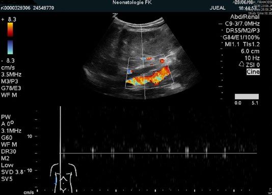
Fig. 4
Abdominal ultrasound examination on DOL 3: absent
flow in the inferior vena cava.11 for hereditary thrombophilias were negative. Sickle cell trait was excluded by hemoglobin electrophoresis at the age of six months. The etiology of RVT remained unexplained in our patient. Chorionamnionitis was the only maternal risk factor (Fig. 1), however, the baby had no signs of a neonatal infection.
12
DISCUSSION This case report describes an infant with the so-called
classical clinical presentation of RVT (macrohematuria,
palpable renal mass, and thrombocytopenia). However,
the full triad has been reported to be only present in
13 % of RVT cases (7). If RVT is suspected, the diagno
sis must be confirmed by imaging. In neonates, the ini
tial method of choice is Doppler ultrasonography (8).
In addition, laboratory markers such as full blood
count and renal function tests should be analyzed.
Coagulation tests, including activated partial throm
boplastin time (aPTT), fibrinogen and INR, are crucial
to exclude an acute hemostatic problem such as DIC
and to acquire base-line values before starting treat
ment.
If bilateral RVT is diagnosed, renal function tests, elec
trolytes and diuresis must be monitored because of
the risk of acute renal failure. In unilateral disease,
creatinine and urea concentrations usually remain nor
mal. Arterial hypertension can develop after RVT and
blood pressure must therefore be monitored regularly.
The role of anticoagulation in RVT is controversially
discussed in the literature. Strategies regarding initia
tion of treatment, route of application, choice of anti
coagulants and treatment duration differ widely due
to a lack of randomized controlled trials (9, 10).13 In cases of isolated unilateral RVT, supportive care with close monitoring is suggested since no benefit has been demonstrated while bleeding complications can occur. In contrast, for patients with unilateral RVT and extension into the IVC, anticoagulation for six weeks to three months should be considered. If both kidneys are affected with renal impairment, either thrombolytic treatment with systemic tissue plasmi nogen activator (tPA) followed by anticoagulation or anticoagulation only for six weeks to three months is generally recommended. Suggested strategies include treatment either with LMWH alone or to start with UFH followed by LMWH. We chose to start with UFH because of its rapid onset of action, its short half life and its reversibility by pro tamine sulfate should side effects (bleeding, heparin induced thrombocytopenia) occur. Disadvantages of anticoagulation with UFH include the need for intra venous administration, frequent blood tests and dose adjustments. Once adequate anticoagulation has been established and the patient is stable, treatment can be changed to subcutaneous LMWH. Major bleeding episodes in children receiving UFH treatment have been described in 1.5 % to 24 % of patients (11). Heparin-induced thrombocytopenia by UFH is relatively rare, occurring in less than 2.5 % of pediatric patients (12).
14 Therapeutic dosing of LMWH is based on anti-FXa levels. The suggested dose for subcutaneously admi nistered enoxaparin in infants < 2 months of age is 1.5 mg/kg 12 hourly. Anti-FXa levels 4 – 6 hours after subcutaneous injection should be between 0.5 – 1 U/ ml. Enoxaparin is easy to administer, even in an out patient setting (12). Although adverse events are con sidered to be rare, several major complications inclu ding major bleeding episodes, hematoma formation at the administration site, gastrointestinal bleeding, and intracranial hemorrhage have been described with an overall incidence of approximately 5 % (13 – 14). Inherited prothrombotic conditions like protein C or S deficiency, antithrombin deficiency, factor V Leiden, and mutation of prothrombin 20210A are more common in newborns with RVT as compared to the general population and should be tested in patients with an unclear origin of RVT. Babies born to mothers with antiphospholipid syndrome or lupus should be screened for lupus anticoagulant. Survival rates of infants with RVT are excellent. However, long-term sequelae, including irreversible renal damage (71 %) and arterial hypertension (19 %) can occur (4). Therefore, nephrology follow-up is required. Importantly, there is a 6.8 % risk for a second episode of venous thrombosis during puberty.
15
RVT should be suspected in newborns with hematuria, CONCLUSION
palpable abdominal mass and/or thrombocyto
p enia,
especially if neonatal or maternal risk factors for
hemostasis imbalance are present. The aim of heparin
treatment is to prevent life-threatening events, throm
bus extension, and long-term complications. However,
there is limited evidence to guide decision-making and
anticoagulation significantly increases the risk of blee
ding.16
REFERENCES 1. Schmidt B, Andrew M. Neonatal thrombosis: report of a
prospective Canadian and international registry. Pediatrics
1995;96:939 – 943 (Abstract)
2. Marks SD, Massicotte MP, Steele BT, et al. Neonatal renal
venous thrombosis: clinical outcomes and prevalence of pro
thrombotic disorders. J Pediatr 2005;146:811 – 816 (Abstract)
3. Kosh A, Kuwertz-Bröking E, Heller C, Kumik K, Schobess
R, Nowak-Göttl U. Renal venous thrombosis in neonates:
prothrombotic risk factors and long-term follow-up. Blood
2004;104:1356 – 1360 (Abstract)
4. Lau KK, Stoffman JM, Williams S, et al. for the Canadian
Pediatric Thrombosis Network. Neonatal renal vein thrombosis:
review of the English-language literature between 1992 and
2006. Pediatrics 2007;120:e1278-e1284 (Abstract)
5. Bidadi B, Nageswara A, Kaur D, Kahn S, Rodrriguez V. Neo
natal renal vein thrombosis: role of anticoagulation and
thrombolysis – an institutional review. Pediatr Hematol Oncol
2016;33:59 – 66 (Abstract)
6. Michelson AD, Bovill E, Andrew M. Antithrombotic therapy in
children. Chest 1995;108:506S-522S (no abstract available)
7. Zigman A, Yazbeck S, Emil S, Nguyen L. Renal vein thrombosis:
a 10-year review. J Pediatr Surg 2000;35:1540 – 1542 (Abstract)
8. Roy M, Turner-Gomes S, Gill G, Way C, Mernagh J, Schmidt
B. Accuracy of Doppler echocardiography for the diagnosis of
thrombosis associated with umbilical venous catheters.
J Pediatr 2002;140:131 – 134 (Abstract)9. Williams MD, Chalmers EA, Gibson BE, Haemostasis and Thrombosis Task Force, British Committee for Standards in Haematology. The investigation and management of neonatal haemostasis and thrombosis. Br J Haematol 2002;119:295 – 309 (Abstract) 10. Monagle P, Chalmers E, Chan A, deVerber G, et al. Antithrom botic therapy in neonates and children. American College of Chest Physicians Evidence-Based Clinical Practice Guidelines (8th Edition) 2008;133:887S-968S (Abstract) 11. Kuhle S, Eulmesekian P, Kavanagh B, Massicotte P, Vegh P, Mitchell LG. A clinically significant incidence of bleeding in critically ill children receiving therapeutic doses of unfrac tionated heparin: a prospective cohort study. Hematologica 2007;92:244 – 247 (Abstract) 12. Schmugge M, Risch L, Huber AR, Benn A, Fischer JE. Heparin- induced thrombocytopenia-associated thrombosis in pediatric intensive care patients. Pediatrics 2002;109:E10 (Abstract) 13. Malowany JI, Monagle P, Knoppert DC, et al. Enoxaparin for neonatal thrombosis: a call for a higher dose for neonates. Thromb Res 2008;122:826 – 830 (Abstract) 14. Romantsik O, Bruschettini M, Zappettini S, Ramenghi LA, Calevo MG. Heparin for the treatment of thrombosis in neo nates. Cochrane Database Syst Rev 2016;CD012185 (Abstract)
concept & design by mesch.ch

SUPPORTED BY

    CONTACT    Swiss Society of Neonatology
               www.neonet.ch
               webmaster@neonet.ch
You can also read