Clinical and serological evaluation of capybaras (Hydrochoerus hydrochaeris) successively exposed to an Amblyomma sculptum-derived strain of ...
←
→
Page content transcription
If your browser does not render page correctly, please read the page content below
www.nature.com/scientificreports

                                                                                                                    Corrected: Author Correction

              OPEN              Clinical and serological evaluation
                                of capybaras (Hydrochoerus
                                hydrochaeris) successively exposed
                                to an Amblyomma sculptum-derived
                                strain of Rickettsia rickettsii
                                Alejandro Ramírez-Hernández 1, Francisco Uchoa2, Maria Carolina de Azevedo Serpa1,
                                Lina C. Binder1, Alessandra Castro Rodrigues3, Matias P. J. Szabó3, Andrea Fogaça4,
                                Celso Eduardo Souza2 & Marcelo B. Labruna 1*
                                Brazilian spotted fever (BSF), caused by Rickettsia rickettsii, is the most lethal tick-borne disease
                                in the western hemisphere. In Brazil, Amblyomma sculptum ticks are the main vector. Capybaras
                                (Hydrochoerus hydrochaeris), the largest living rodents of the world (adults weighing up to 100 Kg),
                                have been recognized as amplifying hosts of R. rickettsii for A. sculptum in BSF-endemic areas; i.e., once
                                primarily infected, capybaras develop bacteremia for a few days, when feeding ticks acquire rickettsial
                                infection. We conducted experimental infections of five capybaras with an A. sculptum-derived strain of
                                R. rickettsii and performed clinical and bacteremia evaluation during primary and subsequent infections.
                                Bacteremia was detected in all capybaras during primary infection, but not in subsequent infections. All
                                animals seroconverted to R. rickettsii (titres range: 64–32,768), and remained seropositive throughout
                                the study. Primary infection resulted in clinical spotted fever illness in four capybaras, of which two had
                                a fatal outcome. Subsequent infections in seropositive capybaras resulted in no clinical signs. Capybaras
                                developed a sustained immune response that prevented a second bacteremia. This condition may
                                imply a high reproduction rate of capybaras in BSF-endemic areas, in order to continuously generate
                                capybaras susceptible to bacteremia during primary infection.

                                The bacterium Rickettsia rickettsii is the etiological agent of Rocky Mountain spotted fever, also known in Brazil
                                as Brazilian spotted fever (BSF), a disease that has been registered in different American countries including
                                Canada, United States, Mexico, Costa Rica, Panama, Colombia and Argentina1,2. This bacterium is transmitted
                                by different tick species throughout Americas [i.e., Dermacentor variabilis, Dermacentor andersoni, Rhipicephalus
                                sanguineus sensu lato (s.l.), Amblyomma cajennense species complex, and Amblyomma aureolatum]2. In Brazil, in
                                the southeastern region, Amblyomma sculptum (a member of A. cajennense species complex) is the main incrim-
                                inated vector, for which capybaras (Hydrochoerus hydrochaeris) and horses act as primary hosts for all parasitic
                                stages3,4.
                                   BSF is the most lethal tick-borne disease in Brazil with increasing numbers of cases and deaths. Between 2007
                                and 2015, 17,117 suspected cases of spotted fever (including other spotted fever group rickettsioses) were reported
                                and 1,245 were confirmed as SFG rickettsioses in 12 Brazilian states from all regions1. Moreover, case-fatality rates
                                have attained values of 30% or higher, which could be associated with low index of suspicion and misdiagnosis by
                                health-care professionals and exposure to particular eco-epidemiological risk factors1.

                                1
                                 Department of Preventive Veterinary Medicine and Animal Health, Faculty of Veterinary Medicine, University
                                of São Paulo, Av. Prof. Orlando Marques de Paiva 87, São Paulo, SP, 05508-270, Brazil. 2Reference Rickettsial
                                Diseases Laboratory, Superintendence for Control of Endemic Diseases, Mogi Guaçu, SP, Brazil. 3Ixodology
                                Laboratory, Faculty of Veterinary Medicine, Federal University of Uberlândia, Uberlândia, MG, Brazil. 4Department
                                of Parasitology, Institute of Biomedical Sciences, University of São Paulo, São Paulo, SP, Brazil. *email: labruna@
                                usp.br

Scientific Reports | (2020) 10:924 | https://doi.org/10.1038/s41598-020-57607-5                                                                    1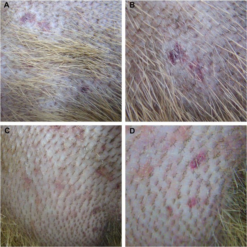
www.nature.com/scientificreports/                                                                    www.nature.com/scientificreports

                                Figure 1. Scheme of the experimental infections conducted on capybaras no. 1 to 5 during the study. Phase
                                I, primary infection of capybara no. 1, and retaining capybara no. 3 as noninfected control; phase II, second
                                infection of capybara no. 1 and primary infection of capybaras no. 2 and 3; phase III, third infection of capybara
                                no. 1 and primary infection of capybaras no. 4 and 5; phase IV, fourth infection of capybara no. 1 and second
                                infection of capybaras no. 4 and 5.

                                     Ticks function as the main reservoirs of R. rickettsii in nature, nonetheless, due to bacterial pathogenic effects
                                on ticks5,6 and some degree of tick refractoriness to bacterial infection7,8, low natural infectious rates (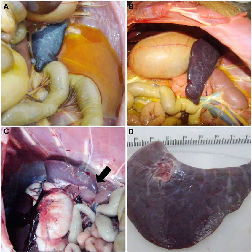
www.nature.com/scientificreports/                                                                     www.nature.com/scientificreports

                                no. 2 and 3 were seven months old (weight: ≈14 Kg), and capybaras no. 4 and 5 were five months old (weight:
                                ≈13 Kg).
                                    Before primary infection, all capybaras were clinically healthy and the indirect immunofluorescence assay
                                (IFA) with paired serum samples taken at a 14-day interval indicated that they were serologically non-reactive
                                (serum dilution 1:64) to crude antigens of R. rickettsii, Rickettsia parkeri, Rickettsia rhipicephali, Rickettsia bellii,
                                Rickettsia typhi and Rickettsia felis, as previously described12,14.
                                    For primary and subsequent infections of capybaras, two separated cotton sleeves (10-cm diameter feeding
                                chambers) were glued on the capybara shaved dorsum, as previously described12,15. These chambers were labelled
                                as chamber A (cranial position on the capybara dorsum) and chamber B (caudal position on the capybara dor-
                                sum).The minimum distance between the two chambers was 5 cm. In all cases, chamber A received 20 males
                                and 20 females (except for the primary infection of capybara 1, which consisted of 10 males and 10 females) of
                                A. sculptum from a R. rickettsii-infected colony. These adult ticks derived from the fifth generation (F5) of a tick
                                colony that was established in the laboratory from a population adapted to capybaras, collected in Itu municipal-
                                ity (São Paulo) in 201216. These adult ticks were previously exposed to rickettsial infection as larvae and nymphs
                                through feeding on guinea pigs in bacteremia that were intraperitoneally inoculated with R. rickettsii strain Itu,
                                as described8. This rickettsial strain was isolated from A. sculptum ticks collected in the same area of origin of the
                                progenitor ticks that generated our tick colony16. The infection rates were ≈30% for the tick batch used in phase
                                I and 50–60% in the batch used during phases II and III (capybaras 2–5). This rate was not calculated in phase IV
                                due to a limited number of available ticks in the colony. Chamber B received uninfected ticks and the results from
                                this chamber will be presented in a subsequent manuscript.
                                    Day 0 (zero) of infection was considered as the day of infestation with infected ticks for the first time (primary
                                infection). Capybara no. 1 were infested again with infected ticks at 120, 248, and 475 days post primary infection
                                (DPI), giving a total of four infection challenges. Capybaras nos. 2 and 3 were each exposed once to infected ticks,
                                whereas capybaras no. 4 and 5 had an additional exposure to infected ticks at 227 DPI (total: two infection chal-
                                lenges). In each infestation with infected ticks, animals were monitored daily during 30 continuous days, for fever
                                (rectal temperature) and other clinical signs. Also, blood samples were collected, within the 30-day following
                                period, from the femoral vein, for the following procedures: (i) with anticoagulant (EDTA) for guinea pig inocu-
                                lation (every 2 days), DNA extraction (every 2 days) and hematology (every 4 days); and (ii) without anticoagu-
                                lant to obtain sera for IFA test (every 6 days). In addition, a skin biopsy from the abdominal region was performed
                                with a 5 mm-punch, every 2 days (within the 30-day following period). These procedures were repeated during all
                                study phases. In the control animal (capybara no. 3 in phase I), only blood was collected for hematology analyses.
                                Prior to each procedure (sample collections and tick feeding chamber preparation) animals were slightly sedated
                                with a mixture of xylazine (0.1 mg/kg) and ketamine (1 mg/kg) by the intramuscular route (IM).
                                    In each infection phase (I to IV) a tick-naïve white New Zealand rabbit was used as control of infected ticks.
                                At the same day of capybara infestation with infected ticks, these rabbits received the same number and batch of
                                infected adult ticks. Formerly, a cotton sleeve (8-cm diameter feeding chamber) was glued on the shaved back of
                                the rabbits as described earlier17. Clinical signs and rectal temperature were registered daily, between 0 and 21
                                days post infestation, and in case of death, necropsy and organ samples (spleen) were collected and frozen. Also,
                                serum samples were collected in a 21-day interval for IFA (described below).

                                Guinea pig inoculation. Anticoagulated blood from capybaras (1.0 ml) was intraperitoneally inoculated
                                into two guinea pigs, for each blood sampling, in order to identify a probable bacteremia in the capybara, as pre-
                                viously described12. For this purpose, guinea pigs were previously anesthetized with a mixture of xylazine (10 mg/
                                kg) and ketamine (100 mg/kg) (IM)18 and a blood sample was collected by intracardiac puncture for IFA analyses
                                (day 0). The clinical signs and rectal temperature of all animals were monitored daily during 21 days. A second
                                blood sample was collected at day 21, as described above, and thereafter guinea pigs were euthanized with sodium
                                pentobarbital (100 mg/kg) by intracardiac injection. A guinea pig was considered febrile if rectal temperature was
                                ≥40 °C for at least two consecutive days. Dead individuals were submitted to necropsy and samples of spleen were
                                frozen at −20 °C for further DNA extraction and real-time PCR for Rickettsia.

                                Hematology tests.         Whole blood samples from capybaras (0.5 ml) were used for estimation of packed cell
                                volume (PCV), red blood cells (RBC) and white blood cells (WBC) total count. PCV was estimated by the micro-
                                hematocrit technique and red and white cells were counted in blood diluted with Gowers and Türk solutions,
                                respectively, using an improved Neubauer chamber, following the procedures described by Madella et al.19.

                                Serology: Indirect immunofluorescence assay (IFA). Sera obtained from capybaras, guinea pigs and
                                rabbits were tested by IFA using R. rickettsii (strain Taiaçu) crude antigen and fluorescein isothiocyanate-labelled
                                sheep anti-capybara IgG (CCZ, São Paulo, SP, Brazil), goat anti-rabbit IgG (Sigma, St. Louis, MO, USA) and
                                rabbit anti-guinea pig IgG (Sigma), respectively, as previously described7,8,14. Samples were initially tested in a
                                1:64 dilution (with PBS) as cut-off and those reactive, further diluted in twofold increments to the endpoint titre,
                                as reported earlier20. In each slide, previously known reactive and non-reactive serum, for each animal species
                                (capybara, rabbits or guinea pig) were included as positive and negative controls, respectively.

                                Real-time PCR analyses of blood and tissue samples.                  DNA from frozen (−20 °C) blood (capy-
                                baras), skin (capybaras) and spleen (guinea pigs and rabbits) samples was extracted using the DNeasy Blood
                                and Tissue kit (Qiagen Inc., Valencia, CA, USA), following the manufacturer’s protocols. Final products were
                                stored at −20 °C for further amplification by polymerase chain reaction (PCR). Extracted DNA samples were
                                used as template to amplify a 147-bp fragment of the citrate synthase gene (gltA) of Rickettsia spp. by a TaqMan
                                real-time quantitative PCR using the primers CS-5 (forward21) and CS-6 (reverse22) and an internal fluorogenic

Scientific Reports | (2020) 10:924 | https://doi.org/10.1038/s41598-020-57607-5                                                                        3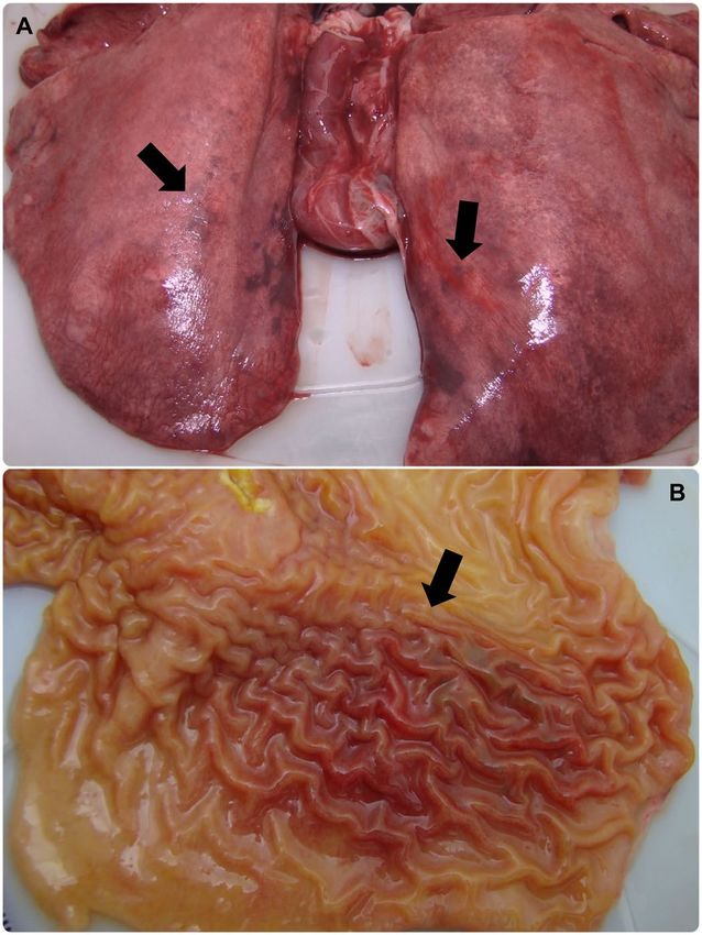
www.nature.com/scientificreports/                                                                    www.nature.com/scientificreports

                                probe (6-FAM d, BHQ- 1) (Integrated DNA Technologies, San Diego, CA), in accordance with reagents and
                                cycling conditions previously reported22. The sensitivity of the technique was determined to be 1 DNA copy of
                                R. rickettsii22. For each reaction, a positive (DNA of Rickettsia vini cultivated in Vero cells) and a negative control
                                (molecular-grade water) were included.
                                    The capybara blood and skin PCR-positive samples were submitted to a second qPCR analysis using the meth-
                                odology described for the first qPCR. The absolute number of rickettsiae per mL of blood or per mg of tissue was
                                determined using a standard curve established with the cycle of quantification (Cq) values of reactions using a
                                dilution series of 102 to 107 copies of a plasmid (pGEM-T Easy, Promega, Madison, USA) containing the 147 pb
                                fragment of gltA. All samples were analyzed in three technical replicates and respective means were calculated.

                                Histopathology and immunohistochemistry. Standard histopathology sections from organs of capyba-
                                ras that died from R. rickettsii infection were cut from formalin-fixed tissues (immersed in 10% neutral buffered
                                formalin for 24 hours and in 70% ethanol afterwards). Then they were embedded in paraffin and stained with
                                hematoxylin-eosin. Immunoalkaline phosphatase staining using naphthol fast red substrate, hematoxylin coun-
                                terstain and a polyclonal rabbit anti-R. rickettsii antiserum (Adolfo Lutz Institute, São Paulo, SP, Brazil) was used
                                for immunohistochemical testing for spotted fever group rickettsiae.

                                Results
                                Capybara infection. Five capybaras were infected during the four study phases (Fig. 1). Capybara 1 was the
                                sole animal that was re-infected three times after primary infection (four infection challenges). Capybaras 4 and
                                5 had one additional infection (two infection challenges). Unexpectedly, capybaras 2 and 3 died during primary
                                infection, impeding further exposure.
                                    As presented in Table 1, during primary infection, capybaras 2, 3, 4 and 5 manifested rectal temperatures
                                higher than 38.5 °C with onset on 8 or 9 DPI. Outstandingly, rectal temperature reached highest values of 39.7
                                and 39.8 °C in capybaras 2 and 3, respectively, which died during the bacteremic period (see below). Also, in both
                                individuals, rectal temperatures around 32 °C (hypothermia) were recorded few hours prior to death. Capybaras
                                2 to 5 presented common clinical signs during primary infection, such as lack of appetite, general and hindlimb
                                weakness (Supplementary Video), and nasal mucous discharge. Moreover, in capybaras 2 and 3, a more severe
                                clinical course was evident with signs such as inappetence, diarrhea, dark urine, prostration, coma, seizures and
                                death (at DPI 18 and 16, respectively). Both individuals also presented skin manifestations such as abdominal
                                and thoracic rash and focal purplish macules with onset at 8 DPI (Table 1 and Fig. 2A–D). The first clinical signs
                                appeared in capybaras no. 2, 3, 5 at DPI 8 and in capybara no. 4 at DPI 9 (mean incubation period: 8.3 days). In
                                contrast, clinical alterations, including rectal temperature >38.5 °C, were absent in capybara 1 during primary
                                infection and further challenges (phases I to IV) and in capybaras 4 and 5 during the subsequent exposure (phase
                                IV). Also, no clinical abnormality was observed in capybara 3 when it was the uninfected control during phase I
                                (Table 1).
                                    All rabbits, infested with the same batch of infected ticks used for infection challenges of capybaras, presented
                                fever. Most of them manifested lack of appetite, auricular, preputial and scrotal vascular abnormalities (i.e. swell-
                                ing, erythema or necrosis). Rabbits from phases I and II were more severely affected, manifested hypothermia
                                and died during the acute phase of the infection; their spleen was shown to contain rickettsial DNA by real-time
                                PCR. Rabbits from phases III and IV seroconverted to R. rickettsii with endpoint titres of 32,768 at 21 days after
                                infestation.
                                    Capybaras 2 and 3, which died during phase II, were submitted to necropsy. On gross examination, both
                                animals exhibited disseminated lesions suggestive of vascular alterations (hyperemia and hemorrhage) in various
                                organs such as spleen, stomach, small and large intestines, liver, lung, kidneys and adrenal glands (Figs. 3A,B,
                                and 4). Spleen enlargement was a prominent feature in both capybaras (Fig. 3A–D). Ascites and jaundice were
                                observed in capybara 3 (Fig. 3A). Moreover, in both capybaras, histopathology revealed a diffuse and predomi-
                                nant lymphohistiocytic vasculitis as well as perivascular edema in multiple tissues, including brain, spleen, gut,
                                kidneys, heart and liver (Fig. 5A–C). Fibrin microthrombi were seen in spleen, liver and heart (Fig. 5C). Examined
                                organs showed marked vascular congestion (spleen, liver, brain, lungs); multifocal necrosis was observed in the
                                spleen, liver, kidneys and heart. Mixed inflammatory cellular infiltrate was observed in kidneys, heart, lungs and
                                liver and severe emphysema in lungs. Immunohistochemical testing for rickettsiae revealed occasional staining
                                of coccobacilli-like structures in blood vessels (Fig. 5D) from heart, spleen and brain.

                                Guinea pig inoculation.          Anticoagulated whole blood collected every two days from each capybara dur-
                                ing infection challenges were inoculated into two guinea pigs simultaneously. During primary infection (first
                                challenge) of capybaras 1 to 5, fever was detected in guinea pigs that were inoculated with blood collected from
                                capybaras at 6 to 16 DPI, being the earliest (6 DPI) in individuals inoculated with blood from capybaras 2 and 3.
                                The proportion of febrile guinea pigs ranged from 9.4 to 61.1% with the highest values (45.0 and 61.1%) in groups
                                inoculated with blood from capybaras 2 and 3. None of the guinea pigs showed fever when inoculated with blood
                                from capybaras 1, 4 and 5 during the subsequent challenges (Table 2).
                                    Auricular and genital vascular signs like erythema, edema and necrosis were recorded in guinea pigs inocu-
                                lated with blood from capybaras 1 to 5 during their primary infection. The proportion of affected animals with
                                these conditions ranged from 3.1 to 61.1%, with the highest frequencies (≥50.0%) in individuals inoculated with
                                blood from capybaras 2 and 3. The above mentioned vascular manifestations were absent in all animals inocu-
                                lated with blood from capybaras 1, 4 and 5 during subsequent challenges (Table 2).
                                    Guinea pig death was recorded in animals inoculated with blood from capybaras 1 to 5 on 6 to 20 DPI of their
                                primary infection. The frequency of dead animals ranged from 3.1 to 27.8%, with the highest values in groups
                                inoculated with blood from capybaras 2 and 3. In addition, Rickettsia DNA was amplified from spleen collected in

Scientific Reports | (2020) 10:924 | https://doi.org/10.1038/s41598-020-57607-5                                                                      4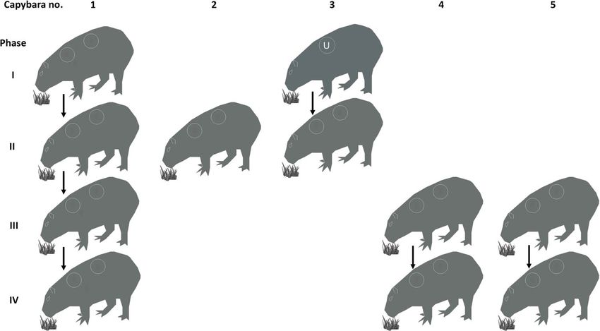
www.nature.com/scientificreports/                                                                                 www.nature.com/scientificreports

          Experimental phases#
          I                          II                                                           III                                                IV
Capybara Rectal           Clinical   Rectal                                                       Rectal                                             Rectal         Clinical
number   Temperature*     Signs╪     Temperature*     Clinical Signs╪                             Temperature*   Clinical Signs╪                     Temperature*   Signs╪
          36.7 (0.59)                36.2 (0.84)                                                  36.1 (1.2)                                         35.8 (1.05)
1                         None                        None                                                       None                                               None
          [35.4–37.9]                [34.9–37.7]                                                  [33.4–38.0]                                        [34.0–38.0]
                                                      Abdominal and thoracic rash (8/12);
                                                      fever (11/13); lack of appetite (12/14);
                                                      nasal mucous discharge (13/+); hindlimb
                                     36.8 (1.76)
2                                                     weakness (14/+); inappetence (14/+);
                                     [32.0–39.7]
                                                      purplish macules (16/+); prostration
                                                      (18/+); coma (18/+); rigid limbs (18/+);
                                                      seizures (18/+); death (18)
                                                      Abdominal and thoracic rash (8/12); fever
                                                      (9/13); lack of appetite (10/14); nasal
          37.1 (0.55)                37.1 (1.87)      mucous discharge (11/+); inappetence
3                         None
          [35.7–38.2]                [32.5–39.8]      (14/+); hindlimb weakness (14/+);
                                                      reluctance to move (15/+); diarrhea
                                                      (15/+); dark urine (15/+); death (16)
                                                                                                                 Lack of appetite (11/15);
                                                                                                  36.8 (0.84)    hindlimb weakness (11/16); nasal 35.8 (1.01)
4                                                                                                                                                                   None
                                                                                                  [35.4–38.7]    mucous discharge (12/15); fever   [34.0–37.5]
                                                                                                                 (13/13); general weakness (14/16)
                                                                                                  37.2 (1.13)    Fever (8/11); mucous feces          36.3 (0.98)
5                                                                                                                                                                   None
                                                                                                  [34.6–39.2]    (11/16); lack of appetite (11/14)   [33.9–38.0]

                                          Table 1. Clinical monitoring of five capybaras (Hydrochoerus hydrochaeris) during one to four exposures
                                          (experimental phases I to IV) to Rickettsia rickettsii strain Itu via infestations with R. rickettsii-infected
                                          Amblyomma sculptum ticks. #Phase I, primary infection of capybara no. 1, and retaining capybara no. 3 as
                                          noninfected control; phase II, second infection of capybara no. 1 and primary infection of capybaras no. 2 and
                                          3; phase III, third infection of capybara no. 1 and primary infection of capybaras no. 4 and 5; phase IV, fourth
                                          infection of capybara no. 1 and second infection of capybaras no. 4 and 5. *Values in °C, shown as: mean
                                          (standard deviation) [range]. ╪Clinical signs (days post infestation with infected ticks: onset/end of clinical
                                          sign). +Death.

                                          all dead guinea pigs. No deaths were recorded in animals inoculated with blood from capybaras 1, 4 and 5 when
                                          they were subsequently infected (Table 2).
                                              Anti-R. rickettsii IgG antibodies (≥1:64 dilution) were detected by IFA in guinea pigs inoculated during pri-
                                          mary infection of capybaras 1 to 5. The frequency of seropositive animals ranged from 9.4 to 50.0%, with high-
                                          est values in animals inoculated with blood from capybaras 2 and 3. Besides, guinea pigs inoculated with 6–18
                                          DPI-capybara blood seroconverted with endpoints titres from 8,192 to 262,144, with higher titres in animals
                                          inoculated with blood from capybaras 2 and 3 and the lowest titres in animals inoculated with blood from capy-
                                          bara 4. Furthermore, none of the guinea pigs inoculated with blood from capybaras 1, 4 and 5 during their subse-
                                          quent challenges presented antibodies to R. rickettsii before and 21 days after inoculation (Table 2).

                                          Hematology. Results were compared with those from capybara 3 during phase I (infested with non-infected
                                          A. sculptum ticks) and reference values (Table 3, Fig. 6). In general, mean PCV values during primary infection
                                          of all infected capybaras were lower than reference. Besides, minimum PCV values of 22.2, 24.4, 25.6, 31.4 and
                                          27.1% in capybaras 1 to 5, respectively, were registered between 12 and 22 DPI (Fig. 6). Conversely, mean values
                                          were higher during subsequent challenges in capybaras 1, 4 and 5 (except for second infection of capybara 1) and
                                          minimum values were within the reference range (Table 3).
                                              Mean RBC counts in capybaras were lower than reference during primary infection of all animals, except
                                          for no. 3 (Table 3). The lowest values were registered during 12–28 DPI in all individuals including capybara 3
                                          (Fig. 6). During subsequent challenges, mean counts of capybaras 4 and 5 were within reference values, except
                                          for capybara 1 in which values were slightly lower, in spite of being higher than the records of primary infection
                                          (Table 3).
                                              Mean WBC counts during primary infection was lower than reference values in capybaras 2 and 3. Besides,
                                          comparing mean counts during primary infection and subsequent challenges in capybaras 1 and 4, lower values
                                          were evident during the first infection (Table 3). The minimum values were registered during primary infection of
                                          capybaras 2 and 3 (phase II), capybara 1 (phase I) and capybara 5 (phase III), with 1.13, 1.38, 1.85 and 2.23 × 10³
                                          cells/mm3, respectively, at 12 DPI (Fig. 6).

                                          Serology. As presented in Fig. 7, capybaras 1, 4 and 5 became first seroreactive to R. rickettsii at 16–18 DPI
                                          and remained seroreactive until the last day of sampling. In capybara 1, antibody titres were followed from 0 to
                                          555 DPI. The first positive sample (≥64 titre) was registered at 16 DPI and peaked with 8,192 at 138 DPI, 18 days
                                          after the second challenge (phase II), and remained high (4,096-8,192) till 238 DPI. The lowest recorded titre was
                                          1,024 at 388 DPI, 140 days after the third challenge. Titres at the day of the 2nd, 3rd and 4th challenges were 2,048,
                                          2,048 and 1,024, respectively (Fig. 7). For capybara 4, the first positive sample was detected with a 1,024 titre at
                                          18 DPI. Posteriorly, it peaked to 8,192 between 46 and 83 DPI, and thereafter, the lowest detected titre was 2,048
                                          at 189 DPI. Before the 2nd challenge (227 DPI), the titre was 4,096. For capybara 5, the first positive sample was

    Scientific Reports | (2020) 10:924 | https://doi.org/10.1038/s41598-020-57607-5                                                                                            5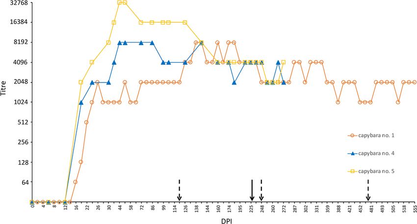
www.nature.com/scientificreports/ www.nature.com/scientificreports
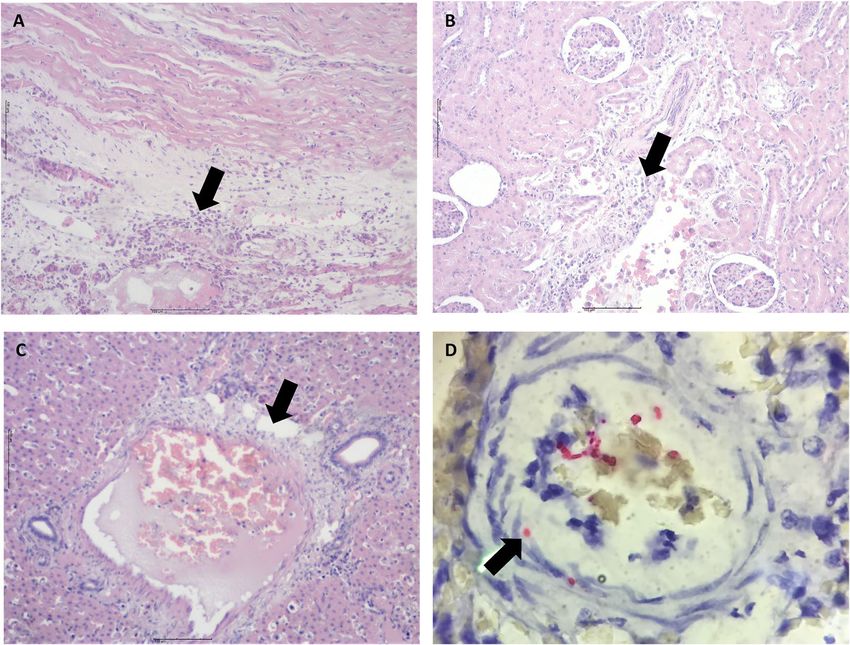
Figure 2. Skin of capybaras no. 2 and 3 during primary infection (phase II) with Rickettsia rickettsii (strain Itu)
via tick exposure. (A,B) Purplish macules in capybara no. 2 (18 DPI). (C,D) Abdominal rash in capybara no. 3
(10 DPI). This figure has been published within the Doctoral Thesis of the first author (A. Ramírez-Hernández),
which is available at the University of São Paulo’s digital library of Theses and Dissertations: https://teses.usp.br/
teses/disponiveis/10/10134/tde-09092019-112817/en.php.
detected at 18 DPI with a 2,048 titre, peaking to 32,768 between 46 and 54 DPI. Then, titre descended to 4,096
(159 DPI), which remained until and after the second challenge. At 251 DPI, 24 days after the second challenge,
capybara 5 titre decreased to 2,048. Lastly, for capybaras 2 and 3, antibodies were detected in the serum sample
before death (18 and 16 DPI, respectively) with titres of 512 and 128, correspondingly (data not graphed).
Real-time PCR from blood and skin samples. Real-time PCR in blood samples collected during pri-
mary infection revealed Rickettsia DNA in samples from capybara 2 on 14, 16 and 18 DPI, capybara 3 on 12, 14
and 16 DPI, and, capybara 5 on 12 and 14 DPI. No rickettsial DNA was detected in the blood of capybaras 1 and 4
during primary infection (Fig. 8). Rickettsia DNA was detected in skin samples from all capybaras during primary
infection, as follows: capybara no. 1 at 16 DPI; no. 2 at 8, 12, 14 and 16 DPI; no. 3 at 10, 12 and 14 DPI; no. 4 at 6,
10, 12 and 14 DPI; no. 5 at 12, 16, 20, 22 and 26 DPI (Fig. 8).
The mean number of rickettsiae per mL of blood in R. rickettsii-positive samples was 7.26E + 04, with the
highest values recorded for capybaras no. 2 at 18 DPI (3.31E + 05) and no. 3 at 16 DPI (9.48E + 04) (Table 4). For
skin samples, the mean number of rickettsia per mg of tissue was 5.24E + 02, with the highest values detected
for capybaras no. 3 on 12 and 14 DPI (3.85E + 03 and 1.18E + 03, respectively) and no. 5 at 26 DPI (1.41E + 03)
(Table 4).
Blood samples and skin biopsies from subsequent infections were not tested because guinea pig inoculations
were negative for the presence of viable rickettsia in capybara blood at all instances.
Discussion
This study evaluated for the first time repeated experimental infections of capybaras with R. rickettsii. We tried to
emulate a natural condition using ticks as the unique infection route for capybaras. Our results of guinea pig inoc-
ulations with capybara blood provided consistent evidence that capybaras developed bacteremia during the pri-
mary infection, but not during subsequent infections when they had already elicited a strong humoral response
to R. rickettsii. In addition, rickettsial DNA was detected in the blood of three of the five capybaras (no. 2, 3 and 5)
by qPCR, confirming the development of bacteremia. Only two published studies performed experimental infec-
tions of capybaras with R. rickettsii. The earliest work published by Travassos et al.11 performed subcutaneous
inoculations of capybaras with guinea pig infected blood samples. More recently, Souza et al.12 infected capybaras
Scientific Reports | (2020) 10:924 | https://doi.org/10.1038/s41598-020-57607-5 6www.nature.com/scientificreports/ www.nature.com/scientificreports
Figure 3. Abdominal cavity and spleen of capybaras no. 2 and 3 after primary infection (phase II) with
Rickettsia rickettsii (strain Itu) via tick exposure. (A) Abdominal cavity of capybara no. 3 with evidence
of ascites, jaundice and spleen enlargement (16 DPI) (B) Spleen enlargement in capybara no. 3 (16 DPI).
(C) Spleen enlargement with apical hemorrhage (arrow) in capybara no. 2 (18 DPI) (D) Enlarged spleen
from capybara no. 2 (18 DPI). This figure has been published within the Doctoral Thesis of the first author
(A. Ramírez-Hernández), which is available at the University of São Paulo’s digital library of Theses and
Dissertations: https://teses.usp.br/teses/disponiveis/10/10134/tde-09092019-112817/en.php.
by two routes, infestations with infected A. sculptum nymphs and intraperitoneal inoculation of homogenate of
guinea pig infected organs. In both studies, capybaras developed bacteremia, animals were not evaluated after the
primary infection, and there were no related clinical signs and fatal outcomes. In addition, the R. rickettsii isolates
used in these previous studies were not from capybara or A. sculptum origin.
It is noteworthy that the R. rickettsii strain used in the present study (strain Itu) was previously isolated from
A. sculptum ticks derived from a population that was sustained by capybaras under natural conditions, in Itu
municipality, a BSF-endemic area of São Paulo state16. In addition, our tick colony of A. sculptum was also derived
from this same BSF-endemic area. Indeed, this information supports strong field applicability of our results,
especially because it was recently demonstrated that the susceptibility of A. sculptum to R. rickettsii varies among
different tick populations, with a clear bias for higher susceptibility to an autochthone R. rickettsii strain that has
already coevolved with a tick population for some time8.
In contrast to the present study, capybaras of the two previous studies did not present clinical alterations dur-
ing the bacteremic period11,12. Herein we showed for the first time that capybaras manifested clinical alterations
during primary infection with strain Itu of R. rickettsii. As expected, no clinical abnormality was observed in the
control animal (capybara 3) infested with non-infected ticks during phase I of the study. While there are no ref-
erence values for normal rectal temperature of capybaras in the literature, in the present study we observed that
rectal temperature of capybara 3 during infestation with noninfected ticks never exceeded 38.2 °C. In addition,
when capybaras 1, 4 and 5 were already immune (seropositive to R. rickettsii) and did not develop bacteremia
during subsequent challenges, their rectal temperature did not exceed 38.0 °C (Table 1). Hence, for convenience,
we adopted in the present study that rectal temperatures >38.5 °C was considered as fever.
A febrile condition was manifested by capybaras 2, 3, 4 and 5 during primary infection, achieving registers
as high as 39.8 °C, which differ from previous studies in which normothermic conditions were registered11,12.
Furthermore, clinical features evidenced by febrile animals including weakness, inappetence, nasal discharge,
diarrhea, nervous disorders and even death (no. 2 and 3), diverge from earlier experimental observations with
Scientific Reports | (2020) 10:924 | https://doi.org/10.1038/s41598-020-57607-5 7www.nature.com/scientificreports/ www.nature.com/scientificreports
Figure 4. Lung and stomach of capybara no. 2 after infection (phase II) with Rickettsia rickettsii (strain Itu)
via tick exposure. (A) Lung of capybara no. 2 with evidence of bilateral disseminated vascular injuries (18
DPI). (B) Stomach of capybara no. 2 with an extended area of hemorrhage in the mucosa (18 DPI). This
figure has been published within the Doctoral Thesis of the first author (A. Ramírez-Hernández), which is
available at the University of São Paulo’s digital library of Theses and Dissertations: https://teses.usp.br/teses/
disponiveis/10/10134/tde-09092019-112817/en.php.
absence of illness11,12. One possible explanation for these differences could be the R. rickettsii strain, which were
not derived from A. sculptum in the two previous studies.
Regarding the afebrile condition of capybara 1, coincident with the shortest bacteremic period when com-
pared with capybaras 2 to 5, we infer a dose-dependent condition for these differences. Before infestations, ran-
dom samples of unfed adult ticks from the same batch of the ticks used for primary infection of capybaras were
tested by PCR (data not shown). Rickettsia rickettsii-infection rates were ≈30% for ticks used in phase I (capybara
1), and 50–60% in ticks used during phases II and III (capybaras 2–5). These differences, in conjunction with the
lower number of ticks used in capybara 1 (half of the ticks used in the remaining capybaras) might have contrib-
uted to the milder infection course of capybara 1. Higher infectious dose rates in capybaras 2 to 5 could be associ-
ated not only with severity of the fever and clinical course, but also with higher proportions of blood-inoculated
guinea pigs presenting fever, vascular injuries, serological responses and even death. Moreover, Rickettsia DNA
was detected in blood of only capybaras 2, 3 and 5.
Differences in infectivity, severity of clinical course, and mortality have been associated with infectious dose
of R. rickettsii in dogs experimentally inoculated with incremental bacterial doses23. Furthermore, Piranda et al.24
observed differences in the severity of illness, onset of fever and bacteremia between dogs inoculated intraperi-
toneally and those exposed to R. rickettsii via tick bite, being more severe in the latter. Piranda et al.24 proposed
that tick-exposed dogs may have received lower infectious doses over several days during adult engorgement in
contrast to one intraperitoneal dose. Thus, in the present study, capybaras infested with tick batches with higher
infection rates might have received higher bacterial doses during a prolonged period of tick feeding.
The clinical signs manifested by capybaras 2 to 5 during primary infection are comparable with the clinical
profile of R. rickettsii infection in susceptible animal hosts (i.e. guinea pigs, rabbits, dogs and humans). As an
example, fever, anorexia, lethargy, weakness, skin rash, diarrhea, prostration, seizures and death have been related
in experimental and natural infections in dogs23–26, and are similar to those related in human cases of BSF27,28.
Correspondingly, gross pathological lesions related to multisystemic vascular disorders observed during necropsy
of capybaras 2 and 3 concur with common findings in susceptible hosts26,29–31. These findings were corroborated
Scientific Reports | (2020) 10:924 | https://doi.org/10.1038/s41598-020-57607-5 8www.nature.com/scientificreports/ www.nature.com/scientificreports
Figure 5. Histopathological and immunohistochemical evaluation in an experimental infection of Capybaras
(Hydrochoerus hydrochaeris) with Rickettsia rickettsii. (A) Inflammation and vasculitis in heart (arrow)
(hematoxylin and eosin staining, objective 4x). (B) Inflammatory cell infiltrate in kidney (arrow) (hematoxylin
and eosin staining objective 4x). (C) Liver with vasculitis and microthrombi (arrow) (hematoxylin and eosin
staining objective 10x). (D) Brain; immunostaining of Rickettsia rickettsii in vessels (red bacili) (arrow),
immunoalkaline phosphatase staining, naphthol fast red substrate with hematoxylin counterstain (objective
100x).
by histopathology, which revealed a multisystemic vascular injury with increased vascular permeability, inflam-
mation, disseminated intravascular coagulation and an ultimate circulatory collapse. These observations are typ-
ical of severe acute rickettsial infection as already described for other host species including humans26,31 and are
underscored by location of Rickettsia in blood vessels of capybaras by immunohistochemistry.
We relied on guinea pig inoculation with capybara blood to determine the bacteremic period in each of the
infected capybaras. The mean bacteremic period was 9.2 days (range: 6–12 days). This result is similar to those
reported by Souza et al.12, when the group of capybaras infected through tick infestation presented a contin-
uous bacteremic period from 6 to 15–18 DPI (total period of 9 to 12 days). Also, they are comparable with
earlier observations made by Travassos et. al.11, with bacteremia lasting from 5 to 11 DPI (6 days) in capybaras
exposed by intraperitoneal inoculation. In addition, similar extents (8–12 days) were registered in diverse studies
with other rodent species in the United States9,32. In contrast, this bacteremic period is shorter than the 26 days
reported for the opossum Didelphis aurita in Brazil10, or the 4 week-period reported for Didelphis virginiana in
the United States33.
In addition to referenced bacteremic periods, it is noticeable that the proportion of guinea pigs with fever,
vascular clinical signs and/or seroconversion to R. rickettsii was higher for those animals that were inoculated
with blood from capybaras 2 and 3, than in those guinea pigs inoculated with blood from capybaras 4 and 5, and
even more if compared with capybara 1. Besides, highest endpoint titres (>131,072) were detected in guinea pigs
inoculated with blood from capybaras 2 and 3 (Table 2). Hence, we can correlate these findings with described
clinical pictures and mortality and associate them with higher bacterial loads, as proposed before.
Detection of Rickettsia DNA in blood was accomplished in capybaras 2, 3 and 5 between 12 and 18 DPI. This
detection period coincided with the deterioration of clinical condition in these animals. Indeed, the highest loads
of Rickettsia per mL of blood were recorded at 18 DPI for capybara no. 2 and 16 DPI for capybara no. 3, exactly
when those animals died. Besides, the DNA detection time points are within the range of bacteremic period,
established by guinea pig inoculation for each capybara. In the study of Souza et al.12, a single sample from one
capybara yielded Rickettsia DNA by real-time PCR at 12 DPI. As stated before, we can infer that differences
between both studies could be related to the R. rickettsii strain and/or the bacterial infectious doses. Regardless,
it must be emphasized that techniques employing the direct detection of rickettsial DNA in bloodstream are not
sensitive enough to determine bacteremic periods, especially in less severe infection courses.
Scientific Reports | (2020) 10:924 | https://doi.org/10.1038/s41598-020-57607-5 9www.nature.com/scientificreports/ www.nature.com/scientificreports
Guinea pig data
Auricular Genital
Fever* vascular signs** vascular signs** Death‡ Seroconversion to R. rickettsii¥
Capybara Endpoint
Number Phase# No. DPI† No. No. No. DPI† No. DPI† titre
1 I 3/32 (9.4) 8, 10 1/32 (3.1) 3/32 (9.4) 2/32 (6.3) 10 3/32 (9.4) 8, 12, 14 8,192
1 II 0/32 (0) 0/32 (0) 0/32 (0) 0/32 (0) 0/32 (0)
2 II 9/20 (45.0) 6–16 10/20 (50.0) 7/20 (35.0) 4/20 (20.0) 6, 10, 16 9/20 (45.0) 6–18 262,144
3 II 11/18 (61.1) 6–16 11/18 (61.1) 9/18 (50.0) 5/18 (27.8) 6, 8, 14, 16 9/18 (50.0) 6, 10–16 131,072
1 III 0/32 (0) 0/32 (0) 0/32 (0) 0/32 (0) 0/32 (0)
4 III 4/32 (12.5) 8–12 3/32 (9.4) 2/32 (6.3) 1/32 (3.1) 8 4/32 (12.5) 10–14 32,768
5 III 6/32 (18.8) 8–16 6/32 (18.8) 5/32 (15.6) 3/32 (9.4) 8, 14, 16 7/32 (21.9) 8–20 65,536
1 IV 0/32 (0) 0/32 (0) 0/32 (0) 0/32 (0) 0/32 (0)
4 IV 0/32 (0) 0/32 (0) 0/32 (0) 0/32 (0) 0/32 (0)
5 IV 0/32 (0) 0/32 (0) 0/32 (0) 0/32 (0) 0/32 (0)
Table 2. Clinical and serological results of guinea pigs (Cavia porcellus) inoculated with blood from capybaras
(Hydrochoerus hydrochaeris) that were submitted to one to four exposures (experimental phases I to IV) to
Rickettsia rickettsii strain Itu via infestations with R. rickettsii-infected Amblyomma sculptum ticks. #Phase
I, primary infection of capybara no. 1; phase II, second infection of capybara no. 1 and primary infection of
capybaras no. 2 and 3; phase III, third infection of capybara no. 1 and primary infection of capybaras no. 4
and 5; phase IV, fourth infection of capybara no. 1 and second infection of capybaras no. 4 and 5. *At least two
consecutive days with rectal temperature ≥40 °C; No. refers to Number of febrile guinea pigs / total Number
inoculated guinea pigs (% febrile guinea pigs). **Vascular abnormalities like edema, erythema and/or necrosis;
No. refers to Number of affected guinea pigs / total Number inoculated guinea pigs (% affected guinea pigs).
†
DPI: days post infestation of capybara with R. rickettsii-infected ticks, when capybara blood was obtained
and inoculated into guinea pigs. ‡Rickettsial DNA was amplified in spleen from all dead animals; No. refers to
Number of guinea pigs that died/ total Number inoculated guinea pigs (lethality rate). ¥Twenty-one days after
inoculation with capybara blood, guinea pigs were tested by immunofluorescence assay for seroconversion to R.
rickettsii antigens; No. refers to Number of guinea pigs that seroconverted/ total Number inoculated guinea pigs
(% seroconversion).
Packed cell volume (%) Red blood cell count (x106 cells/mm3) White blood cell count (x103 cells/mm3)
Phases# Phases# Phases#
Capybara
Number I II III IV I II III IV I II III IV
36.8 (10.83) 35.9 (2.15) 38.7 (2.23) 40.4 (1.49) 2.56 (0.48) 2.75 (0.31) 2.66 (0.49) 2.75 (0.18) 3.46 (1.73) 4.84 (0.75) 4.72 (0.95) 4.95 (0.64)
1
[22.2–50.9] [33.3–39.2] [35.0–41.4] [38.4–42.4] [1.97–3.28] [2.07–3.13] [1.80–3.36] [2.49–2.94] [1.85–6.93] [3.70–5.75] [3.28–6.38] [4.03–5.95]
33.8 (7.27) 2.70 (0.46) 1.97 (1.11)
2 - —
[24.4–40.4] [2.28–3.34] [1.13–3.23]
40.5 (4.46) 34.5 (7.49) 2.91 (0.33) 2.95 (0.36) 4.59 (1.08) 2.71 (1.56)
3
[34.9–48.1] [25.6–42.6] [2.37–3.23] [2.41–3.18] [3.40–6.68] [1.38–4.53]
7.62 (1.78)
35.6 (2.32) 40.6 (1.02) 2.65 (0.35) 3.05 (0.17) 5.89 (1.28)
4 — — [5.48–
[31.4–38.1] [39.2–41.6] [2.21–3.18] [2.78–3.32] [3.43–7.20]
10.55]
31.6 (3.34) 40.8 (1.4) 2.14 (0.42) 3.05 (0.18) 4.98 (2.06) 4.94 (0.85)
5 — —
[27.1–36.0] [38.3–42.4] [1.59–2.81] [2.81–3.29] [2.23–8.73] [3.80–6.28]
Reference
46.6–51.437; 43.3–52.938; 38.4–42.419 3.44–3.9837; 2.82–3.4438; 4.3–4.719 3.96–6.4437; 7.44–15.8238; 3.3–7.319
values*
Table 3. Hematological variables evaluated in five capybaras (Hydrochoerus hydrochaeris) one to four
exposures (experimental phases I to IV) to Rickettsia rickettsii strain Itu via infestations with R. rickettsii-
infected Amblyomma sculptum ticks. Values presented as: mean (standard deviation) [range]. #Phase I, primary
infection of capybara no. 1, and retaining capybara no. 3 as noninfected control; phase II, second infection of
capybara no. 1 and primary infection of capybaras no. 2 and 3; phase III, third infection of capybara no. 1 and
primary infection of capybaras no. 4 and 5; phase IV, fourth infection of capybara no. 1 and second infection of
capybaras no. 4 and 5. *Hematological reference values for capybaras, according to Madella et al.19, Arouca
et al.37, and Van de Heijden et al.38.
Rickettsia DNA amplification was achieved in skin samples from all capybaras during primary infection (6–26
DPI), which is similar with those results registered by Levin et al.34 in skin biopsies from R. rickettsii-infected
guinea pigs (3–22 DPI). It is remarkable that in some capybaras (no. 1 and 4), rickettsial DNA was detected in skin
samples but not in blood samples. In other capybaras (no. 2, 3 and 5) skin samples yielded rickettsial DNA earlier
(no. 2 and 3) or later (no. 5) than blood specimens. Similarly, Levin et al.34 detected Rickettsia DNA in a higher
proportion of ear-skin than in blood samples from R. rickettsii-infected guinea pigs and registered earlier or later
Scientific Reports | (2020) 10:924 | https://doi.org/10.1038/s41598-020-57607-5 10www.nature.com/scientificreports/ www.nature.com/scientificreports
Figure 6. Rickettsia rickettsii antibody titres (IFA) after multiple infections with R. rickettsii (strain ITU) via tick
exposures, in capybaras no. 1, 4 and 5. Dashed arrows indicate 2nd, 3rd and 4th infection of capybara no. 1 at 120,
248 and 475 days post primary infection (DPI), respectively. Straight arrow indicates 2nd infection of capybaras
no. 4 and 5, 227 DPI. This figure has been published within the Doctoral Thesis of the first author (A. Ramírez-
Hernández), which is available at the University of São Paulo’s digital library of Theses and Dissertations:
https://teses.usp.br/teses/disponiveis/10/10134/tde-09092019-112817/en.php.
DNA detections in skin when compared with blood specimens. Those results point out the skin as an important
site for rickettsial proliferation.
During primary infection of all capybaras, reduction in PCV values was perceptible for all individuals, mainly
during 12–22 DPI; also, RBC counts for all capybaras, but no. 3, were below reference values during 12–28 DPI.
Scientific Reports | (2020) 10:924 | https://doi.org/10.1038/s41598-020-57607-5 11www.nature.com/scientificreports/ www.nature.com/scientificreports
Figure 7. Hematological variables evaluated in capybaras (Hydrochoerus hydrochaeris) according to days post
primary infection (DPI) with Rickettsia rickettsii (strain Itu) via tick exposure. Capybara numbers 1 (X), 3
(control) (△), 2 (+), 3 (○), 4 (◇) and 5 (□). This figure has been published within the Doctoral Thesis of the first
author (A. Ramírez-Hernández), which is available at the University of São Paulo’s digital library of Theses and
Dissertations: https://teses.usp.br/teses/disponiveis/10/10134/tde-09092019-112817/en.php.
Souza et al.12 also registered this pattern in tick-infected capybaras with decrease of PCV, RBC and hemoglobin
during 15–21 DPI. Additionally, Keenan et al.23, Piranda et al.24 and Levin et al.26 also registered this hematologi-
cal variation in experimentally infected dogs during the febrile period. Likewise, WBC counts reached the lowest
values in capybaras 1, 2, 3 and 5 during primary infection at 12 DPI, which correlated with the febrile (for cap-
ybaras 2, 3 and 5) and bacteremic periods (all individuals). While previous studies in capybaras did not include
WBC count, these results are comparable with those described by Keenan et al.23 in experimental dogs. However,
leukocytosis was a more common pattern in infected animals, explained mainly by predominant monocyte and
granulocyte responses (monocytosis and granulocytosis)26. Lastly, similar studies must perform thrombocyte
counts due to its variability in infected hosts35. Herein, lack of standardization in manual counts and in blood
smear estimation for capybaras precluded reliable thrombocyte counts.
Quantification of anti-R. rickettsii IgG antibodies through IFA indicates a strong humoral response in all
capybaras. First reactive samples were detected between 16 and 18 DPI and then peaked heterogeneously (26–46
DPI) among convalescent individuals (capybaras 1, 4 and 5) and presented a variable dynamic until the end of the
study (307–555 days). Maximum titres were 8,192 (animals 1 and 4) and 32,768 (no. 5). It is noteworthy that capy-
bara 1-highest titre (8,192) was recorded after the 2nd challenge (phase II), when death of capybaras 2 and 3 was
noted. Capybara 1’s previous highest endpoint (2,048) was observed at 26 DPI (phase I). A probable strong anti-
genic stimulus could explain titre raise in capybara 1 after the second infection, a phenomenon not evidenced in
capybaras 4 and 5. IFA results agree with those of Souza et al.12 in which capybaras infected through tick feeding
showed the first detectable humoral response at 12 DPI and then peaked from 21 to 30 DPI, with mean endpoint
titres varying from 8,192 to 32,768, which remained till the end of the following period (146 DPI).
One main objective of the present study was to evaluate clinical, hematological, immunological and infectious
variables in capybaras after subsequent challenges with R. rickettsii through tick feeding. Data was collected in
capybara 1 during three subsequent challenges at 120, 248 and 475 DPI. Besides, for capybaras 4 and 5, data
was obtained for one subsequent challenge at 227 DPI. As stated before, none of the animals presented fever,
clinical signs or hematological abnormalities during subsequent infections. Also, guinea pigs inoculated with
blood during these phases did not reveal fever, clinical signs or specific antibody responses. Moreover, through
antibody follow-up period, titres ranging from 1,024 to 4,096 were noticeable, previous to each challenge, cor-
roborating a sustained immune response in capybaras, even without recent antigenic stimuli (the maximum
interval between challenges was 227 days). Thus, we can infer that primarily infected capybaras develop a durable
protective immune response that counteract further R. rickettsii challenges and impeded its corporal dissemina-
tion and establishment. Likewise, Keenan, et al.23 conducted an experimental challenge of convalescent dogs with
high R. rickettsii doses, six and twelve months after primary infection, and found no clinical nor hematological
abnormalities.
One drawback of this study could be that the number of R. rickettsii-infected ticks used for primary infection
of capybaras could be much higher than those to which capybaras are usually exposed under natural conditions.
This assumption relies on the fact that ≤1% of the A. sculptum ticks is usually found infected by R. rickettsii
under natural conditions in BSF-endemic areas3,16,21. However, in our study, capybaras were heathy and at good
Scientific Reports | (2020) 10:924 | https://doi.org/10.1038/s41598-020-57607-5 12www.nature.com/scientificreports/ www.nature.com/scientificreports
Figure 8. Molecular detection of Rickettsia rickettsii DNA in blood (X) and skin (○) samples in capybaras
(Hydrochoerus hydrochaeris) according to days post primary infection (DPI) with R. rickettsii (strain Itu) via tick
exposure.
Number of Number of
Capybara rickettsiae per mL rickettsiae per mg
number DPI of blood of skin
1 16 NA 8.84E + 01
8 NA 1.44E + 02
12 NA 2.81E + 02
2 14 4.12E + 04 3.04E + 02
16 2.04E + 04 6.17E + 01
18 3.31E + 05 NA
10 NA 2.17E + 02
12 4.03E + 04 3.85E + 03
3
14 2.24E + 04 1.18E + 03
16 9.48E + 04 NA
6 NA 9.64E + 00
10 NA 1.26E + 01
4
12 NA 1.03E + 02
14 NA 1.02E + 02
12 3.34E + 03 1.91E + 02
14 2.69E + 04 NA
16 NA 4.90E + 02
5
20 NA 1.97E + 02
22 NA 2.73E + 02
26 NA 1.41E + 03
Table 4. Absolute quantification of rickettsiae in Rickettsia rickettsii-positive samples of capybara (Hydrochoerus
hydrochaeris) blood and skin by qPCR. DPI: days post infestation with R. rickettsii-infected ticks NA: Not analyzed
because these samples did not yield rickettsial DNA in the screening qPCR analysis (see Fig. 8).
nutrition before primary infection, what could be major factors contributing to the less severe clinical outcome of
capybara no. 1, which received the least number of infected ticks in this study. On the other hand, if concomitant
diseases and malnutrition are factors contributing to a more severe outcome of R. rickettsii-primary infection of
capybaras under natural conditions, it is yet to be investigated.
In conclusion, the present study confirms that capybaras are susceptible to the strain Itu of R. rickettsii, iso-
lated from an A. sculptum population of capybara origin. In addition, this susceptibility could be dose-dependent
Scientific Reports | (2020) 10:924 | https://doi.org/10.1038/s41598-020-57607-5 13www.nature.com/scientificreports/ www.nature.com/scientificreports
due to evidence of fever, illness and mortality in some of the infected animals that received higher number of
infected ticks. Bacteremic period, hematologic and serologic patterns are similar to those reported previously by
experimental capybara infection with a R. rickettsii strain derived from A. aureolatum ticks (Taiaçu). Finally, it is
confirmed, under experimental conditions, that infected capybaras develop a sustained immune response that
prevents a further bacteremia. This condition may imply a high reproduction rate of capybaras in BSF-endemic
areas, in order to continuously generate capybaras susceptible to bacteremia during primary infection. This
statement relies on the fact that A. sculptum ticks are partially refractory to R. rickettsii, and in the absence of
vertebrate amplifying hosts (e.g. capybaras in bacteremia), R. rickettsii would disappear from the A. sculptum
population after a few tick generations36.
Data availability
The data from this study are available from the corresponding author upon reasonable request.
Received: 5 September 2019; Accepted: 2 January 2020;
Published online: 22 January 2020
References
1. Oliveira, S. V. et al. An update on the epidemiological situation of spotted fever in Brazil. J. Venom. Anim. Toxins Incl. Trop. Dis. 22,
22 (2016).
2. Parola, P. et al. Update on tick-borne rickettsioses around the world: a geographic approach. Clin. Microbiol. Rev. 26, 657–702
(2013).
3. Labruna, M. B. Ecology of Rickettsia in South America. Ann. N. Y. Acad. Sci. 1166, 156–166 (2009).
4. Szabo, M. P., Pinter, A. & Labruna, M. B. Ecology, biology and distribution of spotted-fever tick vectors in Brazil. Front. Cell. Infect.
Microbiol. 3, 27 (2013).
5. Burgdorfer, W. & Brinton, L. P. Mechanisms of transovarial infection of spotted fever Rickettsiae in ticks. Ann. N. Y. Acad. Sci. 266,
61–72 (1975).
6. Niebylski, M., Peacock, M. & Schwan, T. Lethal Effect of Rickettsia rickettsii on Its Tick Vector (Dermacentor andersoni). Appl.
Environ. Microbiol. 65, 773–778 (1999).
7. Soares, J. F., Soares, H. S., Barbieri, A. M. & Labruna, M. B. Experimental infection of the tick Amblyomma cajennense, Cayenne tick,
with Rickettsia rickettsii, the agent of Rocky Mountain spotted fever. Med. Vet. Entomol. 26, 139–151 (2012).
8. Gerardi, M. et al. Comparative susceptibility of different populations of Amblyomma sculptum to Rickettsia rickettsii. Front. Physiol.
10, 653 (2019).
9. Burgdorfer, W. Ecological and epidemiological considerations of Rocky Mountain spotted fever and scrub typhus. In Biology of
Rickettsial Diseases, vol 1. (ed. Walker, D.) 33–50 (CRC Inc. Boca Raton, 1988).
10. Horta, M. C. et al. Experimental infection of opossums Didelphis aurita by Rickettsia rickettsii and evaluation of the transmission of
the infection to ticks Amblyomma cajennense. Vector Borne Zoonotic Dis. 9, 109–118 (2009).
11. Travassos, J. & Vallejo, A. Comportamento de alguns cavídeos (Cavia aperea e Hydrochoerus capybara) às inoculações experimentais
do vírus da febre maculosa. Possibilidade desses cavídeos representarem o papel de depositários transitórios do vírus na natureza.
Mem. Inst. Butantã 15, 73–86 (1942).
12. Souza, C. E. et al. Experimental infection of capybaras Hydrochoerus hydrochaeris by Rickettsia rickettsii and evaluation of the
transmission of the infection to ticks Amblyomma cajennense. Vet. Parasitol. 161, 116–21 (2009).
13. Ueno, T. E. et al. Experimental infection of horses with Rickettsia rickettsii. Parasit Vectors 9, 499 (2016).
14. Pacheco, R. C. et al. Rickettsial infection in capybaras (Hydrochoerus hydrochaeris) from Sao Paulo, Brazil: serological evidence for
infection by Rickettsia bellii and Rickettsia parkeri. Biomedica 27, 364–371 (2007).
15. Labruna, M. B., Ogrzewalska, M., Martins, T. F., Pinter, A. & Horta, M. C. Comparative susceptibility of larval stages of Amblyomma
aureolatum, Amblyomma cajennense, and Rhipicephalus sanguineus to infection by Rickettsia rickettsii. J. Med. Entomol. 45,
1156–1159 (2008).
16. Krawczak, F. S. et al. Rickettsial infection in Amblyomma cajennense ticks and capybaras (Hydrochoerus hydrochaeris) in a Brazilian
spotted fever-endemic area. Parasit. Vectors 7, 7 (2014).
17. Pinter, A., Labruna, M. B. & Faccini, J. L. The sex ratio of Amblyomma cajennense (Acari: Ixodidae) with notes on the male feeding
period in the laboratory. Vet. Parasitol. 105, 79–88 (2002).
18. Gaertner, D. J., Hallman, T. M., Hankenson, F. & Batchelder, M. A. Anesthesia and Analgesia for Laboratory Rodents. In Anaesthesia
and Analgesia in Laboratory Animals (eds. Fish, R. E., Brown, M. J., Danneman, P. J., Karas, A. Z.) 239–297 (Academic Press, 2008).
19. Madella, D. A., Neto, E. J. R., Felisberto, M. E. & Souza, C. Valores hematológicos de capivaras (Hydrochoerus hydrochaeris)
(Rodentia: Hydrochoeridae) de vida livre na regiao de Campinas-SP. Ciência Rural 36, 1321–1324 (2006).
20. Labruna, M. B. et al. Prevalence of Rickettsia infection in dogs from the urban and rural areas of Monte Negro municipality, western
Amazon, Brazil. Vector Borne Zoonotic Dis. 7, 249–255 (2007).
21. Guedes, E. et al. Detection of Rickettsia rickettsii in the tick Amblyomma cajennense in a new Brazilian spotted fever-endemic area in
the state of Minas Gerais. Mem. Inst. Oswaldo Cruz 100, 841–845 (2005).
22. Labruna, M. B. et al. Rickettsia species infecting Amblyomma cooperi ticks from an area in the state of Sao Paulo, Brazil, where
Brazilian spotted fever is endemic. J. Clin. Microbiol. 42, 90–98 (2004).
23. Keenan, K. P. et al. Pathogenesis of infection with Rickettsia rickettsii in the dog: a disease model for Rocky Mountain spotted fever.
J. Infect. Dis. 135, 911–917 (1977).
24. Piranda, E. M. et al. Experimental infection of dogs with a Brazilian strain of Rickettsia rickettsii: clinical and laboratory findings.
Mem. Inst. Oswaldo Cruz 103, 696–701 (2008).
25. Labruna, M. B., Kamakura, O., Moraes-Filho, J., Horta, M. C. & Pacheco, R. C. Rocky Mountain Spotted Fever in Dogs, Brazil.
Emerg. Infect. Dis. 15, 458–460 (2009).
26. Levin, M. L., Killmaster, L. F., Zemtsova, G. E., Ritter, J. M. & Langham, G. Clinical presentation, convalescence, and relapse of
Rocky Mountain spotted fever in dogs experimentally infected via tick bite. Plos One 9, e115105 (2014).
27. Angerami, R. N. et al. Features of Brazilian spotted fever in two different endemic areas in Brazil. Ticks Tick Borne Dis. 3, 346–348
(2012).
28. Angerami, R. N. et al. Brazilian spotted fever: two faces of a same disease? A comparative study of clinical aspects between an old
and a new endemic area in Brazil. Clin. Microbiol. Infect. 15(Suppl 2), 207–208 (2009).
29. Walker, D. H., Crawford, C. G. & Cain, B. G. Rickettsial infection of the pulmonary microcirculation: the basis for interstitial
pneumonitis in Rocky Mountain spotted fever. Hum. Pathol. 11, 263–272 (1980).
30. Randall, M. B. & Walker, D. H. Rocky Mountain spotted fever. Gastrointestinal and pancreatic lesions and rickettsial infection. Arch.
Pathol. Lab. Med. 108, 963–967 (1984).
Scientific Reports | (2020) 10:924 | https://doi.org/10.1038/s41598-020-57607-5 14You can also read