Clinical guidelines for the recognition of melanoma of the foot and nail unit
←
→
Page content transcription
If your browser does not render page correctly, please read the page content below
Bristow et al. Journal of Foot and Ankle Research 2010, 3:25
http://www.jfootankleres.com/content/3/1/25
                                                                                                                                               JOURNAL OF FOOT
                                                                                                                                               AND ANKLE RESEARCH

 REVIEW                                                                                                                                         Open Access

Clinical guidelines for the recognition of
melanoma of the foot and nail unit
Ivan R Bristow1*, David AR de Berker2, Katharine M Acland3, Richard J Turner4, Jonathan Bowling4

  Abstract
  Malignant melanoma is a life threatening skin tumour which may arise on the foot. The prognosis for the condi-
  tion is good when lesions are diagnosed and treated early. However, lesions arising on the soles and within the
  nail unit can be difficult to recognise leading to delays in diagnosis. These guidelines have been drafted to alert
  health care practitioners to the early signs of the disease so an early diagnosis can be sought.

Overview and scope of the guidelines                                                  of the foot exhibiting unusual features. If there is any
Melanoma is a life threatening but potentially treatable                              doubt, a second opinion should be sought. At a local
form of cancer if diagnosed and managed at an early                                   level, foot clinics may wish to establish links with their
stage. Guidelines have been published to assist health-                               local dermatology and oncology services to facilitate
care workers in the recognition of malignant melanoma                                 rapid referral pathways.
of the skin [1]. However, early melanoma arising on the
foot, particularly within the nail unit and on the plantar                            What is a melanoma and how common is it?
surface, can be difficult to recognise. Consequently, this                            A melanoma is a malignant tumour (cancer) arising
can lead to delays in diagnosis. Melanoma arising on the                              from the pigment producing cell of the skin, the mela-
foot carries a particularly poor prognosis when com-                                  nocyte. The number of cases of malignant melanoma
pared to melanoma arising at other body sites [2-4]. As                               worldwide is increasing faster than any other form of
there are no consistent features of an early melanoma,                                cancer amongst Caucasians [5]. When compared to
these guidelines have been drafted to alert health care                               other forms of skin cancer, the disease is relatively
workers to the signs which may suggest melanoma and                                   uncommon [6]. However in the UK, like much of the
therefore warrant a specialist referral. A melanoma                                   world, the incidence of cutaneous melanoma continues
recognised and diagnosed at an early stage can dramati-                               to rise accounting for the majority of skin cancer deaths.
cally increase a patient’s chances of survival.                                       It has been calculated that the lifetime risk for an indivi-
   This guide has been produced as a reference for                                    dual developing the disease is 1:120 for men and 1:95
health care professionals who may be confronted with                                  for women [1]. Currently there are around 8500 new
pigmented and amelanotic lesions on the foot. It has                                  cases annually in the UK with around 1800 melanoma
been split into two sections-melanoma on the skin of                                  related deaths [7]. Cutaneous melanoma can develop on
the foot and melanoma in the nail. The paper is                                       any skin and mucosal surface. The lower limb is the
designed to act as a guide in deciding whether a pre-                                 location of around 30% of all primary cutaneous mela-
senting lesion should be referred on. It is not designed                              nomas, with women are more highly represented in this
to be a diagnostic tool-confirmation of diagnosis can                                 group, and foot and ankle lesions representing around
only be secured though appropriate biopsy, histological                               3-15% of all cutaneous melanomas [8].
examination and specialist interpretation. Furthermore,
it is appreciated that melanoma is not the only malig-                                Who is likely to develop melanoma?
nant skin tumour arising on the foot. However, these                                  There is a relationship between ultra-violet (UV) expo-
guidelines should alert practitioners to any skin lesions                             sure and the development of melanoma on sun exposed
                                                                                      sites. Data has demonstrated that in particular that irre-
* Correspondence: ib@soton.ac.uk                                                      gular and intense exposure to sunlight significantly
1
 School of Health Sciences, University of Southampton, SO17 1BJ, UK
Full list of author information is available at the end of the article                increases the risk of melanoma [9]. However, the

                                        © 2010 Bristow et al; licensee BioMed Central Ltd. This is an Open Access article distributed under the terms of the Creative Commons
                                        Attribution License (http://creativecommons.org/licenses/by/2.0), which permits unrestricted use, distribution, and reproduction in
                                        any medium, provided the original work is properly cited.
Bristow et al. Journal of Foot and Ankle Research 2010, 3:25                                                                          Page 2 of 13
http://www.jfootankleres.com/content/3/1/25

relevance of UV light on non-exposed areas such as the                            Nodular melanoma (NM)
plantar surface of the foot the role is not so clear.                             Nodular melanoma is characterised by a prominent ver-
   Melanoma is a rare occurrence before puberty, but                              tical component to the invasion of the tumour when
shows a gradual increase in incidence from the age of                             viewed under the microscope. This typically corresponds
fifteen, peaking at around the age of fifty. Around 80%                           to a pigmented lesion which may appear nodular to the
of lesions occur between the ages of 20-74 years [10].                            naked eye. This lesion is more often seen in older
White populations have a much greater risk of develop-                            patients.
ing the disease than Hispanics, Asians and Afro-Carib-
beans. Although non-white races overall have a much                               Superficial spreading melanoma (SSM)
lower rate of the disease, they are most likely to develop                        is the most common of the four types so called because
melanoma in acral locations such as the palmar, plantar                           of its radial growth phrase (lateral spread) before
surfaces and nail bed [11-15].                                                    becoming invasive. It may arise de novo or in a pre-
   Melanoma can arise in a pre-existing naevus (mole) or                          existing mole. This type has been most frequently
develop de novo on the skin. The risk of developing                               reported arising on the dorsum of the foot [16].
melanoma can be correlated to the number of naevi
(moles) an individual has. The greater the number-the                             Lentigo maligna (LM)
higher the risk. Dysplastic naevi are atypical moles                              is a type of in situ melanoma, found almost exclusively
which are generally larger than ordinary naevi and tend                           on the face and neck of older adults in the setting of
to have an irregular and indistinct border and irregular                          sun damage. Lentigo maligna may progress to lentigo
colours. Patients with dysplastic naevi are also at a                             maligna melanoma which is a lentigo maligna with an
greater risk of developing melanoma. Recognised risk                              area of dermal invasion.
factors are listed in Table 1.                                                      A small but significant proportion of melanoma lack
                                                                                  pigmentation and are hence labelled amelanotic mela-
Types of melanoma                                                                 noma. Such lesions are more likely to arise on acral
There are four main types of melanoma although not all                            areas such as the feet and be misdiagnosed as other skin
can be specifically classified as one particular type (Figure 1).                 disorders as they maybe fleshy in colour (Figure 2).
                                                                                    A large proportion of melanoma are discovered by
Acral lentiginous melanoma (ALM)                                                  patients and relatives [17]. Unfortunately, for many
This type of melanoma is characterised by having an                               patients, the foot is difficult to see and is seldom
extensive component running as a layer of malignant                               checked. Consequently, changes may not be readily
melanocytes within the basal layer of the epidermis, giv-                         observed or noted by the patient. Chiropodists/Podia-
ing rise to the term “lentiginous”. The term “acral”                              trists can play an important role in screening the foot
defines the location which is of the extremities, namely                          and leg.
the skin of the hands and feet, including the nail unit.                            The prognosis for melanoma corresponds to the histo-
ALM is the only type of MM which arises equally across                            logical (Breslow) thickness of the excised tumour. This
all skin types and is frequently observed in darker skin                          represents a measure of depth of invasion of the tumour
types and represents about half of the melanoma occur-                            into the dermis. For example, a < 1 mm thick lesion has
ring on the hands and feet. In the early stages, the clini-                       a five year survival rate of 95%, whilst a > 4 mm thick-
cal symptoms for this type of melanoma maybe very                                 ness holds a 50% chance of survival at five years. As
subtle such as an ill defined macule or patch of light                            depth of tumour is partly related to its age early identifi-
brown or grey discolouration of the skin.                                         cation of suspect lesions is paramount [18].

Table 1 Recognised risk factors for the development of melanoma
General Risk Factors                                                                         Risk factors for plantar melanoma*
•   Intense and intermittent sunlight and UV radiation exposure                              •   High total naevus body counts
•   High numbers of benign naevi and dysplastic naevi                                        •   Pre-existing naevi on the soles
•   Family history of melanoma                                                               •   History of penetrating injury
•   A personal history of 3 or more severe sunburns                                          •   Exposure to agricultural chemicals
•   Immunosuppression (including organ transplant recipients)
•   Blue or green eye colour
•   Presence of freckles
•   Inability to tan
•   Red hair colour
* Based on a single study identifying a number of risk factors for developing plantar melanoma[44].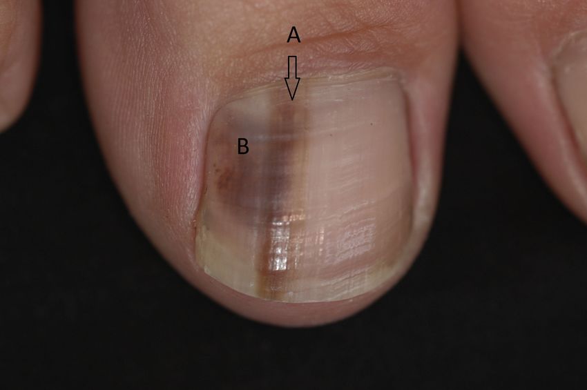
Bristow et al. Journal of Foot and Ankle Research 2010, 3:25                                                     Page 3 of 13
http://www.jfootankleres.com/content/3/1/25

 Figure 1 Various presentations of melanoma on the skin of the foot.

Assessment                                                             accurate measurements can be more objective. The
It is suggested that at an initial appointment details of              examination must be comprehensive and include inter-
any pigmented or solitary lesion arising on the feet is                digital areas and the plantar surface.
recorded in the patient’s notes with a description includ-               When assessing lesions, a history of trauma should
ing location, size, colour and shape. Inclusion of                     not exclude the possibility of a melanoma. Evidence
Bristow et al. Journal of Foot and Ankle Research 2010, 3:25                                                             Page 4 of 13
http://www.jfootankleres.com/content/3/1/25

 Figure 2 Amelanotic melanoma arising on the skin of the foot.

suggests many cases of melanoma are brought to the                         in the periungual areas, beneath or around the nails
attention of the patient by co-incidental trauma and                       [26]. Lack of pigmentation in suspect pedal lesions can
injury. The role of trauma in the aetiology of melanoma                    compound the problem. Many misdiagnoses are made
remains controversial, but it may bring the patient’s                      in favour of more benign conditions such as:
attention to an existing lesion.
  The use of the simple acronym ABCDE [19] is a use-                          •   Ingrowing toe nail
ful tool in remembering the main clinical signs of a                          •   Foot ulcer
potential melanoma (See Table 2) but may miss amela-                          •   Wart/verrucae
notic or smaller lesions [20]. Any mole or solitary vascu-                    •   Tinea Pedis/Onychomycosis
lar lesion whether new or pre-existing which is growing                       •   Bruising
or changing shape or colour should be referred for a                          •   Foreign body
specialist opinion.                                                           •   Sub-ungual haematoma
  The utility of the standard ABCDE system for plantar                        •   Pyogenic granuloma
and nail lesions has been questioned owing to the varia-                      •   Poroma
tion in presentation on the plantar surface and within                        •   Hyperkeratosis-corns/callus
the nail unit compared to other areas of the skin                             •   Necrosis
[21-23]. Moreover, data has highlighted how melanoma                          •   Paronychia
on the foot holds a poorer prognosis than melanoma                            •   Ganglion
elsewhere due to delays in presentation and misdiagno-
sis of the condition [23-25] particularly so when located                     As many of the benign conditions are very common,
                                                                           identifying a rare occurrence of melanoma amongst
                                                                           them can be challenging. In view of the additional diffi-
Table 2 The ABCDE acronym                                                  culties the authors offer an alternative acronym to high-
A Asymmetry. One half of the lesion is not identical to the other.         light potential melanoma on the foot using the acronym
B Border. A lesion with an irregular, ragged or indistinct border.         “CUBED” (Table 3).
C Lesion has more than one Colour present within it.
                                                                              Clinical judgement should identify lesions which
                                                                           appear “unusual” in their form or have atypical features.
D Diameter. The lesion has a diameter of greater than 6 mm.
                                                                           For example, the appearance of a suspicious foot ulcer
E Evolution. Any change in the lesion in terms of size, shape or colour.
                                                                           in a patient without the normal risk factors (neuropathy,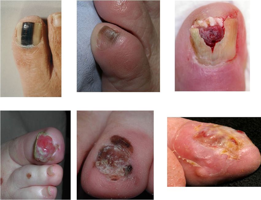
Bristow et al. Journal of Foot and Ankle Research 2010, 3:25                                                                   Page 5 of 13
http://www.jfootankleres.com/content/3/1/25

Table 3 The “CUBED” acronym for foot melanoma
  C    Coloured lesions where any part is not skin colour.
  U    Uncertain diagnosis. Any lesion that does not have a definite diagnosis
  B    Bleeding lesions on the foot or under the nail, whether the bleeding is direct bleeding or oozing of fluid. This includes chronic
       “granulation tissue”.
  E    Enlargement or deterioration of a lesion or ulcer despite therapy
  B    Delay in healing of any lesion beyond 2 months.
Refer when any two features apply.

diabetes etc) should raise concerns as to the correct                  Associated with this drift a small transverse groove will
diagnosis. Furthermore, when individual skin lesions                   often emerge from beneath the nail fold about 2 months
don’t respond to a treatment in the normal, timely man-                after the cause of the bleed. This represents a step dis-
ner the original diagnosis should be re-considered.                    turbance of nail plate production, precipitated by the
  Dermoscopy has been demonstrated to be a useful                      same episode that caused the bleed, but emerging later
adjunct in the visual assessment of pigmented lesions to               as it requires the nail to grow by the length of the prox-
detect potential melanoma on acral skin [27] however,                  imal nail fold before the sign is manifest. Clinical photo-
such equipment requires training and knowledge before                  graphy is of great value in documenting the exact form
use. Readers are referred to the article by Bristow and                and dimensions of pigmented marks within the nail
Bowling [28].                                                          unit. It is best done at the outset, where change over 3
                                                                       months can provide very useful clues. A source of pig-
Nail unit melanoma                                                     ment that clears proximally as it progresses distally will
Like elsewhere on the foot, melanoma of the nail unit                  almost always be subungual blood.
(NUM) is typically diagnosed at a later stage in its evo-                Longitudinal melanonychia reflects melanin pigment
lution than melanoma at most other body sites. Accord-                 created during nail plate generation incorporated within
ingly, the tumours are thicker and there is a worse                    the nail plate as it is formed by the matrix (Figure 5).
prognosis than for other melanoma. A large UK survey                   Subungual bleeding (or subungual haematoma) repre-
of 4 regions demonstrated that NUM represented 1.4%                    sents blood beneath the nail, which in some instances
of melanoma over a 10 year period, giving an incidence                 may be trapped within pockets of nail plate and be car-
of 1 per million of population per year. The 5 year sur-               ried with it as the nail grows. Both longitudinal melano-
vival of this group was 51%, where those with a Breslow                nychia and subungual bleeding have a range of benign
thickness of less than 2.5 mm had a 5 year survival of                 and malignant causes (see Table 4). Clinically they can
88% and those for which the thickness was 2.5 mm or                    be distinguished on a series of points (Table 5), where
greater, had a 44% 5 year survival rate [29].                          some of these points can be clarified with dermoscopy.
                                                                       The dermatoscope is a hand held instrument that com-
Presentation of melanoma in the nail unit                              bines a x10 lens with an internal light source. It can be
There are 2 main patterns of nail unit melanoma                        held directly against the nail plate and periungual skin
(NUM); longitudinal melanonychia and amelanotic                        to examine pigment and other characteristics [31].
tumours (Figure 3). The first may be associated with                   When used in combination with clear jelly, a continuous
alteration of nail plate anatomy in more advanced cases.               medium is established between the light source and the
The latter is almost always associated with nail plate                 reflective pigments of the nail plate by avoiding an air
change. Some NUM may present with features common                      interface. This greatly improves the amount of informa-
to both patterns.                                                      tion available to enable the clinician to analyse the
                                                                       source of pigment [32]. There are occasions when a
Differential diagnosis: Melanoma or haematoma?                         malignancy beneath the nail will bleed such that the
The most common clinical presentation to cause uncer-                  presence of blood does not rule out malignancy and
tainty is subungual bleeding. The history can be of great              associated features need to be considered [30,31]
value. A subungual bleed will normally have arisen                       One of the biological rules of the nail unit is that
within a day or two and may be associated with an epi-                 functioning melanocytes are limited to the matrix and
sode of trauma, or more commonly, a period of vigor-                   nail folds, but not found in the nail bed. This means
ous activity or sport where no trauma is recollected.                  that if pigment change occurs within a structurally nor-
Having been noted, it will not change greatly, although                mal nail or nail bed, with no continuity with the nail
the clinician will note a distal drift with time if they               folds or matrix, then it is not likely to be melanocytic
review over a period of several months [30] (Figure 4).                and hence cannot be a melanoma. This leads to 2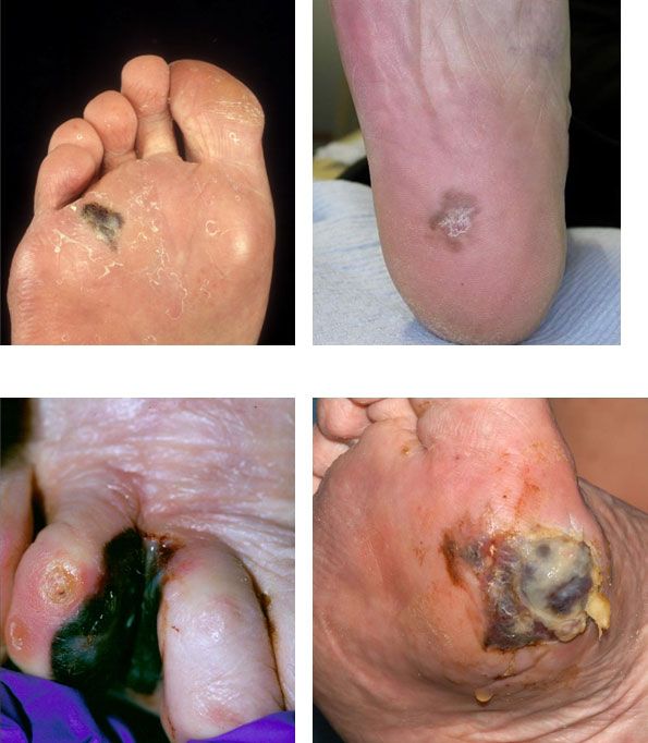
Bristow et al. Journal of Foot and Ankle Research 2010, 3:25                                                   Page 6 of 13
http://www.jfootankleres.com/content/3/1/25

 Figure 3 Various presentations of nail unit melanoma.

simple rules:                                                  the foot. The band is likely to represent a physical dis-
                                                               turbance to nail production associated with the episode
    1. Pigment arising solely within the nail bed with         of trauma which in turn will make the nail less translu-
    normal matrix and nail folds is not likely to be a         cent for a brief zone. This white band is not seen in
    melanoma                                                   melanocytic causes of nail discoloration.
    2. Where melanoma involves the nail bed, there will
    be a history of the disease starting in the nail matrix    What is the likely cause of the longitudinal
    or nail fold.                                              melanonychia?
                                                               The longitudinal melanonychia most likely to represent
  The shape of the outline of the pigmentation is also a       malignancy is that arising as a solitary pigmented streak
useful clue. Blood may present as small irregular pools        in a white person with fair colouring and of middle age
within the nail bed, with adjacent puddles or drops of         or older. In a dark skinned person, benign nail pigmenta-
purplish brown discoloration. By contrast, longitudinal        tion becomes increasingly common with age and is typi-
melanonychia arises as a well organised band of similar        cally found in varying degrees of intensity on several
width throughout the longitudinal axis, arising in the         digits. In all instances, there needs to be careful evalua-
matrix and extending to the distal edge.                       tion to determine the cause of the pigmentation [30,34].
  An anecdotal clinical observation is that traumatic          If no satisfactory benign explanation can be found, then
causes of subungual bleeding are associated with a prox-       they should be reviewed by a Dermatologist to consider
imal white transverse band in many instances [33]. This        the need for biopsy. The most common causes are drugs,
is more common for trauma to digits of the hand than           trauma, fungal infection (Figure 6) and inflammatory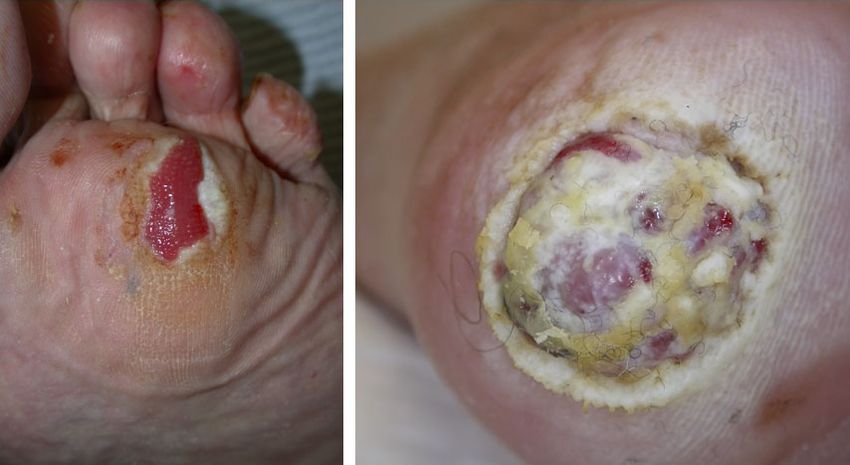
Bristow et al. Journal of Foot and Ankle Research 2010, 3:25                                                       Page 7 of 13
http://www.jfootankleres.com/content/3/1/25

 Figure 4 Subungual haematoma. Demonstration of haematoma by clear nail growth proximally.

diseases such as lichen planus which may be manifest                the melanin within the nail being visible through the
elsewhere on the skin. Both squamous cell carcinoma                 translucent edges of the proximal nail fold as it dwindles
and melanoma would be considered during assessment.                 to a cuticle [35].
In rare instances, the pigment is exogenous, such as that             Evolution of the pigmentation is diagnostically useful,
produced by potassium permanganate. This can be                     but not reliable as a means of ensuring that the source
demonstrated by scraping the surface of the nail. Where             of pigment is benign. Whereas blood may be distin-
there is onycholysis, the same may apply to the undersur-           guished from melanin over a period of a few months,
face of the nail. This is particularly the case where there         the characterisation of a benign or malignant source of
is colonisation by pseudomonas which can lend a green               melanin is less easy. Pigment that does not change is
to black appearance.                                                not necessarily benign, however the longitudinal mela-
   Other details for consideration include the pattern of           nonychia that increases in width or variety of pigment is
the pigment within the longitudinal streak and whether              more likely to represent malignancy than one that is sta-
there is any spread of the pigment onto adjacent skin.              tic. One exception to this is longitudinal melanonychia
Dermoscopy is helpful in both instances and where the               in children where the pigment arises in a subungual
pigment is heterogeneous in both the longitudinal and               naevus which changes as the child matures [34]. Quite
transverse axes (Figure 7), the likelihood of melanoma is           dramatic nail pigmentation can evolve quickly from a
greater [31]. Detection of pigment on the nail folds or             benign lesion and biopsy would rarely be undertaken in
digit pulp can also be easier with dermoscopy. Where                this group. A further exception is the evolution of a pig-
present, it is referred to as Hutchinson’s sign after the           mented streak that comes to be associated with other
surgeon of that name noted it in the early historic                 pigmented streaks on other nails of the hands and feet.
accounts of subungual melanoma and referred to it as a              This indicates a systemic process and is common in
“melanotic whitlow” conferring a poor prognosis. It is to           dark skinned races, those taking certain drugs and in a
be distinguished from the “pseudo-Hutchinsons sign”                 condition termed Laugier Hunziker syndrome. Laugier
which is the appearance of periungual pigment leant by              Hunziker syndrome is increased patchy pigmentation of
Bristow et al. Journal of Foot and Ankle Research 2010, 3:25 Page 8 of 13
http://www.jfootankleres.com/content/3/1/25
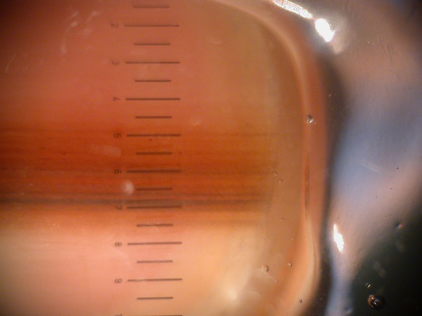
Figure 5 A single nail exhibiting both longitudinal melanonychia and haematoma. A: Longitudinal melanonychia arising in the nail matrix
from the melanocytes. B: Subungual haematoma limited to the nail bed with poorly defined, rounded borders.
Table 4 Causes of melanonychia compared with those of subungual bleeding
Melanonychia Subungual bleeding
Benign racial melanonychia Direct trauma
Laugier Hunziker Indirect microtrauma-end on repetitive trauma
Inflammation Haemorrhagic tendency lowering threshold for effects of trauma. eg
• Lichen planus • warfarin
• Chronic paronychia • leukaemic
• Trauma/friction • thrombocytopaenia
• radiation
Medication e.g. Subungual tumour
• Minocycline • squamous cell carcinoma
• Chemotherapy • wart
• HIV disease or medication • exostosis
• melanoma
• pyogenic granuloma
Addison’s disease
Peutz Jeghers
Subungual naevus
Benign melanocyte activation
Melanoma
Bowen’s disease (in situ squamous cell carcinoma)
OnychomycosisBristow et al. Journal of Foot and Ankle Research 2010, 3:25 Page 9 of 13
http://www.jfootankleres.com/content/3/1/25
Table 5 Features of longitudinal melanonychia compared with those of subungual bleeding-all features are generally
true, but there can be individual exceptions
Melanoncyhia Subungual bleeding
The duration of history is from 3-6 months upwards to 20 years or The duration of history is rarely more than 6 months and is typically shorter
more
A history of trauma is quite common A history of trauma or precipitating activity is quite common
Lateral margins within the nail are mainly straight and longitudinally Lateral margins may be irregular
oriented
Where margins merges with the nail fold, pigment may spread onto Pigment rarely extends from beneath the nail plate
nail fold (Hutchinson’s sign)
There are rarely any detectable transverse features There may be a proximal transverse groove and/or transverse white mark
within the nail
In the absence of clinical tumour, nail plate pigmentation is in Haemorrhage may be broken up into a number of zones
continuity with a single zone
Dermoscopy reveals Dermoscopy reveals
• continuous pigment between proximal nail fold and distal free • Pigment may not be continuous in the longitudinal axis, with clear
edge nail at either the proximal or distal margin
• in the transverse axis, pigment may vary-whereas in the • Pigment may vary in any axis
longitudinal axis it remains largely constant
• There may be longitudinal flecks of darker pigment within the • Droplets of blood may be seen separated from the main zone of
background pigment of the nail pigmentation
• Pigment is mainly brown black • Blood may be seen as a discrete layer of material on the lower aspect
of the nail plate at the free margin
• Pigment may be purple black, with increasing red hues at margins. It
is rarely brown
mucosae of the mouth and/or genitals, associated with non-dermatophytes and may represent a therapeutic
multiple homogenous pigmented longitudinal bands in challenge likely to be surmounted only if the pathogen
the nails. It is common for this problem to present with is known.
one nail in the first instance and hence the value in Levit has used a modification of the ABCD rule devel-
making a proper examination of all nails and other oped for detection of suspicious pigmented lesions on
areas as appropriate [36]. Multiple pigmented bands in the skin and applied it to the nail unit [39]. First is A
dark skinned people may also initially be noted in one for Age, in the 5th to 7th decade of life. B stands for a
nail alone, but are soon detected in others. Band (longitudinal streak) that is brown or black and
measures 3 mm or more. C stands for Change in the
The abnormal nail plate associated with pigment nail band or lack change in the nail morphology in spite
A nail plate that is structurally altered presents a differ- of presumed adequate treatment. D stands for the Digit
ent scenario. Where there is a longitudinal melanony- most commonly involved, which for the foot would be
chia associated with loss of nail integrity this raises the big toe. E stands for Extension of the pigment onto
concern and needs immediate assessment. In other the adjacent skin or nail fold, known also as Hutchin-
instances, the pigment may be broken up or scattered son’s sign and F stands for Family history of melanoma
within a creamy yellow nail plate. Where there is no or dysplastic naevus. All these points are reasonable and
preceding history of longitudinal melanonychia, this may may guide the practitioner to seek advice (Table 6).
represent a pigmented onychomycosis with damage to They may in turn help the dermatologist when deciding
the nail plate. This can be difficult to assess. Unlike mel- to do a biopsy, although all the other points raised in
anocytic pigment which starts in the matrix, the pattern the preceding text would be considered in taking this
of onychomycosis usually extends from the distal free step. However, a final diagnosis of melanoma will
edge with proximal progression. Early reassurance can depend on the histology.
be given if the pigmented change and dystrophic nail
can all be trimmed away with no disturbance of sur- Amelanotic tumour of the nail unit
rounding skin and there is no sign of a more proximal Amelanotic melanoma arises in the nail unit as it is does
origin to the pathology. Suspicion of fungus should at other acral locations, at a rate higher than other body
always be explored by mycological assessment and in sites. The lack of overt pigment appears to delay the
particular culture. There is a wide variety of potential diagnosis further, which in turn affects prognosis [25].
organisms [37,38]. Some of the pigmented fungi are There may sometimes be small pigmented tints to anBristow et al. Journal of Foot and Ankle Research 2010, 3:25 Page 10 of 13 http://www.jfootankleres.com/content/3/1/25 Figure 6 Fungal infection of the nail caused by Fusarium sp. Causing a longitudinal melanonychia otherwise pink or granulomatous mass [31]. The differ- dressings for the last X months and it just wouldn’t ential diagnosis of amelanotic melanoma is considered heal”. Although this article is examining presentation for all pyogenic granuloma, which is a common benign and diagnosis of acral melanoma, squamous cell carci- diagnosis presenting as a vascular nodule. Pyogenic noma can also present this way and hence the value in granuloma is usually found on the fingers or toes, bleeds asking for histological assessment of any lesion that easily and does not readily remit. In Dermatological does not resolve in 2 months, but which oozes or bleeds practice, a pyogenic granuloma would normally be sur- or has no clear diagnosis. Concern is greatest when the gically removed. This provides histology to ensure that tumour causes disturbance of nail integrity as it arises it was not a melanoma at the same time as resolving the in the nail matrix and destroys the specialised nail clinical complaint. In biological terms, pyogenic granu- matrix epithelium such that it can not produce nail. loma has much in common with the granulation tissue In conclusion, NUM is best detected early if all clini- of ingrowing toenail. Amelanotic melanoma presenting cians and patients have a low threshold for asking for as a granulating mass of the nail fold can be interpreted advice early. In particular this means avoiding prolonged as an ingrowing nail. This is a well recognised pitfall in periods of conservative management of change in the podiatry and a potential cause of delayed diagnosis nail or periungual tissues that are limited to one digit which compromises prognosis [40-43]. Where practice and do not respond promptly to appropriate treatment. entails cauterising or simply dressing fleshy granuloma- For less advanced lesions, where there is only altered tous masses of the extremities there is a significant risk pigment, if such pigmentation is limited to a single digit of leaving a malignancy undiagnosed. In the authors’ and cannot confidently be attributed to a single episode experience patients with advanced amelanotic melanoma of subungual bleeding then expert advice should be of the hand or foot often say “they treated it with sought. In all instances, although general practitioners
Bristow et al. Journal of Foot and Ankle Research 2010, 3:25                                                                         Page 11 of 13
http://www.jfootankleres.com/content/3/1/25

 Figure 7 Dermoscopy of the nail plate demonstrating heterogenous streaks in the longitudinal and horizontal axes.

are a good source of general assessment, they typically                       presentation. As a diagnosis of melanoma is relatively
do not have any experience of NUM. We would recom-                            uncommon and can only be made after a full profes-
mend assessment by a Dermatologist.                                           sional assessment and biopsy, practitioners should be
                                                                              cautious and not speculative when giving any advice to
Referral                                                                      the patient about potential diagnoses to prevent any
If a melanoma is suspected, the normal route for referral                     unnecessary alarm and concern. A point to emphasise
would be to a general practitioner. Occasionally, direct                      to all patients is that it is important to know the diagno-
referral to the dermatology department may be possible,                       sis of what is being treated. If that diagnosis is not clear,
but local policies will dictate this. Under current NICE                      or becomes unclear due to unusual clinical response to
guidelines in the UK, patients with suspected melanoma                        development, then both patient and the practitioner
should be seen by a specialist within two weeks of                            need the benefit of a clear diagnosis.

Table 6 The ABCDE of nail melanoma after Levit [39]
  A    Age Range 20-90, peak 5th -7th decades.
  B    Band (nail band): Pigment (brown-black). Breadth > 3 mm. Border (irregular/blurred).
  C    Change: rapid increase in size/growth rate of nail band. Lack of change: failure of nail dystrophy to improve despite adequate treatment.
  D    Digit Involved: Thumb > hallux > index finger > single digit > multiple digits.
  E    Extension: Extension of pigment to involve proximal or lateral nail fold (hutchinson’s sign) or free edge of nail plate.
  F    Family or personal history: Of previous melanoma or dysplastic nevus.
Bristow et al. Journal of Foot and Ankle Research 2010, 3:25 Page 12 of 13
http://www.jfootankleres.com/content/3/1/25
Summary points 8. Soong SJ, Shaw HM, Balch CM, McCarthy WH, Urist MM, Lee JY: Predicting
survival and recurrence in localized melanoma: a multivariate approach.
• Melanoma can occur on any part of the foot, World J Surg 1992, 16:191-195.
including the nail unit, in all ethnic groups and skin 9. Gandini S, Sera F, Cattaruzza MS, Pasquini P, Picconi O, Boyle P, Melchi CF:
types. Meta-analysis of risk factors for cutaneous melanoma: II. Sun exposure.
Eur J Cancer 2005, 41:45-60.
• Early recognition and diagnosis can significantly 10. Ries LA, Wingo PA, Miller DS, Howe HL, Weir HK, Rosenberg HM,
improve prognosis. Vernon SW, Cronin K, Edwards BK: The annual report to the nation on the
• Melanoma of the foot is frequently misdiagnosed, status of cancer, 1973-1997, with a special section on colorectal cancer.
Cancer 2000, 88:2398-2424.
especially when lesions are amelanotic or arise 11. Chang JW, Yeh KY, Wang CH, Yang TS, Chiang HF, Wei FC, Kuo TT,
within the nail unit. Yang CH: Malignant melanoma in Taiwan: a prognostic study of 181
• The use of the “ABCDE” and “CUBED” acronyms cases. Melanoma Res 2004, 14:537-541.
12. Ishihara K, Saida T, Yamamoto A: Updated statistical data for malignant
may improve practitioner’s assessment of unusual melanoma in Japan. Int J Clin Oncol 2001, 6:109-116.
lesions. 13. Al-Maghrabi JA, Al-Ghamdi AS, Elhakeem HA: Pattern of skin cancer in
• Any skin or nail lesion arising on the foot with an Southwestern Saudi Arabia. Saudi Med J 2004, 25:776-779.
14. Muchmore JH, Mizuguchi RS, Lee C: Malignant melanoma in American
unclear diagnosis, which deteriorates or fails to heal black females: an unusual distribution of primary sites. J Am Coll Surg
within two months despite treatment or exhibits 1996, 183:457-465.
unusual features should be reassessed, and referred 15. Bellows CF, Belafsky P, Fortgang IS, Beech DJ: Melanoma in African-
Americans: Trends in biological behavior and clinical characteristics over
if considered appropriate. two decades. J Surg Oncol 2001, 78:10-16.
16. Barnes B, Seigler H, Saxby T, Kocher M, Harrelson J: Melanoma of the foot.
Consent J Bone Joint Surg Am 1994, 76:892-898.
17. Hamidi R, Cockburn MG, Peng DH: Prevalence and predictors of skin self-
Written informed consent was obtained from the patient examination: prospects for melanoma prevention and early detection.
for publication of this case report and accompanying Int J Dermatol 2008, 47:993-1003.
images. A copy of the written consent is available for 18. Büttner P, Garbe C, Bertz J, Burg G, D’Hoedt B, Drepper H, Guggenmoos-
Holzmann I, Lechner W, Lippold A, Orfanos CE, et al: Primary cutaneous
review by the Editor-in-Chief of this journal. melanoma. Optimized cutoff points of tumor thickness and importance
of clark’s level for prognostic classification. Cancer 1995, 75:2499-2506.
19. Malignant Melanoma. [http://www.aad.org/public/publications/pamphlets/
Author details sun_malignant.html].
1
School of Health Sciences, University of Southampton, SO17 1BJ, UK. 2Bristol 20. Strayer S: Diagnosing skin malignancy: Assessment of predictive clinical
Dermatology Centre, Bristol Royal Infirmary, Bristol, BS2 8HW, UK. 3St Johns criteria and risk factors. J Fam Pract 2003, 52:210-218.
Institute of Dermatology, St Thomas’ Hospital, London, SE1 7EH, UK. 21. Albreski D, Sloan SB: Melanoma of the feet: misdiagnosed and
4
Department of Dermatology, Oxford Radcliffe Hospital, Oxford, OX3 7LJ, UK. misunderstood. Clin Dermatol 2009, 27:556-563.
22. Bristow I, Acland K: Acral lentiginous melanoma of the foot: a review of
Authors’ contributions 27 cases. J Foot Ankle Res 2008, 1:11.
The paper was initially drafted by IB and DB. RT, KA and JB reviewed the 23. Metzger S, Ellwanger U, Stroebel W, Schiebel U, Rassner G, Fierlbeck G:
manuscript and made suggested amendments. All authors provided images Extent and consequences of physician delay in the diagnosis of acral
and read and approved the final manuscript. melanoma. Melanoma Res 1998, 8:181-186.
24. Bennett DR, Wasson D, MacArthur JD, McMillen MA: The effect of
Competing interests misdiagnosis and delay in diagnosis on clinical outcome in melanomas
The authors declare that they have no competing interests of the foot. J Am Coll Surg 1994, 179:279-284.
25. Soon SL, Solomon AR Jr, Papadopoulos D, Murray DR, McAlpine B,
Received: 7 June 2010 Accepted: 1 November 2010 Washington CV: Acral lentiginous melanoma mimicking benign disease:
Published: 1 November 2010 the Emory experience. J Am Acad Dermatol 2003, 48:183-188.
26. De Giorgi V, Sestini S, Massi D, Panelos J, Papi F, Dini M, Lotti T: Subungual
References melanoma: a particularly invasive “onychomycosis”. J Am Geriatr Soc
1. Bishop JN, Bataille V, Gavin A, Lens M, Marsden J, Mathews T, 2007, 55:2094-2096.
Wheelhouse C: The prevention, diagnosis, referral and management of 27. Saida T, Miyazaki A, Oguchi S, Ishihara Y, Yamazaki Y, Murase S,
melanoma of the skin: concise guidelines. Clinical Medicine, Journal of the Yoshikawa S, Tsuchida T, Kawabata Y, Tamaki K: Significance of
Royal College of Physicians 2007, 7:283-290. dermoscopic patterns in detecting malignant melanoma on acral volar
2. Hsueh E, Lucci A, Qi K, Morton D: Survival of patients with mealnoma of skin: results of a multicenter study in Japan. Arch Dermatol 2004,
the lower extremity decreases with distance from the trunk. Cancer 140:1233-1238.
Causes Control 1998, 85:383-388. 28. Bristow IR, Bowling J: Dermoscopy as a technique for the early
3. Talley LI, Soong S-j, Harrison RA, McCarthy WH, Urist MM, Balch CM: Clinical identification of foot melanoma: a review. J Foot Ankle Res 2009, 2.
Outcomes of Localized Melanoma of the Foot: A Case-Control Study. J 29. Banfield CC, Redburn JC, Dawber RP: The incidence and prognosis of nail
Clin Epidemiol 1998, 51:853-857. apparatus melanoma. A retrospective study of 105 patients in four
4. Walsh SM, Fisher SG, Sage RA: Survival of patients with primary pedal English regions. Br J Dermatol 1998, 139:276-279.
melanoma. J Foot Ankle Surg 2003, 42:193-198. 30. Braun RP, Baran R, Le Gal FA, Dalle S, Ronger S, Pandolfi R, Gaide O,
5. Lens MB, Dawes M: Global perspectives of contemporary epidemiological French LE, Laugier P, Saurat JH, et al: Diagnosis and management of nail
trends of cutaneous malignant melanoma. Br J Dermatol 2004, pigmentations. J Am Acad Dermatol 2007, 56:835-847.
150:179-185. 31. Phan A, Dalle S, Touzet S, Ronger-Savlé S, Balme B, Thomas L: Dermoscopic
6. Diepgen TL, Mahler V: The epidemiology of skin cancer. Br J Dermatol features of acral lentiginous melanoma in a large series of 110 cases in
2002, 146:1-6. a white population. Br J Dermatol 2010, 162:765-771.
7. UK Skin Cancer mortality statistics. [http://info.cancerresearchuk.org/ 32. Gewirtzman AJ, Saurat JH, Braun RP: An evaluation of dermoscopy fluids
cancerstats/types/skin/mortality/]. and application techniques. Br J Dermatol 2003, 149:59-63.Bristow et al. Journal of Foot and Ankle Research 2010, 3:25 Page 13 of 13
http://www.jfootankleres.com/content/3/1/25
33. Bowling J, McIntosh S, Agnew K: Transverse leukonychia of the fingernail
following proximal nail fold trauma. Clin Exp Dermatol 2004, 29:96-96.
34. Tosti A, Piraccini BM, de Farias DC: Dealing with melanonychia. Semin
Cutan Med Surg 2009, 28:49-54.
35. Baran R, Kechijian P: Hutchinson’s sign: a reappraisal. J Am Acad Dermatol
1996, 34:87-90.
36. Sterling GB, Libow LF, Grossman ME: Pigmented nail streaks may indicate
Laugier-Hunziker syndrome. Cutis 1988, 42:325-326.
37. Parlak AH, Goksugur N, Karabay O: A case of melanonychia due to
Candida albicans. Clin Exp Dermatol 2006, 31:398-400.
38. Perrin C, Baran R: Longitudinal melanonychia caused by trichophyton
rubrum. Histochemical and ultrastructural study of two cases. J Am Acad
Dermatol 1994, 31:311-316.
39. Levit EK, Kagen MH, Scher RK, Grossman M, Altman E: The ABC rule for
clinical detection of subungual melanoma. J Am Acad Dermatol 2000,
42:269-274.
40. Cahill S, Cryer JR, Otter SJ, Ramesar K: An amelanotic malignant
melanoma masquerading as hypergranulation tissue. Foot Ankle Surg
2009, 15:158-160.
41. Gosselink CP, Sindone JL, Meadows BJ, Mohammadi A, Rosa M: Amelanotic
subungual melanoma: a case report. J Foot Ankle Surg 2009, 48:220-224.
42. Lemont H, Brady J: Amelanotic Melanoma Masquerading as an Ingrown
Toenail. J Am Podiatr Med Assoc 2002, 92:306-307.
43. Winslet M, Tejan J: Subungual amelanotic melanoma: a diagnostic pitfall.
Postgrad Med J 1990, 66:200-202.
44. Green A, McCredie M, Giles G, Jackman L: Occurrence of melanomas on
the upper and lower limbs in eastern Australia. Melanoma Res 1996,
6:387-394.
doi:10.1186/1757-1146-3-25
Cite this article as: Bristow et al.: Clinical guidelines for the recognition
of melanoma of the foot and nail unit. Journal of Foot and Ankle Research
2010 3:25.
Submit your next manuscript to BioMed Central
and take full advantage of:
• Convenient online submission
• Thorough peer review
• No space constraints or color figure charges
• Immediate publication on acceptance
• Inclusion in PubMed, CAS, Scopus and Google Scholar
• Research which is freely available for redistribution
Submit your manuscript at
www.biomedcentral.com/submitYou can also read