Clinical outcomes after posterior dynamic transpedicular stabilization with limited lumbar discectomy: Carragee classification system for lumbar ...
←
→
Page content transcription
If your browser does not render page correctly, please read the page content below
Clinical outcomes after posterior dynamic transpedicular
stabilization with limited lumbar discectomy: Carragee
classification system for lumbar disc herniations
Tuncay Kaner, Mehdi Sasani, Tunc Oktenoglu, Murat Cosar and Ali Fahir Ozer

Int J Spine Surg 2010, 4 (3) 92-97
doi: https://doi.org/10.1016/j.esas.2010.06.001
http://ijssurgery.com/content/4/3/92
This information is current as of June 19, 2022.

   Email Alerts     Receive free email-alerts when new articles cite this article. Sign up at:
                    http://ijssurgery.com/alerts

The International Journal of Spine Surgery
2397 Waterbury Circle, Suite 1,
Aurora, IL 60504, Phone: +1-630-375-1432
© 2010 ISASS. All Rights Reserved.
                     Downloaded from http://ijssurgery.com/ by guest on June 19, 2022
Available online at www.sciencedirect.com
SAS Journal 4 (2010) 92–97
www.sasjournal.com
Dynamic stabilization
Clinical outcomes after posterior dynamic transpedicular stabilization
with limited lumbar discectomy: Carragee classification system for
lumbar disc herniations
Tuncay Kaner, MD a, Mehdi Sasani, MD b, Tunc Oktenoglu, MD b, Murat Cosar, MD c,
Ali Fahir Ozer, MD b,*
a
Neurosurgery Department, Pendik State Hospital, Istanbul, Turkey
b
Neurosurgery Department, American Hospital, Istanbul, Turkey
c
Neurosurgery Department, Faculty of Medicine, Canakkale 18 March University, Canakkale, Turkey
Abstract
Background: The observed rate of recurrent disc herniation after limited posterior lumbar discectomy is highest in patients with posterior
wide annular defects, according to the Carragee classification of type II (fragment-defect) disc hernia. Although the recurrent herniation rate
is lower in both type III (fragment-contained) and type IV (no fragment-contained) patients, recurrent persistent sciatica is observed in both
groups. A higher rate of recurrent disc herniation and sciatica was observed in all 3 groups in comparison to patients with type I
(fragment-fissure) disc hernia.
Methods: In total, 40 single-level lumbar disc herniation cases were treated with limited posterior lumbar microdiscectomy and posterior
dynamic stabilization. The mean follow-up period was 32.75 months. Cases were selected after preoperative magnetic resonance imaging
and intraoperative observation. We used the Carragee classification system in this study and excluded Carragee type I (fragment-fissure) disc
herniations. Clinical results were evaluated with visual analog scale scores and Oswestry scores. Patients’ reherniation rates and clinical
results were evaluated and recorded at 3, 12, and 24 months postoperatively.
Results: The most common herniation type in our study was type III (fragment-contained), with 45% frequency. The frequency of
fragment-defects was 25%, and the frequency of no fragment-contained defects was 30%. The perioperative complications observed were
as follows: 1 patient had bladder retention that required catheterization, 1 patient had a superficial wound infection, and 1 patient had a
malpositioned transpedicular screw. The malpositioned screw was corrected with a second operation, performed 1 month after the first.
Recurrent disc herniation was not observed during the follow-up period.
Conclusions: We observed that performing discectomy with posterior dynamic stabilization decreased the risk of recurrent disc herniations
in Carragee type II, III, and IV groups, which had increased reherniation and persistent/continuous sciatica after limited lumbar
microdiscectomy. Moreover, after 2 years’ follow-up, we obtained improved clinical results.
© 2010 SAS - The International Society for the Advancement of Spine Surgery. Published by Elsevier Inc. All rights reserved.
Keywords: Limited discectomy; Carragee classification system; Dynamic stabilization; Lumbar disc herniation
Lumbar disc herniation is a common disease that usually low back pain. Historically, radical discectomy operations
presents itself with low back and leg pain and sometimes were performed; endplates were removed with disc tissue,
with serious neurologic symptoms, as a result of root nerve by use of curettes.2,3 None of these operations prevented
or cauda equina compression. Mixter and Barr1 described a lower back pain and continuous sciatica.4 – 6 The observed
disc excision operation technique for the treatment of sci- rate of continuous or recurrent sciatica was as high as
atica due to disc herniation in 1934; however, they observed 40%.7–9 Notably, the reported rate of recurrent disc herni-
that the operation had not released patients from chronic ation is 25%, and on average, 10% of patients undergo
reoperation because of recurrent pain.8,10,11 After radical
discectomy techniques had been performed for some time,
* Corresponding author: Ali Fahir Ozer, MD, Neurosurgery Department,
American Hospital, Guzelbahce Sk No. 20, 34365 Nisantasi, Istanbul,
subtotal discectomy techniques were developed, involving
Turkey. the removal of disc tissue by use of curettes without the
E-mail address: alifahirozer@gmail.com endplates being touched. The purpose of this modified tech-
1935-9810 © 2010 SAS - The International Society for the Advancement of Spine Surgery. Published by Elsevier Inc. All rights reserved.
doi:10.1016/j.esas.2010.06.001
Downloaded from http://ijssurgery.com/ by guest on June 19, 2022T. Kaner et al. / SAS Journal 4 (2010) 92–97 93
nique was to prevent low back pain without disrupting terior lumbar microdiscectomy in 40 patients. In this study
segmental stability. The standard microdiscectomy tech- dynamic fixation was a non–Food and Drug Administra-
nique, which is still commonly used today, was first de- tion–approved indication for this procedure. We discuss the
scribed in 1977.6,12 Williams13 reported, for the first time, patients’ clinical results as observed at 2 years’ follow-up.
encouraging results after removing minimal intervertebral
disc tissue from a small group of patients who had free disc Materials and methods
fragments compressing the nerve root. Spengler14 described
a less invasive limited discectomy in 1982. In this technique Limited lumbar microdiscectomy with posterior dynamic
only extruded disc fragments and tender disc tissues need to transpedicular stabilization was prospectively performed in
be removed. Curettes were not used in limited discectomy; 40 patients who were grouped according to the Carragee
only disc fragments were removed. classification system as type II, III, or IV between 2004 and
In 2003 Carragee et al8 described a lumbar disc hernia- 2008. These were a consecutive series of patients, and 4
tion classification system, according to the degree of annu- surgeons were involved in this study. All were cases of
lus and the presence of extruded/free disc fragments. They single-level lumbar discopathy. Cases were selected by use
published limited discectomy results, according to disc her- of preoperative magnetic resonance imaging (MRI) and
niation type. In this classification system, they described 4 intraoperative observation.15 The Carragee classification
groups of disc herniation: (1) fragment-fissure herniation system was used in this study, and the Carragee type I
(disc herniation with minimal annular defect and presence (fragment-fissure) group was excluded. The mean postop-
of 1 extruded or sequestered fragment); (2) fragment-defect erative follow-up time was 32.75 months (range, 6 –56
herniation (presence of extruded or sequestered fragments months). In this study 37 patients completed 1 year of
with wide annular rupture; rupture ⬎6 mm); (3) fragment- follow-up and 34 patients completed 2 years’ follow-up.
contained herniation (intact annulus but with 1 or more Clinical results were evaluated by use of a visual analog
fragments below the annulus; such fragments are removed scale (VAS) for leg pain and Oswestry scores (Oswestry
by oblique incision to the annulus); and (4) no fragment- Disability Index [ODI]). Patients’ reherniation rates and
contained herniation (annulus is intact and without free clinical results were evaluated and recorded at 3, 12, and 24
fragments under the annulus). Carragee et al observed high months postoperatively (Table 1). MRI was also performed
rates of recurrent and persistent continuous sciatica after on all patients at the above-mentioned time periods. Recur-
limited discectomy in the latter 3 groups. rent sciatica and persistent symptoms were appreciated clin-
To prevent the risk of failed back syndrome or recurrent ically, and recurrent herniation rates were evaluated accord-
disc herniation, as well as to decrease the frequency of ing to reimaging with MRI.
postoperative sciatica, we performed posterior dynamic Criteria for inclusion in the study were (1) physical
transpedicular stabilization without fusion with limited pos- examination and patient report consistent with sciatica, (2)
Table 1
Preoperative and postoperative patient characteristics: clinical outcomes according to fragment type and annular defect
Fragment-defect group Fragment-contained group No fragment-contained group
All patients (type II) (type III) (type IV)
N 40 (100%) 10 (25%) 18 (45%) 12 (30%)
Mean age (years) 46 48.5 44 47
Mean follow-up (months) 32.75 34.1 31.9 32.9
Gender (F/M) 21/19 4/6 8/10 9/3
VAS
Preoperative 7 7.2 7 6.75
3-month follow-up 2.5 2.7 2.4 2.5
12-month follow-up 1.05 1.5 1 0.8
24-month follow-up 0.5 0.4 0.75 0.37
ODI
Preoperative 62.8 63.8 61.66 63.66
3-month follow-up 24.45 25.8 25.2 22.16
12-month follow-up 11.29 10 12.12 11.27
24-month follow-up 7.5 6 8.5 7.5
Rate of recurrent/persistent sciatica
3-month follow-up 27.7% (11) 30% (3) 22.2% (4) 33.3% (4)
12-month follow-up 8.1% (3/37) 0% 6.25% (1/16) 18.18% (2/11)
24-month follow-up 8.8% (3/34) 0% 6.26% (1/16) 25% (2/8)
Rate of documented reherniation None None None None
Rate of reoperation 1 None None Malpositioned screw was corrected with
a second operation that was
performed 1 mo after the first
Downloaded from http://ijssurgery.com/ by guest on June 19, 202294 T. Kaner et al. / SAS Journal 4 (2010) 92–97
single-level disc herniation that was confirmed with MRI,
(3) non-emergent elective operational cases, (4) patients
who had not had previous operations, (5) patients who were
aged between 18 and 60 years, (5) patients who had a
neurologic deficit and sciatica, and (6) patients with con-
firmed wide-based disc herniation on MRI with predomi-
nant sciatica and back pain. Infection, instability, scoliosis,
insufficient documentation, Carragee type I (fragment-fis-
sure), and malignity were criteria for exclusion.
Surgical technique
All operations were performed in the same hospital by
use of operational microscopy and standard surgical tech-
nique, by 4 neurosurgeons. Single-dose prophylactic anti-
biotics were administered to all patients before incision. A
limited lumbar posterior microdiscectomy procedure was
performed. Discectomy was performed from the interlami-
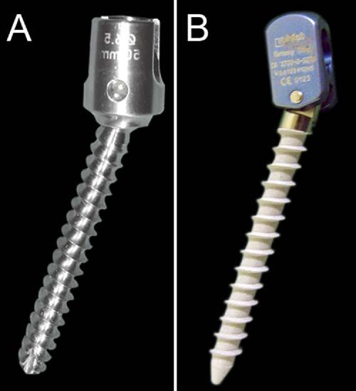
nar space in some patients, whereas in others it was per- Fig. 1. (A) Safinaz dynamic transpedicular screw (Medikon). (B) Cosmic
formed via a small laminotomy, formed by a high-speed dynamic transpedicular screw (Ulrich GmbH & Co. KG).
drill. During the operation, disc type was identified accord-
ing to the Carragee classification system. If the disc type month after the first. We observed that recurrent/persistent
was identified as type I (fragment-fissure), the patient was sciatica rates were significantly lower among Carragee
excluded from the study. When patients were identified as types II, III, and IV. Significant postoperative improve-
type II (fragment-defect), extruded or sequestered disc frag- ments were observed for the ODI and VAS measurements.
ments were removed and the disc space was cleaned of Recurrent disc herniation was not observed during the fol-
loose and easily accessible disc fragments, by use of for- low-up period. The summary of patients’ preoperative and
ceps. Curettes were not used in the disc space, and intact postoperative ODI scores, VAS scores, characteristics, and
annulus parts were left untouched. Because there were no clinical outcomes are provided in Table 1.
annular defects in intraoperatively identified Carragee type III
and IV discs, the annulus was opened with an oblique incision
with a No. 15 blade. Sub-annular fragments were removed Discussion
with rongeurs; such patients were recorded as type III. If there This prospective study relied on the recently developed
were no free disc fragments in the sub-annular space, patie- Carragee classification system. Carragee types II, III, and IV
nts were recorded as Carragee type IV; extensive annulotomy were identified intraoperatively in patients with single-level
was performed in such cases, and protruding disc fragments lumbar disc herniation, and posterior dynamic transpedicu-
were removed. At least 2 surgeons evaluated and recorded the lar stabilization with limited posterior lumbar microdiscec-
state of the annulus and free disc fragments during all opera- tomy was performed. The purpose of this study was to
tions. After discectomy, with the help of lateral intraoperative prevent failed back syndrome by reducing the high rate of
fluoroscopy, posterior dynamic transpedicular stabilization recurrent disc herniation observed in type II patients (de-
was performed for Carragee type II, III, and IV cases, by use scribed as fragment-defect) and to prevent the high rate of
of the Wiltse approach via the inner paravertebral muscle.16 recurrent/persistent sciatica observed in type III and IV
Cosmic (Ulrich GmbH & Co. KG, Ulm, Germany) and patients (fragment-contained and no fragment-contained,
Safinaz (Medikon, Ankara, Turkey) dynamic transpedicular respectively). Ultimately, better clinical results resulted.
screws with rigid rods were used (Fig. 1). This approach yielded lower reherniation rates and vastly
improved clinical results.
Results Unlike previous disc herniation classifications, the Car-
ragee system elaborates a new disc classification according
The most common herniation type in our study was type to the presence of extruded/sequestered or sub-annular frag-
III (fragment-contained), with 45% frequency. The fre- ments and annular stability.8 In their series of 187 cases,
quency of fragment-defect cases was 25%; the frequency of Carragee et al8 reported recurrent/persistent sciatica, reher-
no fragment-contained cases was 30%. The perioperative niation, and reoperation at a frequency of 1.1% among the
complications observed were as follows: 1 patient had blad- type I group. The type I group had small annular fissures
der retention that required catheterization, 1 patient had a and sequestered fragments; they were treated with seques-
superficial wound infection, and 1 patient had a malposi- trectomy. After performing sequestrectomy, we excluded
tioned transpedicular screw. The malpositioned screw was the type I group from our study because of the positive
corrected with a second operation that was performed 1 clinical results. The clinical results and reherniation rates
Downloaded from http://ijssurgery.com/ by guest on June 19, 2022T. Kaner et al. / SAS Journal 4 (2010) 92–97 95
among the other 3 groups were not satisfactory. The rate of disease is one of the major causes of spinal instabi-
recurrent/persistent sciatica and reherniation in the type II lity.28,29,31,32 According to the literature, the rate of insta-
(fragment-defect) group was as high as 27.3%; the reopera- bility in patients with lumbar disc herniation is 20%.31,32
tion rate was significantly high, at 21.2%. The rates of Kotilainen and Valtonen33 treated 190 patients with sin-
recurrent/persistent sciatica, reherniation, and reoperation gle-level lumbar disc herniation by performing lumbar mi-
among types III and IV were 11.9%, 9.5%, and 4.8%, crodiscectomy. During the observation period, 10% of pa-
respectively, and 37.5%, 12.5%, and 6.3%, respectively. In tients complained of sciatica and as many as 29% of patients
comparison to the Carragee type III group, the Carragee had lower back pain. Clinical examination showed various
type IV group had much higher rates of recurrent/persistent signs and symptoms of segmental instability of the lumbar
sciatica. The Carragee type III group had a higher rate of spine in 22% of the surgical patients. In another study
recurrent/persistent sciatica than the Carragee type I group. Kotilainen et al34 reported poor clinical results in patients
Similarly, the rates of reherniation and reoperation observed with protruded disc herniation, as compared with patients
in this study for Carragee types III and IV were not as high with sequestered fragments or prolapse, who displayed bet-
as those observed for the Carragee type II group; however, ter results. According to Kotilainen et al, such results may
the rates were much higher than those observed for the type arise because of segmental instability resulting from the
I group. Because of the study of Carragee et al, it was diffuse nature of disc disease. Frymoyer and Selby31 ac-
surmised that more satisfactory results and decreased rates knowledged that massive central L4-5 disc herniation,
of both recurrent herniation requiring reoperation and re- which is often observed with severe low back pain, presents
current/persistent sciatica would follow a limited posterior a remarkable situation and exhibits a tendency toward de-
lumbar microdiscectomy with posterior dynamic transpe- generative instability. The diffuse/massive-natured disc her-
dicular stabilization. After evaluating the results of 40 cases, nia descriptions of Kotilainen et al and Frymoyer and Selby
after at least 2 years’ follow-up, we observed no recurrent are in line with the Carragee classification of the type IV
disc herniation. Recurrent/persistent sciatica rates were sig- group. In such cases Carragee et al8 removed protruded disc
nificantly lower in all 3 types examined compared with the pieces via extensive annulotomy. They reported a very high
study of Carragee et al and other studies.9,17,18 rate of recurrent/persistent sciatica (37%) in this group. It is
According to the relevant literature, unsatisfactory re- our opinion that the unsatisfactory results of Carragee et al
sults are reported in 38% of patients who undergo lumbar were due to segmental instability; therefore limited discec-
discectomy.7,19 Mochida et al20 confirmed that removing the tomy with posterior dynamic transpedicular stabilization
disc material less aggressively yields better clinical and radio- was performed in our patients.
logic results. Williams13,21 reported, for the first time, encour- The concept of dynamic stabilization was established to
aging results after removing minimal tissue from the interver- control abnormal motion by transferring the weight-bearing
tebral disc space. His clinical success rate was 90%, and the load carried by the spine without performing spinal segment
rate of recurrence was 4% to 9%. Rogers22 described recurrent fusion.35 Thus dynamic stabilizations aim to relieve the pain.35
disc herniation, after removing only disc fragments, in 7 of 33 Some recently published studies showed that dynamic stabili-
patients (21%). In a study by Thome et al,6 recurrent herniation zation (by use of a dynamic pedicular screw–rod system)
was observed in 4 of 42 patients (10%). A recent study by biomechanically provides stability that is similar to that
Barth et al23 compared the results at 2 years’ follow-up for provided by rigid systems.36 Moreover, theoretically, it is
lumbar microdiscectomy and microscopic sequestrectomy. thought that dynamic stabilization systems have advantages
The microdiscectomy patient group presented deterioration in over rigid spinal implants. It is an easier surgery to perform,
functional and radiologic results due to segmental degeneration requires a shorter operative time, and does not have the
at 2 years, whereas sequestrectomy was associated with better associated risks of donor-site pain, pseudarthrosis, and ad-
functional results after 2 years. jacent segment degeneration that fusion surgery entails.37,38
Some clinical and radiologic studies reported that loose- Studies by Schaeren et al39 and Stoll et al40 reported good
ness in the ligaments and facet joint capsules is observed clinical results from the procedure and recommended dy-
with the decrease in disc altitude after disc operations. As a namic stabilization as a safe and effective method of treat-
result, increased load on facet joints may cause segmental ment for patients with degenerative chronic instability.
instability and spondylosis.4 – 6,24,25 Segmental instability in Putzier et al37 reported that disc degenerations showed far
the lumbar spine is one of the reasons for failed back less progression in patients who had nucleotomy with pos-
syndrome. Yorimitsu et al,26 in their follow-up study of terior dynamic system applications than patients who did
more than 10 years, reported the frequency of chronic lower not have dynamic stabilization after 34 months’ follow-up.
back pain, rather than sciatica, after lumbar disc surgery due In our experience, posterior dynamic transpedicular stabili-
to decreased disc space height as 75%. Lumbar instability zation decelerates the degeneration of disc tissue (Fig. 2). It
can be confirmed both clinically and radiologically.27,28 can reduce the occurrence of failed back syndrome. In this
Studies examining clinical instability showed that radio- study dynamic fixation was a non–Food and Drug Admin-
logic findings were not always in agreement with clinical istration–approved indication for this procedure. This study
findings.29,30 Notably, it is thought that degenerative disc and favorable results in the literature have shown that pos-
Downloaded from http://ijssurgery.com/ by guest on June 19, 202296 T. Kaner et al. / SAS Journal 4 (2010) 92–97
2. Carragee EJ, Han MY, Yang B, Kim DH, Kraemer H, Billys J.
Activity restrictions after posterior lumbar discectomy: a prospective
study of outcomes in 152 cases with no postoperative restrictions.
Spine (Phila Pa 1976) 1999;24:2346 –51.
3. Carragee EJ, Spinnickie AO, Alamin TF, Paragioudakis S. A prospec-
tive controlled study of limited versus subtotal posterior discectomy:
short-term outcomes in patients with herniated lumbar intervertebral
discs and large posterior annular defect. Spine (Phila Pa 1976) 2006;
31:653–7.
4. Cinotti C, Postacchini F. Biomechanics. In: Postacchini F, ed. Lumbar
Disc Herniation. Wien: Springer-Verlag; 1999:81–93.
5. Striffeler H, Groger U, Reulen HJ. ‘Standard’ microsurgical lumbar
discectomy vs. ‘conservative’ microsurgical discectomy. A prelimi-
nary study. Acta Neurochir 1991;112:62– 4.
6. Thome C, Barth M, Schare J, Schmiedek P. Outcome after lumbar
sequestrectomy compared with microdiscectomy: a prospective ran-
domized study. J Neurosurg Spine 2005;2:271– 8.
7. Caspar W, Campbell B, Barbier DD, Kretschmmer R, Gotfried Y. The
Caspar microsurgical discectomy and comparison with a conventional
standard lumbar disc procedure. Neurosurgery 1991;28:78 – 86.
8. Carragee EJ, Han MY, Suen PW, Kim D. Clinical outcomes after
Lumbar discectomy for sciatica: the effects of fragment type and
annular competence. J Bone Joint Surg Am 2003;85:102– 8.
Fig. 2. MRI scans in 38 year-old female patient, showing L5 transitional 9. Wera GD, Dean CL, Ahn UM, et al. Reherniation and failure after
vertebrae and disc herniation in Carragee group IV. The patient had severe lumbar discectomy: a comparison of fragment excision alone versus
back pain and sciatica. (A) The preoperative MRI scan shows a moderate subtotal discectomy. J Spinal Disord Tech 2008;21:316 –9.
black disc and diffuse bulging. (B) The postoperative MRI scan after 10. Vaughn PA, Malcolm BW, Maistrelli GL. Results of L4-L5 disc
posterior dynamic transpedicular stabilization shows deceleration in the excision alone versus disc excision and fusion. Spine (Phila Pa 1976)
degeneration process. 1988;13:690 –5.
11. Hu RW, Jaglal S, Axcell T, Anderson G. A population-based study of
reoperations after back surgery. Spine (Phila Pa 1976) 1997;22:2265–71.
12. Caspar W. A new surgical procedure for lumbar disc herniation caus-
terior dynamic transpedicular stabilization can be used in ing less tissue damage through a microsurgical approach. Adv Neuro-
segmental degenerative instability to reduce the risk of surg 1977;4:74 – 80.
failed back syndrome or recurrent disc herniation and to 13. Williams RW. Microlumbar discectomy. A conservative surgical ap-
decrease the frequency of postoperative sciatica and me- proach to the virgin herniated lumbar disc. Spine (Phila Pa 1976)
chanical low back pain. This seems to be a compelling 1978;3:175– 82.
14. Spengler DM. Lumbar discectomy. Results with limited disc excision
argument for a new indication for use of posterior dynamic
and selective foraminotomy. Spine (Phila Pa 1976) 1982;7:604 –7.
transpedicular stabilization. 15. Carragee EJ, Kim DH. A prospective analysis of magnetic resonance
In this prospective study we performed limited lumbar imaging findings in patients with sciatica and lumbar disc herniation.
microdiscectomy in single-level disc herniation patients Correlation of outcomes with disc fragment and canal morphology.
who were grouped according to the Carragee classification Spine (Phila Pa 1976) 1997;22:1650 – 60.
system as type II, III, or IV. To obtain more satisfactory 16. Wiltse LL, Spencer CW. New uses and refinements of the paraspinal
approach to the lumbar spine. Spine (Phila Pa 1976) 1988;13:1008 –12.
clinical results and to decrease the rates of instability due to
17. McGirt MJ, Ambrossi GL, Datoo G, et al. Recurrent disc herniation
segmental degeneration, as well as to reduce the rates of and long-term back pain after primary lumbar discectomy: review of
recurrent disc herniation and recurrent/persistent sciatica outcomes reported for limited versus aggressive disc removal. Neuro-
with failed back syndrome, we also performed posterior surgery 2009;64:338 – 44.
dynamic transpedicular stabilization in the same patients. 18. Häkkinen A, Kiviranta I, Neva MH, Kautiainen H, Ylinen J. Reop-
This approach yielded much improved clinical results. erations after first lumbar disc herniation surgery: a special interest on
residives during a 5-year follow-up. BMC Musculoskelet Disord 2007;
The main deficiency of this study is short follow-up. The 8:2.
study of Carragee et al,8 which served as the cornerstone for 19. O’Sullivan MG, Connolly AE, Buckley TF. Recurrent lumbar disc
our study, had a median follow-up period of 6 years, with protrusion. Br J Neurosurg 1990;4:319 –25.
minimum follow-up of 2 years, and the recurrence of symp- 20. Mochida J, Nishimura K, Nomura T, Toh E, Chiba M. The importance
toms occurred after a long time interval. More clinical studies of preserving disc structure in surgical approaches to lumbar disc
herniation. Spine (Phila Pa 1976) 1996;21:1556 – 63.
need to show the positive results after posterior dynamic trans-
21. Williams RW. Microlumbar discectomy. A 12 year statistical review.
pedicular stabilization with limited lumbar discectomy. Spine (Phila Pa 1976) 1986;11:851–2.
22. Rogers LA. Experience with limited versus extensive disc removal in
patients undergoing microsurgical operations for ruptured lumbar
References discs. Neurosurgery 1988;22:82–5.
23. Barth M, Weiss C, Thome C. Two-year outcome after lumbar micro-
1. Mixter WJ, Barr JS. Rupture of the intervertebral disc with involve- discectomy versus microscopic sequestrectomy. Spine (Phila Pa 1976)
ment of the spinal canal. N Engl J Med 1934;211:210 –5. 2008;33:265–72.
Downloaded from http://ijssurgery.com/ by guest on June 19, 2022T. Kaner et al. / SAS Journal 4 (2010) 92–97 97
24. Kirkaldy-Willis WH, Wedge JH, Yong-Hing K, Reilly J. Pathology 33. Kotilainen E, Valtonen S. Clinical instability of the lumbar spine after
and pathogenesis of lumbar spondylosis and stenosis. Spine (Phila Pa microdiscectomy. Acta Neurochir (Wien) 1993;125:120 – 6.
1976) 1978;3:319 –28. 34. Kotilainen E, Valtonen S, Carlson CA. Microsurgical treatment of
25. Wenger M, Mariani L, Kalbarczyk A, Groger U. Long-term outcome lumbar disc herniation: follow-up of 237 patients. Acta Neurochir
of 104 patients after lumbar sequestrectomy according to Williams. (Wien) 1993;120:143–9.
Neurosurgery 2001;49:329 –35. 35. Sengupta DK. Dynamic stabilization devices in the treatment of low
26. Yorimitsu E, Chiba K, Toyama Y, Hirabayashi K. Long-term out- back pain. Neurol India 2005;53:466 –74.
comes of standard discectomy for lumbar disc herniation: a follow-up 36. Bozkuş H, Senoğlu M, Baek S, et al. Dynamic lumbar pedicle screw-
study of more than 10 years. Spine (Phila Pa 1976) 2001;26:652–7. rod stabilization: in vitro biomechanical comparison with standard
27. Paris SV. Physical signs of instability. Spine (Phila Pa 1976) 1985; rigid pedicle screw-rod stabilization. J Neurosurg Spine 2010;12:
10:277–9.
183–9.
28. Knutsson F. The instability associated with disc degeneration in the
37. Putzier M, Schneider SV, Funk JF, Tohtz SW, Perka C. The surgical
lumbar spine. Acta Radiol 1944;25:593– 609.
treatment of the lumbar disc prolapse. Nucleotomy with Additional
29. Frymoyer JW, Hanley EN, Howe J, Kuhlmann D, Matteri R. A
transpedicular dynamic stabilization versus nucleotomy alone. Spine
comparison of radiologic findings in fusion and in nonfusion patients
(Phila Pa 1976) 2005;30:E109 –14.
ten or more years following lumbar disc surgery. Spine (Phila Pa
1976) 1979;4:435– 40. 38. Strempel A, Neekritz A, Muelenaere P, et al. Dynamic versus rigid
30. Sano S, Yokokura S, Nagata Y, Young SZ. Unstable lumbar spine spinal implants. In: Gunzburg R, Szpalski M, eds. Lumbar Spinal
without hypermobility in postlaminectomy cases. Mechanism of Stenosis. Philadelphia: Lippincott-Williams & Wilkins; 2000:275– 85.
symptoms and effect of spinal fusion with and without spinal instru- 39. Schaeren S, Broger I, Jeanneret B. Minimum four-year follow-up of
mentation. Spine (Phila Pa 1976) 1990;15:1190 –7. spinal stenosis with degenerative spondylolisthesis treated with de-
31. Frymoyer SW, Selby DK. Segmental instability. Rationale for treat- compression and dynamic stabilization. Spine (Phila Pa 1976) 2008;
ment. Spine (Phila Pa 1976) 1985;10:280 – 6. 33:E636 – 42.
32. Morgan FP, King T. Primary instability of lumbar vertebrae as a 40. Stoll TM, Dubois G, Schwarzenbach O. The dynamic neutralization
common cause of lower back pain. J Bone Joint Surg Br 1957;39: system for the spine. A multi-center study of a novel non-fusion
6 –22. system. Eur Spine J 2002;11:170 – 8.
Downloaded from http://ijssurgery.com/ by guest on June 19, 2022You can also read