COMPARATIVE ANALYSIS OF THE QUANTITA- TIVE AND QUALITATIVE METHOD FOR DETER-MINATION OF D - DIMER - Journal of IMAB
←
→
Page content transcription
If your browser does not render page correctly, please read the page content below
https://doi.org/10.5272/jimab.2021271.3539
Journal of IMAB
                                               Journal of IMAB - Annual Proceeding (Scientific Papers). 2021 Jan-Mar;27(1)
ISSN: 1312-773X
https://www.journal-imab-bg.org
                                                 Original article

                                          COMPARATIVE ANALYSIS OF THE QUANTITA-
                                          TIVE AND QUALITATIVE METHOD FOR DETER-
                                          MINATION OF D - DIMER
                                          Irena I. Gencheva
                                          Department of Clinical Laboratory, Clinical Immunology and Allergology,
                                          Medical University - Pleven, Bulgaria.

        SUMMARY                                                 ing D-dimer are formed from plasmin, respectively. Most
        Introduction: D - dimer is a product released dur-      fibrin degradation products contain high molecular weight
ing the process of blood clotting and degradation, which        X-oligomers [1].
can be measured by blood sample analysis. There is usu-                 D-dimer testing is clinically relevant when deep vein
ally the minimal activity of the pro/anticoagulant system       thrombosis (DVT), pulmonary embolism (PE) or disseminated
in the human body, which generates low levels of D-dimer        intravascular coagulation (DIC) are suspected [2, 3].
in healthy individuals. Normal values for plasma D-dimer                False negative and false positive results may occur
are ≤ 0.50 mg / l.                                              in some cases. Due to the frequency of false-negative re-
        Aim: The aim of the present study is to determine       sults, some authors recommend that D-dimer be used only
to what extent the quantitative and qualitative method for      at low probability of pulmonary embolism or deep vein
determination of D - dimer can be interchangeable and what      thrombosis.
is their diagnostic reliability in the normal and pathologi-            It should be noted that there are some physiological
cal area of measurement.                                        and medical conditions that can lead to elevated D-dimer
        Materials and methods: We studied the levels of D-      in patients without pulmonary embolism, deep vein throm-
dimer by two methods - quantitative and qualitative, in 91      bosis, or DIC. These include, but are not limited to, preg-
patients aged 25 to 86 years, of which 59 men and 32            nancy, malignancy, smoking, trauma, infection, or sepsis. In
women. To determine the D-dimer, we used venous blood           addition, elderly patients, immobilized patients, patients
taken in a vacuette containing sodium citrate. We used a        with autoimmune disorders, or those who have recently un-
Roche test for quantitative determination and a Latex ag-       dergone surgery may have an elevated D-dimer [4].
glutination test for qualitative determination.                         In new studies, it is proposed to use corrected age
        Results: It was found that in positive samples above    limits for D-dimer, as D-dimer values †may increase with
0.5 mg/l, there is a very high percentage of coincidence.       age, even in the absence of pathology [5].
There is a discrepancy in the values obtained by the two                There is usually the minimal activity of the pro/an-
methods at the negative values below 0.5 mg/l. We deter-        ticoagulant system in the human body, which generates low
mined the sensitivity, specificity and accuracy of both         levels of D-dimer in healthy individuals. The normal
methods.                                                        plasma D-dimer values are ≤ 0.50 mg/l. A D-dimer above
        Conclusion: The correlation in the results of the two   0.50 mg/l is considered positive [6].
methods is very good, but the quantification of D-dimer is              The sensitivity and specificity of the D-dimer vary
more specific and accurate. We recommend that the value         depending on the type of assay method. However, D-dimer
of 0.5 mg/l should be used as a cut off value for D-dimer.      tests generally have high sensitivity but low specificity.
                                                                There are currently various methods for determining plasma
       Keywords: D-dimer, qualitative, quantitative, cut off    D-dimer levels, both quantitative and qualitative [3, 7].

        INTRODUCTION                                                   PURPOSE
        The D - dimer is a product released during the proc-           Our goal is to determine to what extent the quanti-
ess of blood clotting and degradation, which can be meas-       tative and qualitative method for the determination of D -
ured by blood sample analysis. It is usually released when      dimer can be interchangeable and what is their diagnostic
the blood clot begins to break down.                            reliability in the normal and pathological field of meas-
        Thrombin converts fibrinogen to soluble fibrin by       urement.
splitting of fibrin peptides A and B. Fibrin monomers po-
lymerize spontaneously. Active factor XIII binds two D-do-             PATIENTS AND METHODS
mains and generates a solid fibrin clot. A new plasmin-re-             In our study, we measured the levels of D-dimer in
sistant antigen determinant (“D-dimer”) is obtained. Dur-       the plasma of patients at the University Hospital - Pleven.
ing the degradation of the fibrin clot, fragments contain-      All of them have signed informed consent at the hospital

J of IMAB. 2021 Jan-Mar;27(1)                  https://www.journal-imab-bg.org                                          3539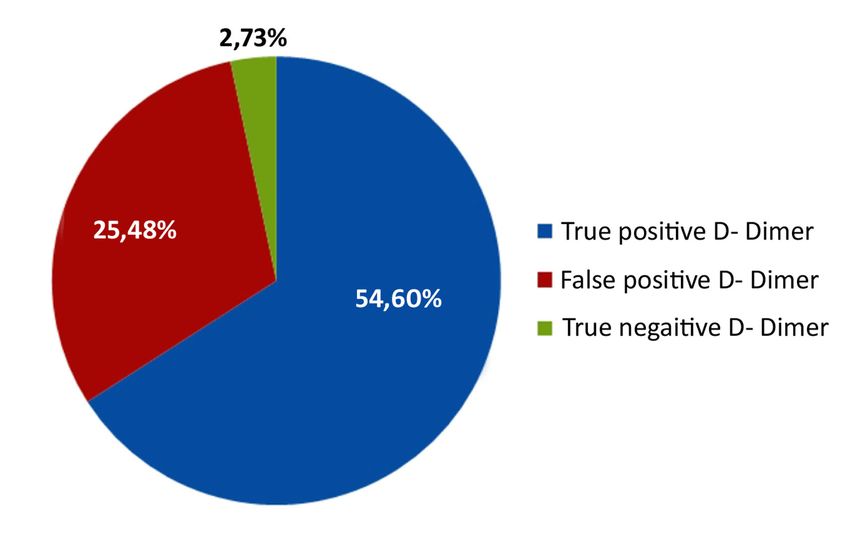
admission. The studied patients were 91, aged 25 to 86          undiluted plasma, but no agglutination with a dilution of
years, of which 59 men and 32 women. The mean age for           1: 2, the result is 0.2 - 0.4 mg/l; in the presence of aggluti-
men was 59 years (SD = 15.4) and for women 64 years (SD         nation with undiluted plasma and with a dilution of 1:2,
= 13.3).                                                        but no agglutination with a dilution of 1:4, the result is
        The patients we selected for the control group in-      0.4 - 0.8 mg / l; in the presence of agglutination with un-
cluded 30 healthy individuals, 15 men and 15 women, aged        diluted plasma and with 1:2 and 1:4 dilutions, but no ag-
31 to 77 years.                                                 glutination with 1:8 dilution, the result is 0.8 - 1.6 mg/l;
        To determine D-dimer, we used venous blood taken        in the presence of agglutination with undiluted plasma and
in a vacuette containing sodium citrate the (blood-antico-      with all dilutions, the result is 1.6 - 3.2 mg/l;
agulant ratio is 9: 1). The blood is mixed well with the an-
ticoagulant, then centrifuged, and the separated plasma is              RESULTS
used for analysis. D-dimer was measured in all plasmas by               In the control group, the levels of D-Dimer were
two methods - quantitative and qualitative. We quantified       measured by a quantitative method, and it was found that
the D-dimer with a Roche test on a biochemical analyzer         the mean value was 0.287 (0.12 - 0.5), and the median was
Cobas 6000, and with a qualitative Latex agglutination          0.295. All measured values were below the cut-off value
test.                                                           of 0.5 mg/l (from reagent manufacturer Roche). This gave
        The Roche assay is an immunoturbidimetric assay         us reason to choose the cut off for our study 0.5 mg/l.
in which latex particles of the same size are encapsulated              During the study of D-dimer in our laboratory was
in monoclonal antibodies (F (ab) 2 fragments) and attached      conducted daily intra-laboratory quality control in two lev-
to the D-dimer epitope. The antigen/antibody complexes          els for both methods.
obtained by adding samples containing D-dimer cause an                  From the 91 samples for D-dimer studied by both
increase in the turbidity of the test reagents. The change      methods, it was found that in the case of positive samples
in absorption over time depends on the concentration of         above 0.5 mg/l, there is a very high percentage of coinci-
D-dimer epitopes in the sample. The precipitate is deter-       dence of the results. We have a 100% match in the results
mined turbidimetrically. Reference value ≤ 0.50 mg/l.           for D-dimer above 1.6 mg/l.
        The qualitative D-dimer test is a latex agglutination           There is a discrepancy in the values obtained by
test using latex particles combined with a highly specific      the two methods for negative D-dimers below 0.5 mg/l. At
D-dimer monoclonal antibody. XL-FDP present in the              these values, it was found that in 28 cases, the qualitative
plasma binds to the coated latex particles, resulting in vis-   test gave a positive result (> 0.2 mg/l) and the quantita-
ible agglutination occurring when the D-dimer concentra-        tive test a negative result (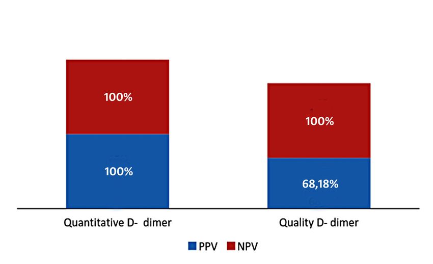
Table 1. Comparative analysis of sensitivity, specificity and accuracy between the two methods for D-dimer
Statistic D – dimer Quantity D – dimer Qualitative
Value 95% CI Value 95% CI
Sensitivity 100% 94.04% to100.00% 100 % 94.04% to 100.00%
Specificity 100% 88.78% to 100.00% 49.68% 32.04% to 65.75%
PPV 100% - 68.18% 65.63% to 70.63%
NPV 100% - 100 % -
Accuracy 100% 96.03% to 100.00% 69.23% 58.68% to 78.49%
Fig. 2. Sensitivity, specificity and accuracy of both methods for the determination of D-dimer
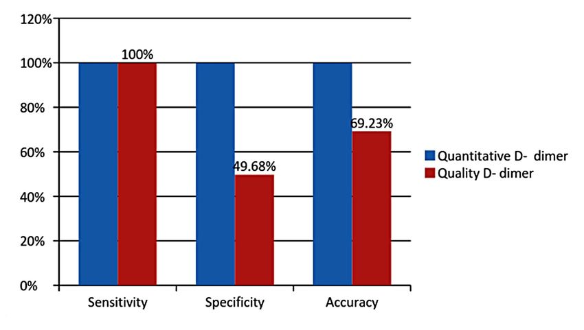
Fig. 3. PPV and NPV of the quantitative and qualitative method for the determination of D-dimer
DISCUSSION must be followed, including the duration of treatment. There
From the obtained results, we can conclude that the is a very good correlation in the results with strongly nega-
results for D-dimer from the two methods are very well cor- tive D-dimers below 0.2 mg/l.
related, especially in the range above 0.5 mg/l. In the area Regarding the results in the range of 0.2 - 0.5 mg/l,
above 1.6 mg/l, there is practically 100% agreement of the there are serious discrepancies. We found that with a qual-
results. The only problem is that above 3.2 mg/l, the qual- ity test, there is a percentage of probability of giving false
ity test cannot give a specific value, which is a serious prob- positive results. In practice, we found that in 25.48% of cases,
lem in patients with high levels of D-dimer in the blood that the qualitative test gave a positive result in samples in which
J of IMAB. 2021 Jan-Mar;27(1) https://www.journal-imab-bg.org 3541the value of the D-dimer is in the range 0.2 - 0.5 mg/l (Fig. In addition, we measured the positive and negative
1). First of all, the problem arises from the difference in the predictive value of the methods. As can be seen in Fig. 3,
reference values of the two tests - the quantitative up to the negative predictive value of both methods is 100%,
0.5 mg/l, and the qualitative up to 0.2 mg/l. The cut off value while the positive predictive value of the quantitative
imposed not only by us but also by most authors for D-dimer method is 100%, and the qualitative - 68.18% [14].
is 0.5 mg/l. The problem would be serious if both methods
work in the same laboratory, and the reference values are CONCLUSION
not unified [8, 9, 10, 11, 12]. In conclusion, we can say that the correlation in the
In terms of sensitivity, both methods are 100% sensi- results of the two methods is very good. The quantification
tive (95% CI- 94.04% to100.00%). As a note, it should be of D-dimer is more sensitive, and specificity is preferred. Es-
noted that with regard to the performance of the qualitative pecially in cases where it is necessary to monitor treatment.
test, the purity of the plates where the reaction between the We recommend that the cut off value for D-dimer be the value
latex reagent and the patient sample takes place must be ob- - 0.5 mg/l. In order to avoid discrepancies in the results, if
served very strictly [8, 13]. The accuracy of the quantitative one laboratory uses both methods for determination of D-
method is 100% (95% CI - 96.03% to 100.00%), while the dimer, we recommend mandatory unification of the reference
qualitative one is 69.23% (95%CI- 58.68% to 78.49%) values.
REFERENCES:
1. Reber G, De Moerloose P. erly. Intern Med J. 2007 Sep; 37(9):607- JD, Jennings I, Kitchen S, Mutch N, et
Standartization of D-dimer testing. In: 13. [PubMed] [Crossref] al. Harmonisation of D-dimer - A call for
Quality in laboratory hemostasis and 6. Legnani C, Palareti G, Cosmi B, action. Thromb Res. 2016; 137:219-220.
thrombosis. Kitchen S, Olson JD, Pres- Cini M, Tosetto A, Tripodi A. Different [Crossref]
ton FE. (Editors). 2nd ed. John Wiley & cut-off values of quantitative D-dimer 11. Ekelund S, Eliasen MM. D-dimer
Sons. 2013. Chapter 13. pp.136-146. methods to predict the risk of venous assays - pitfalls of analytical compari-
[Crossref] thromboembolism recurrence: a post- sons. acutecaretesting.org. May 2016.
2. Thachil J, Lippi G, Favaloro EJ. D- hoc analysis of the PROLONG study. [Internet]
dimer testing: laboratory aspects and Haematologica. 2008 Jun; 93(6):900-7. 12. Linkins L-A, Takach Lapner S.
current issues. Methods Mol Biol. 2017; [PubMed] [Crossref] Review of D-dimer testing: Good, Bad,
1646:91-104. [PubMed] [Crossref] 7. Stegnar M, Bo•iè M. Determina- and Ugly. Int J Lab Hematol. 2017
3. Ginsberg JS, Wells PS, Kearon C, tion of D-dimer by different quantitative May;39 Suppl 1:98-103. [PubMed]
Anderson D, Crowther M, Weitz JI, et al. assays – A harmonization exercise. [Crossref]
Sensitivity and specificity of a rapid Biochemia Medica. 2008; 18(2):216-23. 13. Favresse J, Lippi G, Roy PM,
whole-blood assay for D-dimer in the di- [Internet] Chatelain B, Jacqmin H, Cate HT, et
agnosis of pulmonary embolism. Ann 8. Dempfle CE. D-dimer: standardi- al. D-dimer: Preanalytical, analytical,
Intern Med. 1998 Dec 15;129(12): zation versus harmonization. Thromb postanalytical variables, and clinical ap-
1006-11. [PubMed] [Crossref] Haemost. 2006 Mar;95(3):399-400. plications. Crit Rev Clin Lab Sci. 2018
4. Tripodi A. D-dimer testing in labo- [PubMed] [Crossref] Dec;55(8):548-577. [PubMed] [Crossref]
ratory practice. Clin Chem. 2011 9. Meijer P, Kluft C. The harmoni- 14. Nelson CM, Wright GS, Silbaugh
Sep;57(9):1256-62. [PubMed] [Crossref] zation of quantitative test results of dif- TR, Cota LJ. Improving D-dimer Posi-
5. Harper PL, Theakston E, Ahmed J, ferent D-dimer methods. Semin Vasc tive Predictive Value for Outpatients with
Ockelford P. D-dimer concentration in- Med. 2005 Nov;5(4):321-7. [PubMed] Suspected Deep Vein Thrombosis. Perm
creases with age reducing the clinical [Crossref] J. 2009 Winter;13(1):4-7. [PubMed]
value of the D-dimer assay in the eld- 10. Longstaff C, Adcock D, Olson [Crossref]
Please cite this article as: Gencheva II. Comparative analysis of the quantitative and qualitative method for determina-
tion of D- dimer. J of IMAB. 2021 Jan-Mar;27(1):3539-3542. DOI: https://doi.org/10.5272/jimab.2021271.3539
Received: 02/10/2019; Published online: 22/01/2021
Address for correspondence:
Irena I. Gencheva
Department of Clinical laboratory, Clinical immunology and alergology, Medi-
cal University - Pleven
7, Dame Gruev Str., 5800 Pleven, Bulgaria,
E-mail: gencheva1677@gmail.com
3542 https://www.journal-imab-bg.org J of IMAB. 2021 Jan-Mar;27(1)You can also read