Competitive hydrogen bonding in aspirin-aspirin and aspirin-leucine interactions
←
→
Page content transcription
If your browser does not render page correctly, please read the page content below
Turk J Chem
                                                                                             36 (2012) , 383 – 395.

                                                                                             c TÜBİTAK
                                                                                             doi:10.3906/kim-1112-16

   Competitive hydrogen bonding in aspirin-aspirin and
                                   aspirin-leucine interactions

                     Zeynep YURTSEVER1,3 , Burak ERMAN2 , Ersin YURTSEVER1,∗
                   1
                     Chemistry Department, Koç University, Sarıyer, İstanbul, 34450, TURKEY
                            2
                             Chemical and Biological Engineering Department, Koç University,
                                            Sarıyer, İstanbul 34450-TURKEY
                            3
                              Present address: Department of Biology and Biomedical Sciences,
                              Washington University in St. Louis, St. Louis, MO, 63110-USA
                                               e-mail: eyurtsev@ku.edu.tr

                                                  Received: 09.12.2011

         Aspirin-aspirin and aspirin-leucine interactions are studied by the density functional theory (DFT) and
     high level ab initio calculations with second order Moller-Plesset perturbation theory (MP2). The rotational
     isomers of aspirin are identified by their relative stability both in gaseous phase and in water using the
     polarizable continuum method (PCM). Local minima of aspirin monomers in water are found to be all
     highly populated compared to the gas phase behavior. Homodimers of aspirin form hydrogen bonds with
     bond energies of 10 kcal/mol. Weak hydrogen bonds utilizing phenyl and methyl groups are also found.
     The interaction between aspirin and leucine is stronger with relatively short bond lengths compared to
     homodimeric aspirin interactions. The potential energy surface has several minima with comparable stability.
     This study shows the significance of diverse bonding schemes, which are important for understanding
     complete interaction mechanisms of aspirin.

     Key Words: Competitive hydrogen bonding in aspirin-aspirin and aspirin-leucine interactions

Introduction
Although aspirin has been used to treat inflammatory conditions since the 1880s, the understanding of its
mechanism of action is relatively new. In 1971, Vane discovered that aspirin interferes with the biosynthesis
of prostaglandins. 1,2 This description was improved and detailed more recently by Garavito et al. in the late
1990s. 3−6 The biosynthesis of prostaglandins depends on 2 enzymes, cyclooxygenase1 (COX-1) and cyclooxy-
genase2 (COX-2). Aspirin inhibits both COX-1 and COX-2 by irreversibly acetylating both of the enzymes. 7,8
 ∗ Corresponding   author

                                                                                                                      383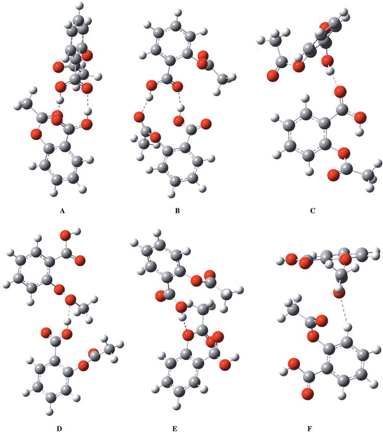
Competitive hydrogen bonding in aspirin-aspirin and..., Z. YURTSEVER, et al.

       Aspirin is thought to act on the arachidonic acid binding region of COX-1 and COX-2 by acetylating
Ser530 of COX-1 and Ser516 of COX-2. Thus, the binding of the acid to the COX pocket is inhibited. The
acetylation efficiency of aspirin is 10-fold higher for COX-1 than for COX-2. 8
      Although the mechanism of action of aspirin has been studied in detail, as briefly outlined above,
an experimentally determined structure of the COX-aspirin complex does not exist. Our knowledge on the
mechanism rests essentially on site-directed mutagenesis studies. Hochgesang et al. postulated that 9 Tyr385
acts as a hydrogen bond donor to position the acetyl group adjacent to the hydroxyl group of Ser530 in COX-2.
The mutation Tyr385Phe (Y385F) was found to decrease the acetylation efficiency significantly, which suggested
that this tyrosine residue was essential for the oxidation of arachidonic acid. There are, however, alternative
possible routes hypothesized for acetylation of Ser530. 7
       Despite multiple biochemical approaches to the problem, mechanism-determining quantum mechanical
calculations have not yet been carried out. The potential energy surface of a single aspirin molecule in the
gas phase was first fully studied by Glaser. 10 That study was carried out almost a decade ago and there are
no further reports of different findings on that issue. Glaser found out from DFT calculations that there are
9 conformational isomers. These isomers are obtained by the coupling of possible rotational motion around 4
rotatable bonds of aspirin (C=O bonds of the carboxyl group and acetoxy group, C-O linkage of the acetoxy
group, and O-H positioning on the carboxyl group).
       Our aim for revisiting the ab initio calculations of aspirin is several fold: firstly, high precision ab initio
calculations are missing in the literature. For any further rational computation of the mechanism of aspirin
activity, a sound characterization of the molecule is necessary. A quantitative basis of the single molecule is
crucial for calculating the properties of modified aspirin. The work by Loll et al. showed that the closely related
bromoaspirin bromoacylates Ser530 of COX-1, while salicylic acid binds to Tyr355 and Arg120. 5 A quantum
mechanical calculation for modifications will lead to predictions that will be invaluable for drug improvement.
Finally, as a prelude to aspirin-amino acid interactions, we studied the complex of aspirin with leucine.
      The crystal structure of the holoenzyme COX-1 was readily available 11 and it was viewed in GOLD 12
for the initial modeling. An aspirin molecule was docked flexibly onto the rigid enzyme and different constraints
were imposed to scan for potential binding sites. The option to dock the small molecule flexibly has allowed us
to maintain an unbiased approach in identifying potentially novel binding sites on COX-1 for aspirin.
      Within the obtained results, we picked the site with the best docking score for aspirin. (The analytical
value of this score is irrelevant, as the scores are subject to significant changes with each imposed constraint.)
This binding site consisted of 10 amino acids with exposed backbones and side chains. An additional filtering
reduced the number to 3 essential amino acids that were within 5 Å of the docked aspirin molecule. (The
reduction from 10 to 3 suggested that the other 7 amino acids merely contributed to the shape and possibly
the stability of the binding pocket.) Out of the 3 amino acids, 2 were leucines and the third was a serine
residue. Because these residues were isolated from the crystal structure, certain parts of the classical amino
acid structure were inaccessible for aspirin, i.e. the amino acids are part of the protein and, as such, bound by
peptide bonds to the adjacent residues. Thus, we used only the accessible parts in our models, which resulted
in structures resembling aldehydes. The end groups that are reduced due to peptide bonding were filled with
H atoms, in order not to add more reactive species to the system.
      For a single aspirin molecule in the gas phase, starting out with all possible combinations of the above-

384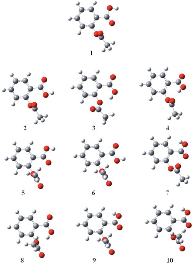
Competitive hydrogen bonding in aspirin-aspirin and..., Z. YURTSEVER, et al.

mentioned rotations, we fully optimized geometries with the same methodology used by Glaser. Then a
polarizable continuum (PCM) was added in order to compare the gas-phase results with those in solution.
In the second step, both the gas phase DFT and the PCM calculations for the aspirin homodimers were carried
out. We were able to characterize 6 basic homodimer structures. Similar to the dimerization calculations,
we tried a large number of coupling schemes between aspirin and leucine moieties, designed based on possible
hydrogen bonds.
      There is growing interest in the computational chemistry of the so-called non-covalent interactions.
Hydrogen bonding and the π -stacking interactions are 2 main elements of this area. Determining π -stacking
energy is a difficult task requiring detailed calculations of the correlation energy, whereas hydrogen bonding can
be studied with relatively simple ab initio or DFT techniques. 13
      Hydrogen bonding is a well-known concept; however, it plays an extremely important role in a large class of
chemical and biochemical problems. A recent article 14 discusses the present state and the future of the hydrogen
bond, stating that many hydrogen bonds can be formed or broken at normal temperatures due to their relative
weakness compared to chemical bonds,. This behavior results in complex dynamics in many chemical problems.
Two important areas where hydrogen bonding plays a significant role are the macromolecular and nucleic acid
interactions, where structural stability is strictly controlled by the strength and number of these bonds. With
highly accurate applications of ab initio and DFT methodologies, as well as molecular simulation techniques,
these interactions can be studied very accurately. In the case of macromolecules, we have previously studied
hydrogen bonding in polyureas and polyurethanes quantum mechanically and correlated physical properties of
such systems to the relative strengths of monomer-monomer interactions. 15−17
       In this work, we present detailed DFT calculations of aspirin molecules, aspirin homodimers, and aspirin-
leucine complexes as prototypes of aspirin-protein interactions. The possibility of various types of hydrogen
bonds and their relative strengths should be an important step in understanding this highly complex problem.

Computational results and discussions
We used Gaussian 09 18 and Molpro 2009 19 for optimizing structures and calculating bond strengths. The
majority of the calculations are carried out by DFT methodology with 3-point exchange functional B3LYP 20
and 6-31g(d) basis set. The combination of BLYP functional and the 6-31g(d) basis set is known to give reliable
bond lengths and angles for stable organic molecules. For more accurate energy calculations, we applied second
order Moller-Plesset theory (MP2) as well as the coupled-cluster calculations with singles, doubles and iterative
triples (CCSD(T)).
       We started by repeating Glaser’s study 10 and locating all of the rotational conformers of aspirin. In
addition to all 9 isomers reported, we found a new isomer that lies at a relatively high energy. The structures
of isomers are given in Figure 1.
      In order to get the correct energy ordering of aspirin isomers, we proceeded with MP2 calculations of
DFT optimized structures and employing correlation-consistent basis sets of cc-pVDZ and cc-pVTZ, as well as
the augmented set of aug-cc-pVDZ. The geometry of each isomer was also optimized within MP2 theory with
cc-pVDZ and aug-cc-pVDZ basis sets. The relative energy ordering for these calculations is given in Table 1,
where the nomenclature used by Glaser is also included.

                                                                                                             385
Competitive hydrogen bonding in aspirin-aspirin and..., Z. YURTSEVER, et al.
Figure 1. Conformational isomers of aspirin.
Our results of energy differences between isomers from DFT calculations match those reported by Glaser
for all 9 isomers. The novel isomer’s energy (numbered 9 in our scheme) is 11.08 kcal/mol above that of the
most stable conformer. Vibrational frequencies of this structure, as well as all the known conformers, have been
computed and they correspond to real minima on the potential energy surface of the ground state. Energy values
reported in Table 1 for MP2 and CCSD calculations are single-point energy values using the DFT optimized
386Competitive hydrogen bonding in aspirin-aspirin and..., Z. YURTSEVER, et al.
geometries. The relative energy ordering does not change drastically upon including the electron correlation.
Only small variations occur for 3 structures with very close energy values, namely 5, 6, and 7. We also used
full geometry optimizations with MP2 formalism. The relative energies of conformers decrease only slightly,
showing that the geometry optimizations with DFT are sufficiently accurate.
Table 1. Comparison of the relative energy of conformers with different methods (kcal/mol).
DFT MP2 CCSD(T)
Conformer Glaser
6-31(d) cc-pVDZ cc-pVTZ Aug-cc-pVDZ cc-pVDZ
1 1a 0.00 0.00 0.00 0.00 0.00
2 2a 0.82 0.82 0.97 1.21 0.84
3 4a 2.96 3.16 3.06 3.80 3.26
4 5a 3.68 4.49 3.54 3.85 4.56
5 1b 5.08 4.93 4.91 4.68 5.31
6 2b 5.25 5.10 5.16 5.00 5.47
7 3a 6.39 5.96 4.88 4.84 6.07
8 4b 6.84 6.59 6.22 6.19 7.01
9 11.08 10.42 9.85 9.64 11.84
10 3b 14.53 13.84 12.69 12.49 14.34
In order to compare the gas-phase results with those in solution we used the polarizable continuum model
(PCM) 21 as implemented in Gaussian 09. This approach does not treat the specific long-range interactions.
Such interactions can be incorporated into the model using the supermolecule approach, where the solvation
shells are formed from individual solvent molecules. However, a proper description of the solvation shell requires
a rather large number of solvent molecules and we decided to proceed with PCM. In this approach, the solvent
is treated as a continuum characterized by its dielectric constant (ε); a cavity containing the solute molecule
is created within this solvent. This cavity is formed from overlapping spheres so that the shape of the solute
molecule can be incorporated into the calculations. There are no solvent molecules within the cavity and the
solvent outside the cavity is polarized due to the presence of the solute. We used water as the biologically relevant
solvent. The PCM calculations, again, preserve the relative ordering of the stability of isomers; however, the
energy differences between isomers decrease considerably both for PCM/DFT and PCM/MP2 calculations as
shown in Table 2. We conclude that the local minima of aspirin in water are all highly populated compared
to the gas phase behavior. These results are again for single point energies from DFT geometries. However,
upon optimizing the geometries within the PCM formalism, we did not detect any significant differences from
the single point results.
It is interesting to note that the internal hydrogen-bonding does not play a significant role in the stability
of conformers. Isomers 3 and 8 display internal hydrogen bonds between the acidic hydrogen and the ester
oxygen of the acetoxy group. These bonds are between 1.80 and 1.85 Å and they form 6-membered rings.
Conformer 4 also has a hydrogen bond but in this case the carbonyl oxygen of the acetoxy group is involved.
The H. . . O distance is still 1.85 Å but now a 7-membered ring is formed. These conformers are definitely not
the most stable ones, since steric effects cancel out the additional stability of intramolecular hydrogen bonds.
387Competitive hydrogen bonding in aspirin-aspirin and..., Z. YURTSEVER, et al.
Table 2. Comparison of the gas phase and solvated (PCM) conformational energies. (kcal/mol).
Conformer DFT/6-31(d) MP2/aug-cc-pVDZ
Gas Phase In water Gas Phase In water
1 0.00 0.00 0.00 0.00
2 0.82 0.32 1.21 0.65
3 2.96 0.92 3.80 1.92
4 3.68 2.47 3.85 2.65
5 5.08 3.17 4.68 2.71
6 5.25 3.52 5.00 3.10
7 6.39 4.46 4.84 2.96
8 6.84 4.33 6.19 3.64
9 11.08 7.15 9.64 5.78
10 14.53 9.22 12.49 7.32
The vibrational spectra of all isomers were calculated within DFT as described above. The spectra
display the same characteristics except for the isomers with internal hydrogen bonding. The most pronounced
shifts for these cases were observed in the O-H stretching frequency of the carboxylic acid, which was reduced
by 50-100 wave numbers for structures 3, 4, and 8 compared to other non-hydrogen bonded isomers. Unlike the
inter-molecular hydrogen bonds we studied, 15−17 C=O stretching was not strongly affected by intra-molecular
interactions.
Aspirin homodimer
A recent crystal structure study discusses the issue of the “elusive” form of the aspirin. 22 The regular crystal
packing of aspirin (form I) has double hydrogen bonds formed by alternating carboxylic acid and acetoxy groups.
Form II dimers are connected by utilizing the hydrogen atoms from either methyl or phenyl groups. Since the
second type of bonding should be considerably weaker than standard bonds using the carboxylic acid hydrogen,
this form was not observed for some time. This polymorphism has also been studied by a mixture of ab initio
and classical methods. 23
The dimerization of aspirin occurs due to the formation of various inter-molecular hydrogen bonds. In
order to study both the strongly bonded aspirin dimers and also their relatively weak analogs, we examined the
conformational space in detail by optimizing a large set of initial structures. A complete search of the aspirin-
aspirin conformational phase space is not an easy task due to the existence of 10 conformational isomers. The
rotations of the methyl group also introduce additional degrees of freedom to the search of possible dimer
structures. However, some of the coupling schemes of these isomers cannot lead to hydrogen bonding and they
can safely be discarded. Finally we proceeded to select all isomer-isomer pairs, which may result in a hydrogen-
bonded structure, not just through carboxylic acid and acetoxy groups but also through hydrogen atoms from
the methyl and phenyl groups.
It is clear that the lowest energy conformers should have 2 hydrogen bonds. In the main pattern, either
2 carboxylic acid groups form a dimer or the carboxylic acid connects to an acetoxy group. The first type
388Competitive hydrogen bonding in aspirin-aspirin and..., Z. YURTSEVER, et al.
can form one-dimensional chains that result in the standard crystal packing of aspirin. The remaining part
of the phase space of aspirin-aspirin dimer consists of various funnels, where a large number of local minima
form various basins. These different local minima correspond to small structural variations of a stable dimer
conformer. Most of the variations are due to the relatively free rotations around C-O and C-C bonds.
Similar to the single molecule calculations, we used the B3LYP exchange functional with 6-31g(d) basis
set for the geometry optimizations. After optimizing a large number of possible coupling schemes, we selected
the basic types of dimer conformers where we were able to characterize 6 unique structures. A summary of the
binding energy of homodimers is given in Table 3.
Table 3. Interaction energy (kcal/mol) for aspirin-aspirin homodimers.
Dimer DFT/6-31g(d) MP2/aug-cc-pvdz PCM/DFT/6-31g(d)
A –18.76 –20.34 –18.55
B –17.10 –17.66 –16.24
C –8.99 –12.21 –8.05
D –8.62 –11.47 –7.73
E –5.72 –9.97 –6.60
F –2.56 –6.19 –3.30
The interaction energies were calculated with the super molecule approach and then corrected for the
basis-set-superposition-error (BSSE) by the counterpoise method. As this was a rather small basis set, BSSE
corrections were about 25% of the interaction energy of the strongly bound cases and they could go up to 50%
for the weaker bonds.
The global minimum of the aspirin-aspirin dimer was designated as type A (Figure 2). In this structure,
2 phenyl rings were perpendicular to each other. Double hydrogen bonds were formed by the carboxylic acid
groups. The O. . . H distance was 1.7 Å, which is exceptionally short. The dihedral angle between acid groups
was around 20 ◦ . The next lowest lying conformer (B) was only 1.7 kcal/mol less stable than conformer A. One
of the hydrogen bonds was similar to that in conformer A, i.e. between 2 carboxylic acid groups. The other
bond was between the hydrogen of the carboxylic acid and the carbonyl oxygen of the acetoxy group. The
distances were 1.70 and 1.80 Å, respectively.
Both of these structures were stable as they include double hydrogen bonds. The most stable dimer
with a single hydrogen bond was the conformer C, where 2 carboxylic acid groups formed a single hydrogen
bond. The free hydrogen in the carboxylic acid formed an internal hydrogen bond with the ester oxygen of
the acetoxy group. As expected, the interaction energy was almost exactly half of that of the most stable
conformer A. The hydrogen bond length was found to be 1.80 Å. A close-lying conformer D could be described
in terms of a hydrogen bond formation between the carboxylic acid hydrogen and the carbonyl oxygen of the
acetoxy group similar to that of conformer B with the bond length of 1.77 Å. These calculations show that
interactions between 2 carboxylic acid groups or carboxylic acid-acetoxy groups are of similar strength, around
8-9 kcal/mol, and have bond lengths of 1.7-1.8 Å. The conformer E was 3.0 kcal/mol less stable than those with
single hydrogen-bonded structures. Here the carboxylic acid hydrogen interacts with the ester oxygen of the
acetoxy group with a bond length of 1.91 Å.
389Competitive hydrogen bonding in aspirin-aspirin and..., Z. YURTSEVER, et al.
Figure 2. Conformations of aspirin dimers.
The final conformer F was the least stable one, with 2.6 kcal/mol interaction energy. As described by
Vishweshwar et al., 23 form II of aspirin has methyl C-H. . . O and phenyl C-H. . . O bonds. Conformer F had
2 weak hydrogen bonds: the first one was between the phenyl hydrogen and the carbonyl oxygen of the acid
390Competitive hydrogen bonding in aspirin-aspirin and..., Z. YURTSEVER, et al.
group at 2.43 Å and the second one was between the methyl hydrogen and the carbonyl oxygen of the acetoxy
group at 2.63 Å.
MP2 calculations strongly enhanced the interaction energies especially for the weaker conformers, but
it is known that MP2 favors long range interactions. Therefore, the actual bond energies should fall between
these 2 sets of values. Even though the augmented basis set of aug-cc-pVDZ is a reasonably large one, BSSE
corrections were not negligible and should not be ignored.
Zero-point (ZPE) corrections to the interaction energies were computed from frequency calculations.
ZPE corrections were calculated for both supermolecule and fragments separately, scaled by 0.9804. Then the
total ZPE correction was obtained from the differences between ZPE corrected total energies. In all structures,
these corrections were between 0.6 and 1.6 kcal/mol. These corrections are found to be the same order as in
aspirin-leucine complexes and hence the values reported are without the ZPE corrections.
Overall, we can safely conclude that hydrogen bonds between carboxylic acid and acetoxy groups are
of similar strength, with around 10 kcal/mol bond energies and 1.7-1.8 Å length. If 2 simultaneous bonds are
formed, then the bond energies can be treated as additive quantities. BSSE corrections are significant for both
methods studied. Finally ZPE corrections are small compared to bond energies and do not affect the stability
of the hydrogen bonded complexes.
Aspirin-leucine complexes
Similar to our studies of the dimerization, we tried a large number of possible coupling schemes between aspirin
and leucine moieties. Leucine was specifically chosen due to the abundance of leucine residues near the aspirin
binding pocket of COX-1. These are LEU 92, 93, 99, 112, 115, 117, 123, 531, 534, and 535 as can be seen in
the crystal structure given by the Protein Data Bank file 1PTH.PDB. Both aspirin and leucine can donate and
receive hydrogen atoms in the bonding, resulting in many different types of conformers. We were able to locate
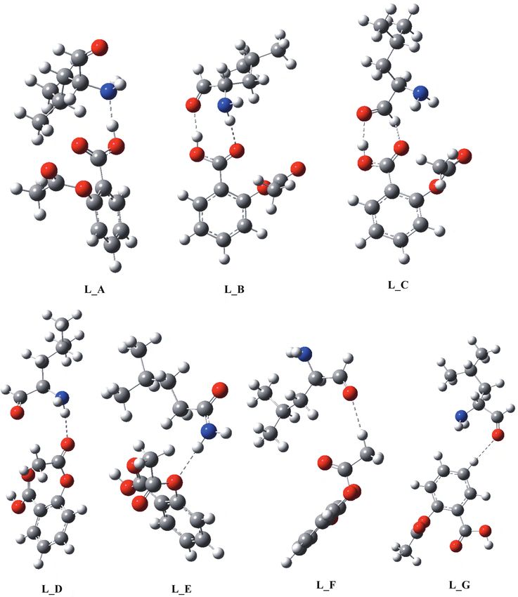
7 different structures for the aspirin-leucine supermolecule. The structures are given in Figure 3 and the BSSE
corrected interaction energies in the gas phase are given in Table 4.
The global minimum on the potential energy surface belongs to the structure L A, where the acidic
hydrogen of aspirin was bonded to the nitrogen of leucine. The hydrogen bond length (1.68 Å) was slightly
shorter than those in the dimers of aspirin. The second conformer L B displayed a hydrogen bond utilizing
the acidic hydrogen of aspirin bonded to the oxygen of the amino acid. At the same time, a carbonyl oxygen
of the aspirin formed a relatively long hydrogen bond with both amine hydrogen atoms. One of these bonds
was 1.78 Å, while the second one was around 2.05 Å. The final conformer that had similar stability to L C
probably belongs to the same group of local minima with L B. Here the N-H..O distance was too large to have
a significant effect on the stability. If the acetoxy group of the aspirin is involved in the hydrogen bonding,
then the supermolecule was considerably less stable than all conformers described above. Conformer L D had a
hydrogen bond between the carbonyl oxygen in the acetoxy group and the amine hydrogens. The bond lengths
were now 2.14 Å. Similarly, the conformer L E now bonded the ester oxygen in the acetoxy group and the amine
hydrogen with the distance again being around 2.15 Å. In both cases the BSSE corrected interaction energies
were between 2 and 4 kcal/mol. Finally, we tried to locate structures where methyl and phenyl hydrogens were
involved. Conformers L F and L G were such cases with weak interactions in the order of 1 kcal/mol.
391Competitive hydrogen bonding in aspirin-aspirin and..., Z. YURTSEVER, et al.
Figure 3. Aspirin-leucine complexes.
MP2 results showed stronger interactions compared to DFT calculations. As we discussed in the dimer-
ization of aspirin, this increase in energy is mostly due to the fact that MP2 favors long distance interactions (for
392Competitive hydrogen bonding in aspirin-aspirin and..., Z. YURTSEVER, et al.
these supermolecules there are more long-range hydrogen bonds than aspirin dimers), whereas DFT underesti-
mates them. Still, MP2 results for the last 2 conformers were well within the error bounds of the calculations,
showing that such bonds might be stable at room temperature. The stabilities of these conformers in water,
again calculated within the PCM formalism, display slight deviations from their gas phase counterparts. How-
ever, they are not significant enough to warrant a separate discussion. All of these structures have all positive
eigenvalues of the Hessian and therefore correspond to real minima. As in the aspirin-aspirin complexes, ZPE
corrections are not included in Table 3.
Table 4. Interaction energies of aspirin-leucine heterodimers (kcal/mol).
Structure DFT/6-31g(d) MP2/aug-cc-pvdz
LA –14.63 –16.37
LB –11.46 –12.45
LC –10.33 –13.95
LD –3.58 –6.04
LE –2.29 –5.36
LF –1.30 –3.62
LG –0.45 –2.07
Conclusions
Our computational studies show that dynamics of aspirin-amino acid complex can be understood in terms of
the number, type and strength of hydrogen-bonding. There are isomers of the aspirin molecule that exhibit
intramolecular hydrogen bonding although they are not the most stable ones. However, all these isomers are
populated at room temperature, especially when in solution.
There are competing hydrogen bonds between aspirin-aspirin and aspirin-leucine molecules. The most
stable bonds involve the acetoxy and carboxylic acid groups of aspirin and the amine group of leucine. However,
there are relatively weak isomers where methyl and phenyl hydrogens are also involved in hydrogen-bond
formation. These unusual methyl- or phenyl-coordinated hydrogen bonds in biological systems have been
proposed before and were recently detected by NMR spectroscopy. 24,25
In the case of the aspirin-aspirin complexes, hydrogen bonds through carboxylic acid groups are about
8-9 kcal/mol in an additive nature, that is if there are 2 such bonds, the total stability reaches 20 kcal/mol.
For the aspirin-leucine complexes, the hydrogen bonds through amine groups are considerably stronger with
15 kcal/mol. In all cases, there is a variety of structures with weaker hydrogen bonds. These local minima
are accessible at room temperature and so understanding (the mechanisms of) aspirin-amino acid interactions
should include all these possible structures. Among those interactions, a particularly significant one is obtained
in the inhibition of the protein cyclooxygenase, COX, as discussed above. Different mechanisms have been
suggested for the inhibitory role of aspirin, 7 and a precise evaluation of the hydrogen bonding established by
aspirin is crucial for a better understanding of the mechanism.
393Competitive hydrogen bonding in aspirin-aspirin and..., Z. YURTSEVER, et al.
Acknowledgments
B. Erman and E. Yurtsever would like to acknowledge the support given by Koç University.
References
1. Mitchell, J. A.; Akarasereenont, P.; Bishopbailey, D.; Wood, E. G.; Thiemermann. C.; Vane, J. R. Brit. J. Pharm.
1993, 109, P2-P2.
2. Vane, J. R. Nature 1994, 367, 215-216.
3. Trummlitz, G.; Van Ryan, J.; Klein, C. T.; Clayton, G.; Powers, R.; Garavito, R. M. Annals of the Rheumatic
Diseases 2004, 63, 361-362.
4. Garavito, R. M., Scientific American 1999, 280, 108-108.
5. Loll, P. J.; Garavito, R. M.; Carrell, H. L. Acta Crystallographica Section C-Crystal Structure Communications
1996, 52, 375-377.
6. Loll, P. J.; Picot, D.; Garavito, R. M. Nature Structural Biology 1995, 2, 637-643.
7. Silverman, R. B. The Organic Chemistry of Drug Design and Drug Action. 2 ed. MA. Elsevier, 2004.
8. Kalgutkar, A. S.; Crews, B. C.; Rowlinson, S. W.; Garner, C.; Seibert, K.; Marnett, L. J. Science 1998, 280,
1268-1270.
9. Hochgesang, G. P.; Rowlinson, S. W.; Marnett, L. J. J. Amer. Chem. Soc. 2000, 122, 6514-6515.
10. Glaser, R. J. Org. Chem. 2001, 66, 771-779.
11. Sidhu, R. S.; Lee, J. Y.; Yuan, C.; Smith, W. L. Biochemistry 2010, 49, 7069-7079.
12. Discovery Studio Visualizer v2.5.5.9350 Copyright 2005-2009 Accelrys software Inc.
13. Hobza, P. (ed) Phys. Chem. Chem. Phys. 2008, 10, 2561-2561.
14. Buckingham, A. D.; Del Bene, J. E.; McDowell, S. A. C.; Chem. Phys. Lett. 2008, 463, 1-10.
15. Yılgör, E.; Yurtsever, E.; Yılgör, I. Polymer 2000, 41, 849-857.
16. Yılgör, E.; Yılgör, I.; Yurtsever, E. Polymer 2002, 43, 6551-6559.
17. Yılgör, E.; Yurtsever, E.; Yılgör, I. Polymer 2002, 43, 6561-6568.
18. Gaussian 09, Revision A.1, Frisch, M. J.; Trucks, G. W.; Schlegel, H. B.; Scuseria, G. E.; Robb, M. A.; Cheeseman,
J. R.; Scalmani, G.; Barone, V.; Mennucci, B.; Petersson, G. A.; Nakatsuji, H.; Caricato, M.; Li, X.; Hratchian,
H. P.; Izmaylov, A. F.; Bloino, J.; Zheng, G.; Sonnenberg, J. L.; Hada, M.; Ehara, M.; Toyota, K.; Fukuda,
R.; Hasegawa, J.; Ishida, M.; Nakajima, T.; Honda, Y.; Kitao, O.; Nakai, H.; Vreven, T.; Montgomery, Jr., J. A.;
Peralta, J. E.; Ogliaro, F.; Bearpark, M.; Heyd, J. J.; Brothers, E.; Kudin, K. N.; Staroverov, V. N.; Kobayashi, R.;
Normand, J.; Raghavachari, K.; Rendell, A.; Burant, J. C.; Iyengar, S. S.; Tomasi, J.; Cossi, M.; Rega, N.; Millam,
N. J.; Klene, M.; Knox, J. E.; Cross, J. B.; Bakken, V.; Adamo, C.; Jaramillo, J.; Gomperts, R.; Stratmann, R. E.;
Yazyev, O.; Austin, A. J.; Cammi, R.; Pomelli, C.; Ochterski, J. W.; Martin, R. L.; Morokuma, K.; Zakrzewski, V.
G.; Voth, G. A.; Salvador, P.; Dannenberg, J. J.; Dapprich, S.; Daniels, A. D.; Farkas, Ö.; Foresman, J. B.; Ortiz,
J. V.; Cioslowski, J.; Fox, D. J. Gaussian, Inc., Wallingford CT, 2009.
394Competitive hydrogen bonding in aspirin-aspirin and..., Z. YURTSEVER, et al.
19. MOLPRO, version 2009.1, a package of ab initio programs, Werner, H. J.; Knowles, P. J.; Lindh, R.; Manby, F.
R.; Schütz, M.; Celani, P.; Korona, T.; Mitrushchenkov, A.; Rauhut, G.; Adler, T. B.; Amos, R. D.; Bernhardsson,
A.; Berning, A.; Cooper, D. L.; Deegan, M. J. O.; Dobbyn, A. J.; Eckert, F.; Goll, E.; Hampel, C.; Hetzer, G.;
Hrenar, T.; Knizia, G.; Köppl, C.; Liu, Y.; Lloyd, A. W.; Mata, R. A.; May, A. J.; McNicholas, S. J.; Meyer, W.;
Mura, M. E.; Nicklass, A.; Palmieri, P.; Pflüger, K.; Pitzer, R.; Reiher, M.; Schumann, U.; Stoll, H.; Stone, A. J.;
Tarroni, R.; Thorsteinsson, T.; Wang, M.; Wolf, A. 2009.
20. Becke, A. D. J. Chem. Phys. 1996, 104, 1040-1046.
21. Tomasi, J.; Mennuci, B.; Cammi, C. Chem. Rev. 2005, 105, 2999-3090.
22. Payne, R. S.; Rowe, R. C.; Roberts, R.J. ; Charlton, M. H.; Docherty, R. J. Comp. Chem. 1999, 20, 262-273.
23. Vishweshwar, P.; McMahon, J. A.; Oliveira, M.; Peterson, M. L.; Zaworotko, M. J. J. Amer. Chem. Soc. 2005,
127, 16802-16803.
24. Krimm, S. Science 1967, 158, 530-531.
25. Horowitz, S.; Yesselman, J. D.; Al-Hashimi, H.M.,Trievel, R. C. J. Biol. Chem. 2011, 286, 18658-18663.
395You can also read