Concours FNS d'images scientifiques 2020/21 SNF-Wettbewerb für wissenschaftliche Bilder 2020/21 SNSF Scientific Image Competition 2020/21 ...
←
→
Page content transcription
If your browser does not render page correctly, please read the page content below
1
    Concours FNS d’images scientifiques 2020/21
    SNF-Wettbewerb für wissenschaftliche Bilder 2020/21
    SNSF Scientific Image Competition 2020/21
The hidden face of science                                                                       2

Science is by no means a cold and disinterested activity. It is something done by real people,
with passion and devotion, involving trial and error and happy accidents. The contemporary
practice of scientific research is revealed in the 680 works that were submitted by scientists
in Switzerland to the 2020 and 2021 editions of the SNSF Scientific Image Competition.
The Biel/Bienne Festival of Photography is exhibiting a selection of the works that were
awarded prizes by the jury and those chosen by popular vote, along with an electronic music
piece set to scientific images and videos, presented by Witold Langlois, founder and curator
of METEO.

La face cachée de la science
Loin d’une pratique distanciée et froide, la science est affaire de femmes et d’hommes,
de passions et de dévouement, de tâtonnements et d’heureux hasards. La pratique
contemporaine de la recherche se dévoile au long des 680 œuvres soumises par les
scientifiques de Suisse aux éditions 2020 et 2021 du Concours FNS d’images scientifiques.
Les Journées photographiques de Bienne en exposent une sélection, les œuvres
primées par le jury et choisies lors d’un vote public, ainsi qu’une mise en musique
électronique d’images et de vidéos scientifiques proposée par Witold Langlois, fondateur et
curateur de METEO.

Die verborgene Seite der Wissenschaft
Die Wissenschaft ist keine abgehobene, unnahbare Disziplin, sondern entspringt der
menschlichen Natur, der Leidenschaft und Hingabe, dem Trial and Error und zuweilen
auch glücklichen Fügungen. Einen Einblick in die aktuelle Forschungspraxis geben die
680 Arbeiten, welche die Schweizer WissenschaftlerInnen im Rahmen der Ausgaben 2020
und 2021 des SNF-Wettbewerbs für wissenschaftliche Bilder eingereicht haben.
An den Bieler Fototagen werden eine Auswahl davon, die von der Jury ausgezeichneten
und im Rahmen einer Publikumswahl gekürten Werke sowie von Witold Langlois
(Gründer und Kurator von METEO) präsentierte, mit elektronischer Musik hinterlegte
wissenschaftliche Bilder und Videos gezeigt.
3
    SNSF Scientific Image Competition 2020
    Concours FNS d’images scientifiques 2020
    SNF Wettbewerb für wissenschaftliche Bilder 2020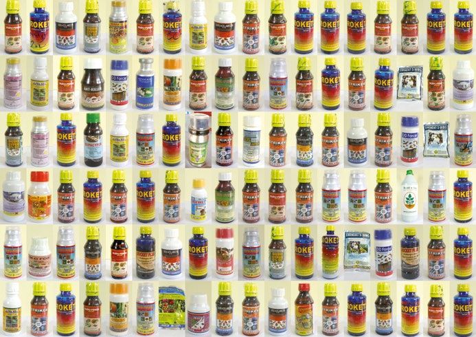
5
    Foot pad of an Asian           As part of a study on influencing factors on elephant foot health, I
                                   took pictures of the feet of more than 150 Asian elephants living in
    elephant                       European zoos. This picture shows a right front foot of a ten-year-
                                   old male from below. In the middle of the picture is the foot pad with
    Paulin Wendler                 its natural grooves working like the treads of our shoes. The brighter
                                   areas on the lateral and upper edges are the soles of the five-foot
                                   nails. When lifted (as for this picture), this foot has a circumference
                                   of 117 cm, and it expands when bearing weight.

                                   COMMENT OF THE JURY
                                   This stunning picture forces viewers to slow down, immersing
                                   them in a dazzling view of organic structures of unknown scale
                                   from something they have never seen before: the underside of an
                                   elephant foot. These intricate lines form a landscape sculpted by
                                   evolution. Their sophisticated subtlety contrasts with the image
                                   usually associated with the lumbering, heavy and almost archaic
                                   mammal they belong to. These physiological structures contradict
                                   contemporary assumptions that evolution’s efficiency consists of
                                   making organisms faster.

                                   THE AUTHOR
                                   Paulin Wendler (GER), born in 1992, is a veterinarian based in
                                   Germany. During her studies of Veterinary Medicine at Leipzig
                                   University, she pursued her interest in zoo and wild animal medicine.
                                   During her doctoral studies at the Clinic for Zoo Animals, Exotic Pets
                                   and Wildlife of the University of Zurich, she conducted a project on
                                   foot health of Asian elephants in European zoos. She is currently
                                   working in a veterinary practice for small animals and horses in
                                   Brunswick (GER).

    First prize
    Category 1 – Object of Study   SNSF Scientific Image Competition 2020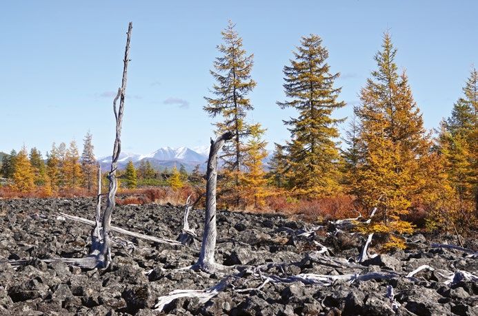
Pesticides from Smallholder farmers are not very strong on safe pesticide use. This
compilation displays pesticide bottles of mostly 100 ml, obtainable
6
mystery shopping in by any person in agro dealer shops in Uganda. Almost all of the
displayed products are highly or moderately hazardous to human
Uganda health. Nevertheless, sales staff do not explain safety labels or
Curdin Brugger otherwise indicate potential dangers. In our research we study how
agro dealers (don’t) advise smallholder farmers and see how good
their own knowledge and awareness is regarding the safe use of
pesticides.
We recruited local farmers and sent them to buy pesticides, all
using the same description of a fall armyworm outbreak in maize.
This image displays all the products obtained in different shops
around the country. Safety labels are supposed to indicate the
level of hazard and protective measures, usually as a red or yellow
band at the bottom of the bottle. Looking closely, you can see some
unreadable pictograms and an array of wrong colours.
COMMENT OF THE JURY
The slightly random repetition in this typology of poisons echoes in
its chosen arrangement the situation faced by farmers: numerous
substances, as interchangeable as they are dangerous; highlighted
by the colour yellow, universal symbol for hazardous materials.
Familiar in art, the serial representation finds a captivating use in
this social science research project.
THE AUTHOR
Curdin Brugger (CH), born in 1993, is a master’s student in
Epidemiology at the Swiss Tropical and Public Health Institute
in Basel. He is motivated to improve public health in developing
countries, with a focus on social, behavioural and occupational
determinants of health. His current research investigates the impact
of pesticide use on the health of small-scale farmers in Uganda.
Distinction of the jury
Category 1 – Object of Study SNSF Scientific Image Competition 2020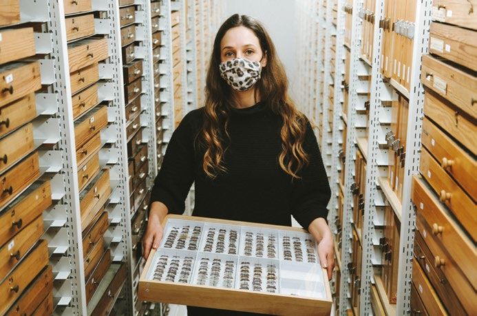
7
    Sunwalk                        In my research, I focus on public spaces and new ways of delivering
                                   mobility in Chinese cities. This shot was taken while I was
    Jérôme Gapany                  conducting ethnographic fieldwork in south-eastern Fujian Province,
                                   where daily lives are shaped by rapid urban change, sometimes in
                                   uncanny ways.
                                         Here, a woman holding an umbrella tries to hide from the sun
                                   while walking on a running track. Summer heat in south-eastern
                                   Chinese cities is an increasing challenge with rapid urbanisation.
                                   In this image, I tried to capture a fleeting moment of solitude in a
                                   densely populated urban landscape.

                                   COMMENT OF THE JURY
                                   At first sight, this very strong composition appears to mix sport
                                   and fashion; but actually it beautifully illustrates the paradox
                                   encountered by the researcher: that studying overpopulated cities
                                   involves the depiction of isolated people.

                                   THE AUTHOR
                                   Jérôme Gapany (CH), born in 1990, is a Swiss ethnographer and PhD
                                   candidate at the University of Lausanne. He holds a Master in Asian
                                   Studies from the Geneva Graduate Institute and the University of
                                   Geneva. His current research focuses on mobility infrastructures
                                   in Fuzhou city, China. His interest in photography stems from
                                   experiences in China, Taiwan, and South Korea, where he picked up
                                   his camera to document everyday politics and street life.

    Distinction of the jury
    Category 1 – Object of Study   SNSF Scientific Image Competition 2020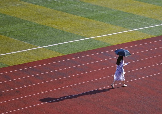
Fisherwoman in                          Norine and her lovely zebrafish. Her project is a great example of
                                        how model organisms could help us to fight diseases. Zebrafish
                                                                                                                   8

the lab                                 is a great model for studying developmental diseases since it
                                        develops externally, very fast and its organs are transparent! This
Kaan Mika                               allows scientist to observe morphological defects at early stages
                                        in the development. In order to overexpress a gene, one can inject
                                        messenger RNA directly into zebrafish eggs, or use morpholinos in
                                        order to knock down a specific gene. Moreover, with the CRISPR/
                                        Cas9 system it is possible to delete a region, induce a specific
                                        mutation or even insert a gene cassette in a desired location in the
                                        genome. Therefore, using model organisms like zebrafish allows
                                        scientists to elucidate the molecular mechanisms involved in some
                                        human diseases.

                                        COMMENT OF THE JURY
                                        A lively portrait which makes the researcher seem very approachable
                                        and relatable, a clear challenge to the usual representation of
                                        scientists in their lab. Instead of stereotypical white lab coats,
                                        vivid colours interact at all levels, while the scientist’s strong self-
                                        expression contrasts with the standardisation inherent in the
                                        research carried out with model animals such as zebrafish.

                                        THE AUTHOR
                                        Kaan Mika (Turkey), born in 1989, is a molecular biologist pursuing
                                        a PhD at University of Lausanne. He works with a famous model
                                        organism called Drosophila melanogaster and he investigates the
                                        olfactory system of these flies. He is a self-taught photographer and
                                        he uses Instagram to promote science, collaborating with scientists
                                        around Lausanne.

First prize
Category 2 – Women and Men of Science   SNSF Scientific Image Competition 2020
9
    Keep calm and                           This photo portraits my colleague and dear officemate Yuan Xiao
                                            during the final rush towards the completion of her PhD studies.
    write on                                Yuan wrote her thesis in an impressively short amount of time and
                                            brilliantly defended her PhD shortly after. I found her total dedication
    Stefano Danzi                           to it both bemusing and inspiring. It even led her to change her
                                            desktop background image to “Keep calm and write on”.

                                            COMMENT OF THE JURY
                                            A moment of intimacy captured on camera with empathy, a
                                            spontaneous movement showing a moment of relaxation during
                                            intense intellectual work – natural and relatable.

                                            THE AUTHOR
                                            Stefano Danzi (IT, 1991) is a Materials science PhD at ETH Zurich.
                                            He obtained MSc in Materials engineering and nanotechnology from
                                            Politecnico di Milano and spent one year as a visiting student at
                                            the Massachusetts Institute of Technology (MIT) before moving to
                                            Switzerland. While not being a photographer whatsoever, he enjoys
                                            street photography as a graphical approach to storytelling.

    Distinction of the jury
    Category 2 – Women and Men of Science   SNSF Scientific Image Competition 2020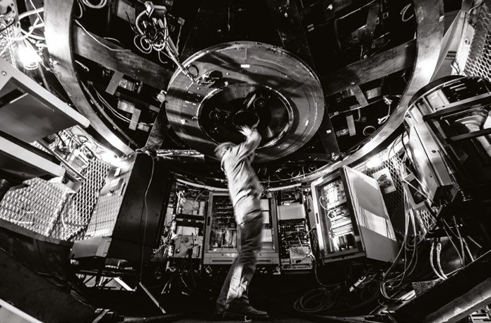
The astronomer                          To install new equipment to the telescope, it is often necessary to
                                        remove older equipment, granting a unique view into the telescope’s
                                                                                                                10

Nicolas Blind                           mirrors and internal mechanics. Until the 80s, astronomers would
                                        still sit at night in the so-called telescope cage (pictured here) to
                                        perform visual observations. With the advent of photo-sensitive
                                        electronic devices, the astronomer’s eye was progressively replaced
                                        by today’s detector arrays, which are able to count individual
                                        photons. This is therefore a rare view of an astronomer looking into
                                        the telescope focus.

                                        COMMENT OF THE JURY
                                        The choice of black and white photography avoids the expected
                                        aestheticisation of technical apparatus. A strong composition which
                                        reveals, as emphasised by the slight blur, a very rare moment in the
                                        work of an astronomer: tinkering with the hard mechanics needed to
                                        explore ethereal outer space.

                                        THE AUTHOR
                                        Nicolas Blind (FR), born in 1984, is an optical engineer working at
                                        Geneva Observatory (CH). He obtained his PhD in high angular
                                        resolution instrumentation for astronomy at UGA (Grenoble), and
                                        already contributed to several instruments of the European Southern
                                        Observatory (ESO). While he works daily with state of the art
                                        detectors, able to count individual photons, his photographic work
                                        focuses on analog processes (more specifically, original processes
                                        from the 19’s century, like wet plate collodion or cyanotype), with a
                                        pinch of digital when necessary.

Distinction of the jury
Category 2 – Women and Men of Science   SNSF Scientific Image Competition 2020
11
A Surrogate Mother‘s The photo shows the room of a surrogate mother in Kiev, Ukraine.
In the last few years, surrogacy – carrying a child for others – has
Room in Kiev become an important industry in the country. Especially poor women
see it as an opportunity to earn within nine months a sum equalling a
Anika König ten-year income. But at the same time, surrogacy is stigmatised and
many women hide their pregnancies from their families and friends.
Once the pregnancy becomes visible, they move to flats in the city
provided by surrogacy agencies. These flats are very impersonal and
look as if the surrogates were constantly on the go: they keep all
their belongings in plastic bags.
The photo shows all the belongings of a surrogate whom
I interviewed for my anthropological research on transnational
surrogacy. The towel which was hung up to dry on a laundry rack
was the most personal item in the room. During one of my visits, she
was transferred to the hospital and vacated the room within minutes,
leaving no trace.
COMMENT OF THE JURY
The strength of the picture lies in the obvious absence of its
object. It reveals the invisibility of surrogate mothers in society
by not showing them, the bags emphasising their precarious and
ephemeral status. The composition shows a deep understanding of
what photography can achieve, both carrying respect and empathy
for its subject while maintaining a distance, similarly to quality works
known from photojournalism.
THE AUTHOR
Anika König (D) is a senior lecturer at the Department of Ethnology
at the University of Luzern. She obtained an MA in Social and
Cultural Anthropology from Freie Universität Berlin and Sociology
from Technische Universität Berlin, and a PhD in Anthropology from
the Australian National University. Her current research focuses on
transnational gestational surrogacy commissioned by Swiss and
German intended parents.
First prize
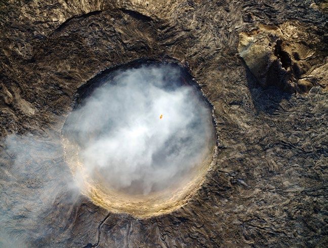
Category 3 – Places and Tools of Science SNSF Scientific Image Competition 2020Siberian trees witness This picture shows several hundred-year-old living and relict larch
(Larix cajanderi Mayr) trees on the extensive, 56 km-long lava flow of
12
1000 years of climate the Anyui volcano in northeastern Siberia (around 68°N and 166°E).
While the volcanic complex in Russia’s far Northeast is mainly
change unexplored, it provides a unique dendrochronological archive for
Andreas Rigling the development of multi-centennial to millennial long, temperature-
sensitive tree-ring chronologies, in one of the world’s most remote
regions where high-resolution paleoclimatic evidence is still
missing. The picture was taken during the first international and
interdisciplinary Anyui-Expedition in September 2019, a joint venture
of the University of Cambridge (United Kingdom), the Universities
of Krasnoyarsk and Yakutsk (both Russia), and the Swiss Federal
Research Institute WSL (Switzerland). Photo credit: Andreas Rigling,
Swiss Federal Research Institute WSL, Switzerland.
COMMENT OF THE JURY
A clear, honest photographic document set in a dramatic landscape;
the image condenses the passing of time over millennia, projecting
us into a future impacted by climate change.
THE AUTHOR
Andreas Rigling (CH), born in 1964, is a forest and dendro-
ecologist at the Swiss Federal Institute for Forest, Snow and
Landscape Research, and adjunct professor at ETH Zürich. Among
others, he uses tree rings to analyse current and reconstruct past
environmental conditions. He is a dedicated and passionate hobby
photographer with a focus on nature and specifically forest topics
and he uses photography as an important communication tool for
science, teaching and knowledge transfer.
Distinction of the jury
Category 3 – Places and Tools of Science SNSF Scientific Image Competition 202013
Sequential sequencing 142 years after Eadweard Muybridge’s invention, the principle of
chronophotography remains essential in understanding human
Reto Togni movement. The image not only builds on a rich cultural heritage of
sequence photographs, nowadays best known in extreme sports, but
also illustrates cutting-edge scientific processes that reveal internal
working principles of the human body. Shown is a gait trial using a
unique moving fluoroscope that tracks and follows the movements
of a study participant while taking a series of x-ray images to analyse
bone kinematics. By merging a series of six photographs taken
during the trial, the image not only shows but also follows the maxim
of novelty through continuous reinterpretation and repurposing of
methods that are more than a century old.
COMMENT OF THE JURY
A playful mise en abyme of chronophotography, depicting by means
of a digital collage the motion of an apparatus that is itself taking
sequential X-ray images of the legs of a person walking. It brings
into view the actual processes involved in a scientific experiment,
rather than solely its results.
THE AUTHOR
Reto Togni is a PhD candidate at the Laboratory for Movement
Biomechanics at ETH Zurich. He holds a BA in Industrial Design and
a MA/MSc in Innovation Design Engineering. Mundane everyday
things have the capacity to shape, change who people are, what
they do, how they view the world and are viewed by others. As
a developer of such everyday objects, he disentangles these
relationships, analyses their components, prototypes interventions
and designs new practices.
Distinction of the jury
Category 3 – Places and Tools of Science SNSF Scientific Image Competition 2020Transparency in A rotating Lego model, which was made transparent by x-ray
computed tomography. The coloured structures are traces of
14
science the emission activity of a radioactive substance. These were not
visualised with x-rays, but with SPECT, another medical imaging
Peter von technique. By combining the two different signals, the exact position
Niederhäusern of a contrast medium can be determined. This is important for
observing the effects of medical treatments. The film was created for
the calibration of a novel 3D reconstruction algorithm. Lego bricks
are well suited to this purpose because they are cost-effective and
their dimensions are well known. Innovative and low-cost solutions
like this can help to achieve scientific goals, too.
COMMENT OF THE JURY
A mesmerising loop which puzzles the viewers, challenging their
sense of orientation until they finally recognise the familiar shape of
a popular toy. A vivid illustration that state-of-the-art research can
proceed through a joyful bricolage of serendipity and simplicity, here
revealing a behind-the-scenes moment, when an apparatus is merely
calibrated before its actual use.
THE AUTHOR
Peter von Niederhäusern, born and raised in Schwarzenburg BE,
studied Biomedical Engineering (M.Sc.) at the universities of Bern
and Basel and finished his master’s thesis in the domain of eye
diseases. His current Ph.D. studies involve the development of novel
visualization methods for nuclear medicine specialists. Apart from
applied research, Peter is also interested in sports, the marvels of
the universe, and all things involving technology and research and
their implication for society.
First prize
Category 4 – Videos SNSF Scientific Image Competition 202015
The digital This video illustrates the computational creation of a prototypical
zebrafish embryo. What does the average embryo look like and
prototypical embryo how can we find out? We use modern optical microscopy to create
3D reconstructions of living zebrafish embryos. With the help of
Marvin Albert computational algorithms we then warp the embryos such that
differences in their pose and shape are eliminated. Finally, a 3D
representation of the prototypical zebrafish embryo results from
overlaying and averaging all of the obtained images.
Details: the zebrafish embryos were 36 hours old when their
images were taken using multi-view light sheet fluorescence
microscopy. The image contrast represents the expression
pattern of a chemokine receptor protein, visualising among other
structures the eye, the gill arches, vasculature and neuronal bundles.
Computational processing of the images was performed on a high
performance computer cluster and took two days.
COMMENT OF THE JURY
A very aesthetic presentation of the scientific method, based on an
intuitive way of explaining visually a very complex scientific project:
creating virtual lifeforms based on real observations.
THE AUTHOR
Marvin Albert (born 1989, German) is a biophysicist at the University
of Zurich. He creates 3D computer models of animal development.
He obtained his PhD from the European Molecular Biology
Laboratory (EMBL) in Heidelberg, where he specialised on extracting
quantitative information from microscopy images to study cell
movement and tissue growth.
Distinction of the jury
Category 4 – Videos SNSF Scientific Image Competition 202017
SNSF Scientific Image Competition 2021
Concours FNS d’images scientifiques 2021
SNF Wettbewerb für wissenschaftliche Bilder 202119
Computed tomography Amid the COVID-19 pandemic, post-mortem computed tomography
(PMCT) examination of lung disease is becoming increasingly
of suspected important as a relatively rapid means of making suspicious findings
visually apparent. This study focuses on visual communication of
COVID-19 pneumonia findings and the development of new visual display modes using
Eloisa Aldomar, Zurich the volume rendering technique (VRT). The 3D reconstruction shows
a PMCT of the lungs of a 30-year-old man with classic features of
University of the Arts COVID-19 pneumonia. Extensive tissue compaction (with higher
density values in the CT data) is not shown. Under proper lighting,
the spatial visualisation of the outer part of the lungs enables
examination of morphologic abnormalities. The image has been
enhanced using both warm and cold light sources.
COMMENT OF THE JURY
This stunning technical image offers an extremely insightful post-
mortem view of the lung of a COVID-19 patient. Its particularity,
however, lies in the fact that we are not dealing with a microscopic
image here but with a very accurate data visualisation based on
VRT. The image emphasises the great educational potential of an
aesthetically sensitive translation of data, using colour very sparingly
and efficiently.
THE AUTHOR
Eloisa Aldomar, born in 1977, is a designer based in Switzerland.
She obtained her Bachelor (2017) and Master (2019) of Arts ZFH in
design with a specialisation in scientific and knowledge visualisation
from ZHdK, Zurich. She completed her bachelor and master theses
in cooperation with the Institute of Forensic Medicine UZH, Zurich.
First prize
Category 1 – Object of Study SNSF Scientific Image Competition 2021Reconstruction tomographique d’une Computer-Tomographie einer Lungen- 20
suspicion de pneumonie COVID-19 entzündung mit Verdacht auf C ovid-19
Eloisa Aldomar, Université des Arts de Zurich Eloisa Aldomar, Zürcher H
ochschule der
Künste, Zürich
Moyen relativement rapide de faire Mit der Covid-19-Pandemie hat die
apparaître visuellement des observations postmortale Computertomographie
suspectes, la tomodensitométrie post- (PMCT) eine neue Wichtigkeit erlangt,
mortem (TPM) des maladies pulmonaires weil sie problematische Befunde bei
revêt une importance croissante dans le Lungenkrankheiten schnell sichtbar
contexte de la pandémie de COVID-19. Cette machen kann. Die Studie befasst sich
étude se concentre sur la communication mit der visuellen Kommunikation von
visuelle des résultats et le développement Befunden und der Entwicklung von neuen
de nouveaux modes d’affichage visuel visuellen Möglichkeiten, basierend auf der
utilisant la technique de rendu volumique Visual Rendering Technique (VRT). Diese
(VRT). Cette reconstruction 3D est issue 3D-Rekonstruktion zeigt die PMCT der
d’une TPM des poumons d’un homme de 30 Lunge eines 30-jährigen Mannes mit den
ans présentant des symptômes classiques klassischen Auffälligkeiten einer Covid-
de la pneumonie COVID-19. La fibrose 19-Lungenentzündung. Eine grossflächige
étendue des tissus (révélée par des valeurs Gewebeverdichtung (mit höheren
de densité plus élevées des données Dichtewerten in den CT-Daten) ist nicht
tomographiques) n’apparaît pas. Sous un ersichtlich. Mit der richtigen Helligkeit ist es
éclairage approprié, la visualisation spatiale möglich, durch die räumliche Visualisierung
de la partie externe des poumons permet den äusseren Bereich der Lunge hinsichtlich
d’examiner les anomalies morphologiques. morphologischer Veränderungen zu
L’image a été rehaussée en utilisant untersuchen. Das Bild wurde mit warmen
différentes sources de lumière chaude et und kalten Lichtquellen bearbeitet.
froide.
Kommentar der Jury │
Commentaire du jury │ Dieses eindrückliche Bild ermöglicht einen
Cette étonnante image technique offre une aufschlussreichen Blick auf die Lunge
vue post-mortem des poumons d’un patient eines verstorbenen Covid-19-Patienten. Das
COVID-19 extrêmement révélatrice. Sa Besondere ist, dass wir es nicht mit einem
particularité réside toutefois dans le fait qu’il mikroskopischen Bild zu tun haben, sondern
ne s’agit pas d’une image micrographique, mit einer Visualisierung basierend auf VRT.
mais d’une visualisation très précise de Deutlich zeigt sich auch der pädagogische
données tomographiques. L’utilisation Wert, den eine ästhetisch feinfühlige
économe et efficace des couleurs souligne Umsetzung von Daten hat, hier zum Beispiel
le potentiel éducatif élevé d’une traduction durch den zurückhaltenden und effektiven
esthétiquement sensible des données. Einsatz von Farben.
1er prix Erster Preis
Catégorie 1 – L’objet d’étude Kategorie 1: Forschungsobjekt21
Empty memory The image “Empty memory” was recorded by a sophisticated neutron
imaging detector. The detector is used in advanced applications
Markus Strobl, to observe the interior structures and crystallographic features of
materials and specimens which define their mechanical performance.
Paul Scherrer Institute In the image shown here, the detector merely recorded the empty
beam, without a sample. Thus, it is the performance of the detector
itself that is the subject of the specific measurement usually taken
as reference for sample data. But the image also reveals the traces
of high-intensity beams probed in the past through what might be
called a memory effect. As a result, what you see is an image of
an empty beam formed primarily by the “memory” of previously
recorded empty beams. Together with a distinct pattern of rows of
dead pixels, without memory, the image constitutes an abstraction of
emptiness and remembered shapes for the viewer to interpret.
COMMENT OF THE JURY
A poetic way of dealing with abstraction, as revealed by the title.
This image, which at first glance resembles a modern painting, is
intended to show that every phenomenon leaves traces, even on
scientific instruments. The scientist seems to be touched both by the
aesthetics of what he sees and by the fact that even the recording of
a beam leaves a memory. The jury appreciates this beautiful image,
together with the moving narrative about emptiness.
THE AUTHOR
Markus Strobl, born in 1972, is a scientist whose research focuses on
neutron scattering and imaging for materials research. He obtained
his PhD from the Technical University of Vienna and has designed
and built several neutron instruments. He holds an affiliated
professorship at the University of Copenhagen and leads the neutron
imaging activities at the Paul Scherrer Institute. In the past, he also
contributed works to and curated exhibitions of visual arts in Vienna,
Berlin and Genoa.
Distinction of the jury
Category 1 – Object of Study SNSF Scientific Image Competition 2021Mémoire vide Leere Erinnerungen 22
Markus Strobl, Institut Paul Scherrer Markus Strobl, Paul Scherrer Institut
L’image «Mémoire vide» a été capturée Das Bild „Leere Erinnerung“ wurde von
par un système d’imagerie neutronique. einem hochmodernen „Neutron Imaging
De tels détecteurs sont utilisés dans le Detector“ aufgenommen. Das Instrument
cadre d’applications sophistiquées afin wird normalerweise verwendet, um
d’observer les structures intérieures et les innere Strukturen und kristallographische
caractéristiques cristallographiques de Merkmale von Materialien und Präparaten zu
matériaux et de spécimens qui définissent beobachten. Das gibt Aufschluss über ihre
leurs performances mécaniques. Cependant, mechanischen Eigenschaften. In diesem Bild
sur l’image présentée ici, le détecteur a zeichnete der Detektor nur den Strahl selbst
uniquement enregistré le faisceau vide auf. So wird die Leistung des Detektors zum
sans échantillon. Par suite, ce sont les Zweck der Messung, die sonst als Referenz
performances du détecteur lui-même qui für Probedaten gemacht wird. Gleichzeitig
font l’objet des mesures spécifiques servant werden auch die Spuren von hochintensiven
habituellement de référence aux données Strahlen vergangener Messungen sichtbar.
d’échantillonnage. Mais l’image révèle Es handelt sich um einen Memory-Effekt.
également les traces de faisceaux de haute Deshalb sieht man hier das Bild eines leeren
densité observés dans le passé, traces Strahls, der aus der Erinnerung von anderen
induites par ce qui pourrait être appelé leeren Strahlen entstanden ist. Zusammen
un effet mémoire. Au final, on contemple mit dem charakteristischen Muster der toten
l’image d’un faisceau vide principalement Pixels ist das Bild eine Abstraktion der Leere
formé par la «mémoire» des faisceaux und von Formen, die aus der Erinnerung
vides préalablement enregistrés. En raison entstanden sind und auf verschiedene Weise
du dessin distinctif formé par les rangées interpretiert werden können.
de pixels morts et sans mémoire, cette
image constitue une abstraction de vides Kommentar der Jury
et de formes mémorisées qui est laissée à Im Titel zeigt sich ein poetischer Umgang
l’interprétation de l’observateur. mit Abstraktion. Das Bild, das auf den ersten
Blick einem modernen Gemälde ähnlich
Commentaire du jury sieht, zeigt, dass alle Phänomene Spuren
Comme le titre le révèle, il s’agit là d’une hinterlassen, sogar auf wissenschaftlichen
manière poétique de traiter l’abstraction. Geräten. Der Forscher erfasst die Ästhetik
Ressemblant à première vue à une peinture des Bildes und staunt über die Tatsache,
moderne, cette image vise à mettre en dass sogar ein Strahl eine Spur hinterlässt.
lumière que chaque phénomène laisse Die Jury ist beeindruckt von der Schönheit
des traces, même sur les instruments des Bildes und der treffenden Beschreibung
scientifiques. Le scientifique semble à la fois der Leere.
touché par l’esthétisme de ce qu’il voit et
par le fait que même l’enregistrement d’un
faisceau de neutrons laisse des traces. Le
jury apprécie la beauté de cette image et sa
façon émouvante de mettre en scène le vide.
Mention du jury Auszeichnung der Jury
Catégorie 1 – L’objet d’étude Kategorie 1: Forschungsobjekt23
Dissection of a stink These days, the brown marmorated stink bug, Halyomorpha halys, is
a major invasive insect pest in European horticulture. By analysing
bug’s gut the bug’s gut microbiota, we hope to find entomopathogenic bacteria
which could be used as biological control agents. To this end,
Samuel Cia, ETH stink bugs had first to be caught and dissected. This image shows
Zurich the dissected digestive tract of an adult H. halys, including fore-,
mid- and hind guts as well as the insect’s red-coloured symbiont-
inhabiting crypts. The gut dissection and image were made under
a dissecting microscope. Neither the organs nor the image were
artificially coloured.
COMMENT OF THE JURY
A beautiful and naturally coloured image of a very obscure field
of research. While the jury liked the fragility and poetry the image
conveys, this research into the gut microbiota of small bugs
also points to an epistemological paradox: the careful study of
entomopathogenic bacteria is being done primarily to find ways of
controlling populations of the stink bug, which is considered a pest
in horticulture.
THE AUTHOR
Samuel Cia was born in 1992 and studied agricultural sciences
at ETH Zurich. He obtained his master’s through a joint research
project of ETH Zurich and Agroscope in Wädenswil on the invasive
pest H. halys and its gut microbiome. After gaining experience
in research groups specialising in breeding, microbiology and
horticulture, he is now working at Agroscope testing varieties of
pome fruit.
Distinction of the jury
Category 1 – Object of Study SNSF Scientific Image Competition 2021Dissection des intestins d’une punaise Sezierung des Darms einer Wanze 24
Samuel Cia, ETH Zurich Samuel Cia, ETH Zürich
Communément appelée punaise diabolique Die Marmorierte Baumwanze (Halyomorpha
ou punaise marbrée, l’Halyomorpha halys), auch als Stinkkäfer bekannt, ist
halys constitue de nos jours une espèce ein invasives Insekt, das im Obst- und
extrêmement invasive pour l’horticulture Gartenbau in Europa grosse Schäden
européenne. En analysant son microbiote anrichtet. In der Darmflora der Wanze hoffen
intestinal, l’équipe de recherche espère wir Bakterien zu finden, die für die Wanze
identifier des bactéries entomopathogènes schädlich sind und als biologisches Mittel
qui pourraient être utilisées comme agents zu ihrer Bekämpfung eingesetzt werden
de lutte biologique. A cette fin, les punaises können. Zu diesem Zweck mussten die Tiere
ont tout d’abord dû être capturées et gefangen und seziert werden. Im Bild sieht
disséquées. Sur cette photo apparaissent man den Verdauungstrakt einer adulten
les parties antérieures, intermédiaires Wanze mit Vorder-, Mittel- und Hinterdarm
et postérieures des intestins disséqués sowie die rotgefärbte Krypte, in der
d’une H. halys, ainsi que les cryptes de endosymbiotische Mikroorganismen leben.
couleur rouge abritant le microbiote de Die Sezierung und das Bild wurden unter
l’insecte. La dissection des organes et einem Seziermikroskop gemacht. Weder
l’image correspondante ont été réalisées die Organe noch das Bild wurden farblich
au microscope et sans aucune coloration nachbearbeitet.
artificielle.
Kommentar der Jury
Commentaire du jury │ Ein farblich nicht nachbearbeitetes Bild
Une magnifique image aux teintes naturelles grosser Schönheit aus einem unbekannten
qui illustre un domaine de la recherche Forschungsgebiet. Der Jury gefiel die
encore très obscur. Si le jury a aimé la Zartheit und Poesie, die das Bild ausstrahlt.
fragilité et la poésie qu’elle dégage, les Gleichzeitig weist die Forschung auf ein
recherches effectuées sur le microbiote interessantes epistemologisches Paradox
intestinal de petits insectes mettent aussi hin: Das Studium entomopathogener
en lumière un paradoxe épistémologique. Bakterien im Darm der Wanze dient dazu,
Cette étude minutieuse des bactéries neue Mittel zur Bekämpfung derselben
entomopathogènes est en effet Wanze zu finden, weil diese im Obst- und
principalement poursuivie afin de découvrir Gartenbau als Schädling gilt.
des moyens de contrôler les populations de
punaises, qui sont considérées comme des
parasites au regard de l’horticulture.
Mention du jury Auszeichnung der Jury
Catégorie 1 – L’objet d’étude Kategorie 1: Forschungsobjekt25
Alpine root diversity The leaves and flowers we observe in alpine grasslands are merely
the tip of an iceberg, as roots and rhizomes account for around
Patrick Möhl, 80 per cent of grassland biomass. A look below ground reveals
an astonishing diversity of root structures with different colours,
University of Basel thicknesses and branching patterns. Recent technical advances
have allowed us to capture the full range of root structures in situ,
from tiny root hairs to thick rhizomes. We can monitor root growth
and tackle important questions about how alpine plants respond to
climate change. The roots shown here were scanned directly through
a transparent PVC (polyvinyl chloride) tube at a soil depth of 0–20
centimetres in a typical alpine grassland at 2,500 metres above sea
level. After recovering from a five-week summer drought, alpine
plants produced more roots.
COMMENT OF THE JURY
This image focuses on the soil and on what we usually do not
see: a nice, surprising view of the underground hidden network
that connects plants. The image depicts the material evidence of
structures still poorly studied that enable plants and multi-species
communities to communicate via the soil.
THE AUTHOR
Patrick Möhl, born in 1990, studied biology as an undergraduate and
is now a PhD candidate in ecology at the University of Basel. His
work addresses how climate change affects alpine plants, with a
focus on their small but very abundant roots. He has always enjoyed
photography and is amazed that the scientific methods used to
monitor root growth can also produce inspiring imagery.
Distinction of the jury
Category 1 – Object of Study SNSF Scientific Image Competition 2021Diversité racinaire alpine Wurzeldiversität in den Alpen 26
Patrick Möhl, Université de Bâle Patrick Möhl, Universität Basel
Les feuilles et les fleurs des prairies alpines Die Gräser und Blumen einer alpinen Wiese
ne constituent que le sommet de l’iceberg sind nur die Spitze des Eisbergs, da etwa
dans la mesure où racines et rhizomes 80% der Biomasse einer Wiese aus Wurzeln
représentent près de 80% de leur biomasse. und Rhizomen bestehen. Ein Blick unter
Si l’on plonge sous la surface du sol, une die Oberfläche zeigt eine grosse Vielfalt
surprenante variété de structures racinaires von Wurzeln: Sie weisen unterschiedliche
offrant différentes couleurs, épaisseurs Farben, Dicken und Verzweigungsmuster
et ramifications s’offre alors au regard. auf. Neuste technische Entwicklungen
De récentes avancées technologiques machen es möglich, die ganze Bandbreite
permettent de saisir in situ ces structures von Wurzelstrukturen vor Ort abzubilden,
dans toute leur complexité, des racines les von feinsten Wurzelhaaren bis zu dicken
plus fines de l’épaisseur d’un cheveu aux Rhizomen. So können wir das Wachstum
épais rhizomes. Dédiée à la surveillance de von Wurzeln überwachen und bekommen
la croissance des racines, cette technique Hinweise darauf, wie alpine Pflanzen auf
permet aussi d’aborder d’importantes den Klimawandel reagieren. Die Wurzeln
questions quant à la manière dont les in diesem Bild wurden direkt mit einer
plantes alpines s’adaptent au changement transparenten PVC-Röhre in einer Bodentiefe
climatique. Les racines exposées ici ont été von 0-20 cm gescannt. Es handelt sich um
scannées directement à travers un tube de eine typische alpine Wiese auf 2500 Meter
PVC (polychlorure de vinyle) transparent à über Meer. Nachdem sie sich von einer
une profondeur de 0-20 centimètres dans fünfwöchigen Trockenperiode erholt hatten,
une prairie alpine typique à 2500 mètres intensivierten die alpinen Pflanzen das
d’altitude. Après s’être remises d’une Wurzelwachstum.
sécheresse estivale de cinq semaines, les
plantes alpines produisent plus de racines. Kommentar der Jury
Das Bild zeigt uns, was normalerweise unter
Commentaire du jury der Erdoberfläche verborgen bleibt – ein
Cette image se focalise sur le sol et ce que dichtes Wurzelgeflecht, das die Pflanzen im
l’on ne voit habituellement pas: une vue Untergrund miteinander verbindet. Auf dem
esthétique et surprenante du mystérieux Bild manifestieren sich kaum erforschte
réseau souterrain qui relie les plantes. Elle Strukturen, die es möglich machen, dass
offre un reflet matériel de ces structures Pflanzen und Pflanzengruppen im Boden
toujours trop peu étudiées qui permettent miteinander kommunizieren.
aux plantes et aux communautés multi-
espèces de communiquer par l’intermédiaire
du sol.
Mention du jury Auszeichnung der Jury
Catégorie 1 – L’objet d’étude Kategorie 1: Forschungsobjekt27
The cube This image was acquired by a scanning electron microscope at 2,700
times magnification. This cube comes from a microbial mat collected
Clément Pollier, in the Laguna de los Cisnes (Tierra del Fuego, Chile) as part of my
master’s thesis in geology. The microbial mats are mainly composed
University of Geneva of cyanobacteria responsible for the precipitation of carbonates. The
origin of this cube is not fully understood. It probably corresponds
to a resistant form of metabolic dormancy (also called “gemmule”)
produced by cyanobacteria when they are subjected to stressful
environmental conditions (in this case, strong salinity). When
conditions improve, a new cyanobacterium forms from this gemmule
in a process similar to germination.
COMMENT OF THE JURY
A stunning view of an unexplained phenomenon: square microbial
mats found, for example, in the formation process of strombolites.
The picture has a very strong visual and conceptual impact. The
black-and-white colouring makes it dramatic. The surprising
geometry found in natural systems is fascinating and symbolises
the doubt humans feel when other-than-human agents produce
structures with right angles.
THE AUTHOR
Clément Pollier, born in 1998, is a master’s student in
geomicrobiology at the University of Geneva, where he obtained his
Bachelor in Earth and Environmental Sciences and was also awarded
an Excellence Master Fellowship. He is doing his thesis on the
microbialites of the Laguna de los Cisnes (southernmost Chile).
Distinction of the jury
Category 1 – Object of Study SNSF Scientific Image Competition 2021Le cube Der Würfel 28
Clément Pollier, Université de Genève Clément Pollier, Universität Genf
Cette image a été réalisée au moyen d’un Das Bild wurde mit einem
microscope à balayage électronique à Rasterelektronenmikroskop mit 2700-facher
un grossissement de 2700 fois. Ce cube Vergrösserung aufgenommen. Der Würfel
provient d’un tapis microbien prélevé stammt aus einem Biofilm, den ich in
dans la Laguna de los Cisnes (Terre de der Laguna de los Cisnes (Feuerland,
Feu, Chili) dans le cadre de mon mémoire Chile) für meine Masterarbeit in Geologie
de géologie. Les tapis microbiens sont gesammelt habe. Diese mikrobiellen
principalement composés de cyanobactéries Matten bestehen aus Cyanobakterien,
qui sont responsables de la précipitation die für die Kalkausfällung verantwortlich
des carbonates. L’origine de ce cube n’est sind. Der Ursprung des Würfels ist nicht
pas entièrement élucidée. Il correspond ganz geklärt. Wahrscheinlich handelt
certainement à une forme résistante de es sich um einen umweltresistenten
repos métabolique (également appelée metabolischen Ruhezustand (Gemmula),
«gemmule») produite par les cyanobactéries in den die Cyanobakterien eintreten, wenn
lorsqu’elles sont soumises à des conditions sie Stressfaktoren ausgesetzt sind (in
environnementales stressantes (une forte diesem Fall ein hoher Salzgehalt). Wenn sich
salinité dans le présent cas). Lorsque die Umgebungsbedingungen verbessern,
les conditions s’améliorent, une nouvelle entsteht in einem Prozess, der einer
cyanobactérie se forme à partir de cette Keimung ähnelt, ein neues Cyanobakterium
gemmule lors d’un processus similaire à la aus der Gemmula.
germination.
Kommentar der Jury
Commentaire du jury Ein fantastisches Bild eines unerklärten
Image étonnante d’un phénomène Phänomens: eine würfelförmige mikrobielle
inexpliqué, les tapis microbiens cubiques Struktur aus einem Biofilm, der zum
apparaissent par exemple lors du processus Beispiel beim Entstehungsprozess von
de formation des stromatolites. De par l’effet Stromatolithen auftritt. Das Bild hat
dramatique du traitement noir et blanc, elle eine starke visuelle und konzeptionelle
exerce un fort impact visuel et conceptuel. Wirkung. Dass es in Schwarz und Weiss
La surprenante géométrie des systèmes ist, verleiht ihm eine gewisse Dramatik.
naturels s’avère fascinante et symbolise Die für natürliche Systeme überraschende
les doutes ressentis par l’Homme lorsque Geometrie ist faszinierend und symbolisiert
des agents non humains produisent des den Zweifel, den Menschen verspüren, wenn
structures à angles droits. nicht-menschliche Wesen Strukturen mit
rechten Winkeln produzieren.
Mention du jury Auszeichnung der Jury
Catégorie 1 – L’objet d’étude Kategorie 1: Forschungsobjekt29
Surveying alpine For Lukas and me, our research at Furka was always teamwork. This
image is emblematic of that spirit. We each do our task alone, but
solifluction at Furka we are connected through the telescope of a surveying instrument.
Being connected allows us to work together as a team. Even if
Armin Rist and there is only one person in focus here, as is so often the case in
Lukas Munz, research, we can only be successful together. The picture shows a
field operation on the Blauberg near the Furka Pass. We use a total
University of Bern station and a reflector to measure the positions of points marked
in the terrain in order to determine the rate of movement of the
frost-induced, downhill soil substrate displacement – so-called
solifluction. The unusually early onset of winter took us by surprise,
but we were still able to carry out our survey to a large extent.
Comment of the jury
This image tells us a story about the teamwork, solidarity and trust
needed during scientific explorations in extreme environments.
The framing is strong, with the telescope used here as a device
– normally not even intended to take pictures – that connects
the remotely operating researchers in their collaborative tasks.
Reversing the first impression of a cross-hairs sight where the other
becomes a target, it represents the importance of mutual trust in
science in a spectacular way.
The author
Lukas Munz studied geography at the University of Bern. He wrote
his master’s thesis on the hydrothermal regime of the soil as a
factor in alpine solifluction. He is currently doing his doctorate at the
Mobiliar Lab for Natural Risks at the University of Bern’s Institute of
Geography. His research focuses on flood risks, risk awareness and
risk communication
First prize
Category 2 – Women and Men of Science SNSF Scientific Image Competition 2021Surveillance de la solifluction alpine au col Alpine Solifluktion an der Furka 30
de la Furka Armin Rist und Lukas Munz, Universität Bern
Armin Rist et Lukas Munz, Université de
Berne
Pour Lukas et moi, les recherches que Für Lukas und mich war die Forschung
nous effectuons à Furka ont toujours an der Furka eine Teamarbeit. Das Bild
constitué un travail d’équipe. Cette photo versinnbildlicht dieses Gefühl. Wir machen
est emblématique de cet état d’esprit. Nous unsere Arbeit beide alleine, sind aber
accomplissons nos tâches respectives durch das Teleskop des Messinstruments
seuls, mais nous sommes connectés verbunden. So können wir als Team arbeiten.
par l’intermédiaire de l’instrument de Auch wenn im Bild nur eine Person im Fokus
surveillance que constitue le télescope. Le steht, ist eine gute Zusammenarbeit – wie
fait d’être reliés nous permet de travailler so oft in der Forschung – der Schlüssel
en équipe. Même si une seule personne zum Erfolg. Das Bild entstand während
apparaît ici sous l’objectif, nous pouvons einer Feldstudie auf dem Blauberg in der
uniquement réussir ensemble – comme Nähe des Furkapasses. Wir verwenden ein
c’est souvent le cas dans la recherche. Cette Tachymeter und einen Reflektor, um die
photo représente une opération de terrain Position von markierten Punkten zu messen.
menée sur le Blauberg à proximité du col Damit wollen wir die Solifluktion bestimmen,
de la Furka. Nous utilisons une station also die im Oberboden durch Frost
intégrale et un réflecteur pour mesurer la entstehenden Bewegungen. Der unerwartet
position de points repérés au sol afin de frühe Wintereinbruch überraschte uns,
déterminer la vitesse du glissement du aber wir konnten einen Grossteil unserer
substrat, ou solifluction, induit par le gel. Messungen trotzdem durchführen.
L’arrivée inhabituellement précoce de l’hiver
nous avait pris par surprise, mais nous avons Kommentar der Jury
néanmoins pu mener à bien une grande Das Bild zeigt den Wert von Teamwork,
partie de notre étude. Solidarität und Vertrauen während
wissenschaftlichen Experimenten in
Commentaire du jury extremen Wetterverhältnissen. Das Teleskop,
Cette image témoigne du travail d’équipe, das normalerweise in der Fotographie nicht
de la solidarité et de la confiance qui sont zum Einsatz kommt, setzt einen strengen
nécessaires aux explorations scientifiques Rahmen, der die beiden Forscher in ihrer
effectuées dans les environnements kollaborativen Arbeit verbindet. Durch die
extrêmes. Le cadrage est affirmé: le Umkehrung des ersten Eindrucks – die
télescope – qui n’est normalement Person befindet sich nicht im Fadenkreuz
aucunement destiné à prendre des eines Visiers – wird auf spektakuläre Weise
photos – est utilisé ici comme un appareil die Wichtigkeit von gegenseitigem Vertrauen
qui relie les chercheurs éloignés lors in der Wissenschaft aufgezeigt.
de l’accomplissement de leurs tâches
communes. Inversant la première impression
d’un regard croisé où l’autre devient cible,
elle souligne de manière spectaculaire
l’importance que la confiance mutuelle revêt
pour les sciences.
1er prix Erster Preis
Catégorie 2 – Les femmes et les Kategorie 2: Männer und Frauen
hommes de la science der Wissenschaft31
Science during a The picture shows Melissa standing in one of the many aisles of
the Entomological Collection at ETH Zurich (EEC). She studies how
pandemic diet shapes butterflies’ distribution, morphology and behaviour,
especially butterflies that feed on poisonous plants. Here she holds
Fabienne Meier, ETH a drawer of butterflies of the genus Luthrodes, some of which feed
Zurich on toxic plants that have been shown to cause neurodegenerative
diseases when consumed by humans. Understanding how these
tiny insects are able to feed on poisonous plants with impunity is
one of Melissa’s primary research goals. The COVID-19 pandemic
has made Melissa’s scientific fieldwork impossible, but she is able
to use the historical holdings of the EEC to continue her research.
She has used specimens from the collection to identify the historical
distribution of species which are now considered rare, extracted
their DNA to understand their evolutionary relationships and
examined their wing patterns to investigate how diet influences
defensive traits.
COMMENT OF THE JURY
A snapshot that shows research work altered by the COVID-19
pandemic. Instead of fieldwork, activities shift towards studies of
dead specimens in a closed environment. Still, science goes on.
THE AUTHOR
Fabienne Meier, born in 1995, is studying environmental science at
ETH Zurich towards a master’s degree with a focus on forest and
landscape management. Besides her studies she works at the EEC,
assisting in archiving the Palearctic insect holdings. In her free time,
she enjoys nature photography.
Distinction of the jury
Category 2 – Women and Men of Science SNSF Scientific Image Competition 2021La science durant une pandémie Wissenschaft während der Pandemie 32
Fabienne Meier, ETH Zurich Fabienne Meier, ETH Zürich
Cette photographie représente Melissa dans Auf dem Bild steht meine Teamkollegin
l’une des nombreuses ailes de la Collection Melissa in der entomologischen Sammlung
entomologique de l’ETH Zurich (EEC). Elle der ETH Zürich. Sie untersucht, wie die
étudie comment le régime alimentaire Ernährung von Schmetterlingen die
des papillons influe sur leur répartition, Verbreitung, die Morphologie und das
leur morphologie et leur comportement, Verhalten beeinflusst; insbesondere bei
en particulier lorsqu’ils se nourrissent de Schmetterlingen, die sich von giftigen
plantes toxiques. Elle tient ici un tiroir rempli Pflanzen ernähren. Hier trägt sie die
de Luthrodes, dont certains basent leur Schublade mit Schmetterlingen der Gattung
alimentation sur des espèces végétales Luthrodes. Einige dieser Schmetterlinge
qui, du fait de leur toxicité, provoquent des ernähren sich von giftigen Pflanzen, die in
maladies neurodégénératives lorsque des Menschen neurodegenerative Erkrankungen
êtres humains les consomment. Comprendre auslösen. Sie will in ihrer Forschung
comment ces insectes fragiles peuvent insbesondere herausfinden, wie es
se régaler en toute impunité de plantes möglich ist, dass das Gift keine negativen
vénéneuses constitue l’un des principaux Auswirkungen auf diese kleinen Insekten
objectifs de recherche de Melissa. Dans hat. Die Covid-19-Pandemie verunmöglichte
l’impossibilité de se rendre sur le terrain du die Arbeit im Feld, aber dank der grossen
fait de la pandémie, elle peut exploiter les Insektensammlung an der ETH konnte
fonds historiques de l’ECC pour poursuivre Melissa ihre Forschung fortsetzen. Sie
ses travaux scientifiques. Elle a ainsi hat die historische Verbreitung der heute
utilisé des spécimens de cette collection seltenen Arten bestimmt und ihre DNA
pour identifier la répartition historique analysiert, um evolutionäre Beziehungen
de papillons désormais considérés zwischen verschiedenen Arten zu verstehen.
comme rares, extrait leur ADN pour mieux Zudem hat sie die Flügelmuster studiert, um
appréhender leurs relations évolutionnaires herauszufinden, wie sich die Ernährung auf
et examiné les dessins de leurs ailes pour Verteidigungseigenschaften auswirkt.
étudier dans quelle mesure leur régime
alimentaire influe sur leurs caractéristiques Kommentar der Jury
défensives. Eine geglückte Momentaufnahme, die zeigt,
wie sich die Forschung durch Covid-19
Commentaire du jury verändert hat. Die Arbeit im Feld wurde
Un instantané qui reflète la manière dont la durch das Studium von präparierten
pandémie de COVID-19 affecte la recherche. Exemplaren im geschlossenen Raum ersetzt.
Les activités se déplacent des travaux de Und trotzdem: Die Forschung geht weiter.
terrain à l’étude de spécimens morts dans un
environnement clos. Mais la science poursuit
son chemin.
Mention du jury Auszeichnung der Jury
Catégorie 2 – Les femmes et les Kategorie 2: Männer und Frauen
hommes de la science der WissenschaftYou can also read