Consideration of steroids for endodontic pain
←
→
Page content transcription
If your browser does not render page correctly, please read the page content below
Endodontic Topics 2002, 3, 41–51 Copyright C Blackwell Munksgaard
Printed in Denmark. All rights reserved ENDODONTIC TOPICS 2002
1601-1538
Consideration of steroids for
endodontic pain
J. GORDON MARSHALL
Up to 80% of endodontic patients who report with preoperative pain continue to experience some level of pain
following the endodontic procedure. Since endodontic pain is often associated with chronic inflammation, the
presence of bacterial by-products, influx of primed immune cells and activation of the cytokine network and
other inflammatory mediators, pain may be reduced by administration of glucocorticoid steroids. This review
will include the pharmacology, pharmacodynamics and purported mechanisms of actions of steroids as well as
their indications for endodontics, contraindications, dosages and side-effects.
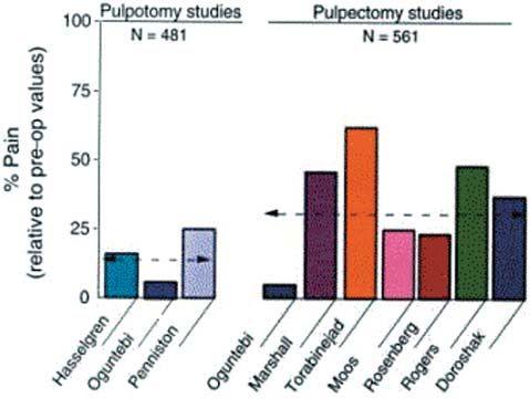
Endodontic post-treatment pain continues to be a cacy of a wide variety of analgesics for their ability to
significant problem facing the dental profession (1, ameliorate dental pain (8, 9, 11, 16–18). Most of
2). For those patients presenting with preoperative these studies have used an oral surgical third molar
pain, it has been reported that up to 80% of this extraction model that evaluated pain of an acute in-
population will continue to report pain after endo- flammatory nature. In contrast, endodontic pain is
dontic treatment, with pain levels ranging from mild often associated with chronic inflammation, the pres-
to severe (3–5). Many studies (3–13) have demon- ence of bacterial by-products, influx of primed im-
strated that endodontic treatment either in the form mune cells and activation of the cytokine network and
of pulpotomy or pulpectomy is efficacious in reducing
post-treatment pain (for details, see article by P. Ro-
senberg in this issue of Endodontic Topics). As can be
seen (Fig. 1) the pain relief afforded by endodontic
treatment is effective but rarely immediate and com-
plete. Post-treatment pain is usually mild in nature
rarely lasts longer than 72 h and is usually well man-
aged with non-steroidal anti-inflammatory agents
(NSAID) or acetaminophen.
However, some patients will continue to have pain
at moderate to severe levels that persists for several
days even after appropriate endodontic treatment.
For example, in patients who present for treatment
with a diagnosis of a symptomatic necrotic tooth, 47–
60% may expect moderate to severe pain in the first
24 h post treatment and 16–24% of patients at 72 h Fig. 1. Effect of pulpotomy on pulpectomy procedures on
postoperatively (14, 15). From these studies, it is reducing post-treatment endodontic pain (100% Ω pre-oper-
ative level of pain). From: Hargreaves KM & Baumgartner
evident that post-treatment analgesic intervention is
JC. Endodontic Therapeutics. In: Walton R, Torabinejad
required in a variable percentage of endodontic cases. M, eds. Principles and practice of endodontics. Philadelphia:
Numerous clinical studies have evaluated the effi- WB Saunders, pp 530–544, 2002 (20).
41Marshall other inflammatory mediators (19). The chronicity of albumin and corticosteroid-binding globulin (CBG pulpal and periapical inflammation may permit or transcortin), account for most of these steroid sprouting of nociceptor terminals and thus change binding sites. Only the unbound portion of cortico- the peripheral anatomy of the pain system (21). It is steroid is free to enter cells and mediate effects; thus, possible that the efficacy of analgesics differs when 90% of total plasma concentration of steroids is not comparing the pain associated with acute postopera- pharmacologically active. Cortisol has a biologic half- tive inflammation due to oral surgery, with pain as- life of approximately 90 min. sociated with the more persistent inflammatory pro- Chemical modifications of the cortisol molecule cess observed in endodontic pain patients. have produced a number of synthetic glucocorticoids Various classes of drugs have been studied for the with greater biologic half-lives, greater anti-inflamma- management of post-treatment endodontic pain (3, tory properties and less mineral corticoid activity 4, 9, 11, 13, 14). These include non-narcotic anal- (Table 1). The adrenal cortex produces approximately gesics comprising NSAID’s and acetaminophen, 10 mg/day of cortisol in the non-stressed adult (22). opioids and steroids. The purpose of this article is to Under severe stress, this level may be increased more consider the use of steroids specifically glucocortico- than 10 fold. The metabolism of steroids takes place ids in the management of endodontic pain. This re- in hepatic and extrahepatic sites and involves sequen- view will focus on the pharmacology, purported tial additions of oxygen or hydrogen molecules fol- mechanisms of actions of steroids, as well as their in- lowed by conjugation to form water-soluble deriva- dications for endodontics, contraindications, dosages tives that are excreted in urine. Little biliary or fecal and side-effects. The history of steroid use in endo- excretion of steroids is seen in human beings. dontics will be covered as well as a critical evaluation Glucocorticoids inhibit the production by multiple of the research done to date on the effects of gluco- cells or factors that are important in producing the corticoids on endodontic post-treatment pain. inflammatory response. This inhibition is a result of The adrenal cortex synthesizes fat-soluble cortico- the effect of glucocorticoids on gene transcription steroids from cholesterol. These steroids contain 21 that produces a decrease in the release of vasoactive carbon atoms in a four membered hydrocarbon ring and chemoattractive factors, decreased secretion of system. Corticosteroids comprise glucocorticoids and lipolytic and proteolytic enzymes, decreased extrava- mineral corticoids. This review focuses on the gluco- sation of leukocytes to areas of tissue injury, and ulti- corticoids since they act at multiple sites to inhibit im- mately decreased fibrosis (22). Glucocorticoids also mune and inflammatory reactions. In humans, cortisol produce profound effects on the immune response by is the primary glucocorticoid that is continuously syn- inhibition of cytokine production, specifically inter- thesized and secreted from the adrenal cortex. This feron g, granulocyte/monocyte colony stimulating process is under the control of the hypothalamus and factor (GM-CSF), interleukins 1, 2, 3, 6 (IL-1, IL-2, anterior pituitary. Along with the adrenal cortex, they IL-3, IL-6) and tumor necrosis factor a (TNF a) make up the hypothalamic-pituitary-adrenal (HPA) (Table 2). Thus, the pharmacological effects of gluco- axis, a system that regulates glucocorticoid levels (22). corticoids oppose many of the inflammatory pro- The hypothalamus produces corticotropin-releasing cesses that are known to occur during periapical in- hormone (CRH), which travels to the anterior pitu- flammation. Indeed, the ability of glucocorticoids to itary via the hypothalamic-hypohyseal portal system decrease periapical inflammation after endodontic and stimulates the release of adrenocorticotrophic hor- treatment has been demonstrated in a dog model by mone (ACTH) by pituitary corticotropes. ACTH, a both Holland (23), and Smith et al. (24). Glucocort- peptide of 39 amino acids, is the main regulator of cor- icoids bind non-covalently with specific receptor pro- tisol secretion. In turn, glucocorticoids inhibit ACTH teins in target tissues to regulate the expression of secretion via direct and indirect actions inhibiting corticosteroid–responsive genes. These receptors (CRH) neurons resulting in decreased CRH release, have high specificity and affinity for natural or syn- and via direct effects on corticotropes. thetic glucocorticoids. Once formed, the cytoplasmic Cortisol and synthetic glucocorticoids circulate in hormone-receptor complex becomes activated and the blood with 90% or more reversibly bound to enters the cell nucleus where it directs the transcrip- plasma proteins. Two circulating plasma proteins. tion of specific mRNA. The newly formed mRNA is 42
Steroids for endodontic pain

processed within the nucleus and then translocates to           served in the saline group at 1 day, but this difference
the ribosomes to serve as a template for newly syn-             was not statistically significant. Thus, steroid treat-
thesized proteins that are responsible for the biologic         ment to endodontic patients results in significant re-
effects (25). Examples of steroid-induced protein syn-          ductions in pulpal concentrations of PGE2, with a
thesis include lipocortin 1, a 37-kDa protein that has          trend towards a reduction in cytokine levels.
antiphospholipase A2 activity (26). Lipocortins pre-              Glucocorticoids also induce synthesis of kinase II
vent the synthesis of arachadonic acid and thereby re-          or angiotensin converting enzyme (ACE), which can
duce the biosynthesis of both cyclo-oxygenase and               lead to a reduction of bradykinin. Bradykinin has four
lipoxygenase products, including prostaglandins,                main pro-inflammatory actions including vaso-
leukotrienes and thromboxane related substances                 dilation, increased vascular permeability, leukocyte
(25). From this perspective, lipocortins represent one          chemoattraction, and nociceptor activation (28).
of the body’s natural ‘NSAID-like’ proteins. Gluco-             Bradykinin activates sensory nociceptors and elicits
corticoids also inhibit the induction of the gene cod-          release of substance P, neurokinin A, and calcitonin
ing for COX-2 in monocytes (26).                                gene-related peptide (CGRP) via receptors B1 and
   Reductions in pulpal levels of both PGE2 and IL-8            B2. The pain of acute inflammation appears to be me-
in cases of untreated irreversible pulpitis have been           diated by B2 receptors, whereas the pain of chronic
demonstrated after the administration of the gluco-             inflammation appears to involve an increased number
corticoid Depo-Medrol (27). Forty patients with a di-           of B1 receptors (29). Reduction of bradykinin levels
agnosis of irreversible pulpitis randomly received, in a        and postoperative pain by the administration of gluc-
double blind fashion, an intraosseous injection (IO)            ocorticoids has been demonstrated by Hargreaves &
of either 40 mg of methylprednisolone or sterile sa-            Costello (30) using microdialysis probes in the oral
line. No endodontic treatment was performed, the                surgery model.
teeth were extracted at either 1 or 3 days after IO in-           Glucocorticoids have also been shown to produce
jection and pulp tissue was removed. Enzyme                     a protein termed ‘vasocortin’, which has the ability to
immunoassay of the pulp tissue demonstrated a sig-              suppress edema that is not suppressed by NSAIDs
nificantly lower concentration of PGE2 at 1 day post            (22). Nitric oxide synthase is inducible by pro-inflam-
steroid injection. Mean pulpal concentrations of IL-8           matory cytokines resulting in increased nitric oxide
in the steroid group were only 17% that of levels ob-           production. Nitric oxide may increase blood flow and

  Table 1. Pharmacokinetics and relative potencies of corticosteroids
  Corticosteroid               Plasma t1/2        Tissue t1/2       Anti-               Naπ-retaining         Equivalent
                               (minutes)          (hours)           inflammatory        potency               dose (mg)*
                                                                    potency
  Cortisol                       90                8–12              1                     1                  20
  Cortisone                      30                8–12              0.8                   0.8                25
  Prednisone                     60               12–36              4                     0.8                25
  Prednisolone                 200                12–36              4                     0.8                 5
  6a-Methylprednisolone        180                12–36              5                     0.5                 4
  Fludrocortisone              200                 8–12             10                  125                   **
  Triamcinolone                300                12–36              5                     0                   4
  Betamethasone                100–300            36–54             25                     0                   0.75
  Dexamethasone                100–300            36–54             25                     0                   0.75

  * Dosages are approximate and apply to oral or intravenous administration, as glucocorticoid potencies may vary greatly
     following intramuscular administration.
  ** This agent is not used for glucocorticoid effects.

                                                                                                                        43
Marshall
Table 2. Effects of glucocorticoids on components of inflammatory/immune responses (adapted from Schimmer
and Parker (22), and Barnes (26))
Factor Glucocorticoid effect on
gene transcription Comments
Arachadonic Acid and metabolites Increased transcription Inhibition via glucocorticoid induction of lipocortin
(prostaglandins and leukotrienes) of lipocortin 1 that inhibits phospholipase A2
(macrophages, monocytes, fibroblasts)
Interleukin IL-10 Increased transcription Anti-inflammatory cytokine secreted by macrophages
inhibits transcription of many pro-inflammatory
cytokines, chemokines and inflammatory enzymes
Cytokines: IL-1, IL-2, IL-3, Decreased transcription Cytokines exert multiple pro-inflammatory effects
IL-4, IL-5, IL-6, IL-11, production and release blocked by glucocorticoids
12, IL-13, TNF-a, GM-CSF (macrophages, monocytes, lymphocytes, endothelial
cells)
Interleukin (IL)-receptor antagonist Increased transcription Cytokine that block the binding of IL-1 to its
receptors, glucocorticoids increase synthesis
Chemokines: IL-8, RANTES, Decreased transcription Chemokines attract inflammatory cells to site of
MIP-1a, MCP-1, MCP-3, -4, eotaxin inflammation, synthesis inhibited by glucocorticoids
Inducible form of Nitric Oxide Decreased transcription Nitric Oxide synthase may increase blood flow and
Synthase (iNOS) plasma exudation and amplify inflammatory
response, potently inhibited by glucocorticoids
Cyclo-oxygenase 2 (COX-2) Decreased transcription Glucocorticoids inhibit the induction of the gene
coding for COX-2 in monocytes and macrophages
Tachykinins Repression of the Glucocorticoids may inhibit neurogenic
preprotachykinin-A inflammation by decreasing tachykinins which may
gene, reduced expression amplify inflammatory responses
of tachykinin
receptors, increased
expression of neutral
endopeptidase which
degrades tachykinins
Bradykinin Increased transcription Glucocorticoids suppress bradykinin levels by
of Kinase II or increased degradation of bradykinin via induction
angiotensin converting of ACE synthesis. Bradykinin produces
enzyme (ACE) vasodilation, increased vascular permeability,
nociceptor activation, and leukocyte attraction
plasma exudation, thus amplifying the inflammatory cyclo-oxygenase, which converts arachadonic acid to
response. Glucocorticoids produce a potent inhi- prostaglandin endoperoxides. Glucocorticoids could
bition of nitric oxide synthase, leading to a decrease in therefore be presumed to have greater anti-inflamma-
inflammation (26). Glucocorticoids may also inhibit tory and possibly greater analgesic properties than the
neurogenic inflammation by inhibition of the release NSAIDs in pain conditions where multiple inflamma-
of neuropeptides (26). tory mediators are present and contribute to the de-
In contrast to the multiple sites of action and velopment of inflammation and pain.
multiple anti-inflammatory effects of glucocorticoids, Several studies have reported that glucocorticoids
the anti-inflammatory and analgesic effects of suppress postoperative edema in oral surgery patients
NSAID’s are much more selective. NSAIDs are be- (18, 31–33). It may be conjectured that in an endo-
lieved to act primarily via inhibition of the enzyme dontic pain model a reduction in periapical edema in
44Steroids for endodontic pain
a relatively non-compliant area might lead to a reduc- therefore prevents manifestations of adrenal insuf-
tion in pain (3). ficiency. NSAIDs should not be used with steroids
Most of the effects mediated by glucocorticoids are due to increased GI adverse effects.
not immediate since time is required for changes in The anti-inflammatory properties of glucocortico-
gene expression and protein synthesis to occur; thus, ids were first appreciated and utilized as an adjunct to
steroid actions many only be apparent after several endodontic therapy almost half a century ago (39–
hours or even days after administration. This delayed 41). Glucocorticoids have been used as a pulp-cap-
onset of action has been shown in a study by Nobu- ping agent (42), as an intracanal medicament either
hara et al. (34), where administration of systemic alone or in combination with antibiotics/antihistam-
dexamethasone significantly reduced the numbers of ines (13, 39, 40, 43, 44), and systemically (3, 4, 9,
PMN’s in periapical tissue after endodontic over-in- 14, 41, 45–50) as a means to decrease pain and in-
strumentation but not until 48 h postoperatively. flammation in endodontic patients. The following
Glucocorticoids have been used in endodontics for section will critically evaluate this research and then
their potent anti-inflammatory effects. They may have make recommendations for the use of glucocorticoids
widespread effects on many organ systems but these in endodontics; these recommendations will include,
effects are typically only seen at supraphysiological indications, contraindications, case selection, agent,
doses given over a long-term period, usually more dosage, and route of administration.
than 2 weeks. It has been stated that ‘A single dose of Several studies have evaluated the intracanal admin-
glucocorticoid, even a large one, is virtually without istration of steroids in endodontic patients. Wolfsohn
harmful effects, and a short course of therapy up to (39) in 1954 first reported on the use of a steroid
1 week) in the absence of specific contraindications, is as an intracanal medicament. His uncontrolled report
unlikely to be harmful’ (22). This has been demon- included 79 cases of either acute serous or acute sup-
strated in an in vivo study by Czerwinski et al. (35) parative pulpitis. After instrumentation, 0.5 mL of
who concluded that single large doses (2 mg/kg) of hydrocortisone was placed into the canal(s) followed
dexamethasone were essentially without harmful side- by a temporary filling. Patients reported their pain at
effects. This dosage is 10–25 times the amount advo- 24, 48 and 72 h. The author concluded that the use
cated for endodontic pain attenuation. of hydrocortisone resulted in the reduction and elim-
Glucocorticoids are contraindicated in patients with ination of severe secondary inflammatory reactions in
systemic fungal infection and known hypersensitivity the periodontal membrane following treatment.
to the drug. Steroids should be used with caution in Blitzer (40) recommended the use of TACT (terra-
patients with ulcerative colitis, pyogenic infection, di- mycin, antihistamine, cortril (25 mg/cc hydrocorti-
verticulitis, peptic ulcer, renal insufficiency, hyperten- sone) and tetracyn) as an intracanal medicament in
sion, osteoporosis, pregnancy, diabetes mellitus, ocu- cases where polyantibiotic therapy had failed. He re-
lar herpes, acute psychosis and history of tuberculosis ported on 51 cases, of which only two were failures,
(36). Psychological disturbances can occur during and concluded that hydrocortisone aids materially in
glucocorticoid therapy. These reactions are reversible reducing inflammation in the periapical tissues.
and range in severity from mild (euphoria, insomnia, Ehrmann (43) reported on the use of Ledermix
or nervousness) to pronounced (manic-depressive or (triamcinolone and dimethylchlorotetracycline in a
schizophrenic psychosis). The frequency and severity water soluble cream) for pulp capping, exposures, and
of adverse effects are correlated to the dose and dur- as an intracanal medicament in cases with pericemen-
ation of therapy (37). Drug interactions associated titis. He concluded that Ledermix stopped the pain
with the corticosteroids appear to be minimal. Cer- associated with pericementitis. Langeland et al. (44)
tain drugs decrease blood levels of the steroid via in- used Ledermix as an intracanal medicament in cases
creased metabolic clearance; these include phenobar- of continued postoperative pain after pulpal extir-
bital, phenytoin, rifampin, and ephedrine. Short-term pation or canal instrumentation. They reported pain
steroid use can produce a reversible suppression of relief in these cases within minutes to a few hours
endogenous cortisol production. Williamson et al. after the placement of Ledermix. In a double-blind
(38) have shown that this suppression is compensated clinical study of 50 consecutive patients with vital
for by adequate amounts of synthetic steroid and pulps, another study compared the use of 0.1 cc/ca-
45Marshall nal of dexamethasone (4 mg/cc) to sterile saline 0.1 patients with interappointment pain were included in cc/canal as an intracanal analgesic (51). After com- this study. Previously instrumented teeth were re- plete instrumentation, the intracanal medicament was opened without anesthesia, and medicated with either placed in the canal(s) followed by a temporary filling. Kenacomb (nystatin 100 000 units/g, neomycin Patients recorded their preoperative pain levels as well 2.5 mg/g, gramicidin 0.25 mg/g, triamcinolone 1.0 as post treatment pain at 24, 48 and 72 h. The mg/g in aqueous cream base) or placebo (aqueous authors reported that the dexamethasone group had cream) and coronally sealed with Cavit. Patients re- significantly less pain at 24 h when compared to pla- corded pain as none, mild, moderate and severe at 1, 2, cebo (P ⬍ 0.05). No postoperative infections were 4, 8, 12, and 24 h post medication. Results showed that noted in either group. the intracanal use of corticosteroid-antibiotic medi- Chance et al. (52) in a double-blind study com- cation significantly reduced the mean pain score at all pared the effect of intracanal meticortelone (pred- time periods when compared to placebo (P ⬍ 0.001). nisolone acetate 2.5%) vs. saline on post treatment Several other studies have evaluated the effects of pain in 280 patients. The preoperative pulpal and systemically administered steroids for treating endo- periapical diagnosis was recorded for each patient; dontic pain patients. The systemic administration of a however, pretreatment pain levels were not noted. corticosteroid to alleviate pain and inflammation in After complete instrumentation, the intracanal solu- endodontic patients was first reported by Stewart & tion was placed in the canal via three applications of Chilton (41) in 1958. The authors reported on 107 a saturated paper point followed by a cotton pellet patients who presented for endodontic treatment and temporary filling. Patients reported their pain at with severe infection and swelling or had a postopera- 24 h post treatment. The results indicate that the tive ‘flare-up’ (see article by R. Walton in this issue of corticosteroid was effective in significantly reducing Endodontic Topics for more information on flare-ups). the incidence of pain in vital teeth when compared After canal instrumentation and temporization, the to saline (P ⬍ 0.05). There was no difference in pain patients received a combination of corticosteroid, an- incidence in necrotic teeth when comparing the two tihistamine, and antibiotic to be taken orally. This solutions. combination included metreton (2.5 mg prednisone, Rogers et al. (13) compared the pain-reducing effi- 2 mg chlorophenpyridamine) 1 tablet TID for 2 days cacy of dexamethasone and ketorolac when used as and penicillin 250 mg TID for 3 days. Conclusions an intracanal medication, with oral ibuprofen and a were that the use of corticosteroid-antihistamine-anti- placebo. Forty-eight patients were included in the biotic therapy before or after conservative endodontic study. Following instrumentation of the vital pulps, therapy appeared to be very helpful in reducing acute two groups received either 0.1 mL dexamethasone symptoms. (4 mg/mL) or 0.1 mL ketoralac tromethamine (30 Stewart (54) evaluated the effect of oral dexametha- mg/mL) as an intracanal medicament. The other two sone either alone or in combination with oral penicil- groups received no intracanal medication and either lin on endodontic post treatment inflammation. ibuprofen (600 mg) or an oral placebo. Patients re- Teeth with vital or necrotic pulps were included. After corded their pain pretreatment and post treatment at endodontic instrumentation and temporization, pa- 6, 12, 24 and 48 h after therapy initiation on a visual tients were divided into four groups; a control group analog scale. At the 12-h period, both dexamethasone that received no medication, an antibiotic group that and ketoralac provided statistically better pain relief received phenethicillin 250 mg TID for 3 days, a ster- than placebo (P ⬍ 0.05). At the 24-h period, only ke- oid group that received dexamethasone 0.75 mg BID toralac had better pain relief than placebo. There for 2 days, and an antibiotic/steroid group that re- were no differences at 6 and 48 h. No differences ceived both phenethicillin and dexamethasone. In his were seen between groups in the amount of post- summary, the author stated that ‘dexamethasone is treatment pain medication required. capable of minimizing postoperative inflammation Negm (53) in a randomized, double-blind study and consequent edema and pain thus substantially re- determined the effect of a corticosteroid-antibiotic ducing requirements for analgesics.’ combination compared to placebo for the treatment In a randomized, prospective, double-blind, pla- of post treatment pain in vital teeth. A total of 480 cebo controlled study by Marshall & Walton (3), the 46
Steroids for endodontic pain

effect of intramuscular injection of dexamethasone on     effect of four different doses of dexamethasone on
post treatment endodontic pain was compared to pla-       post treatment endodontic pain. All 106 patients in-
cebo. After endodontic instrumentation and/or ob-         cluded in the study presented with pretreatment pain.
turation, patients received an IM injection of 1.0 mL     Endodontic instrumentation and/or obturation were
of dexamethasone (4 mg/mL) or 1.0 mL of sterile sa-       performed after which patients received a randomized
line. Pain levels were recorded preoperatively and at     intraoral intramuscular injection of placebo (1 mL
4, 24 and 48 h post treatment. Results indicated that     sterile saline), or one of four doses of dexamethasone
dexamethasone significantly reduced pain incidence        (2 mg/mL, 4 mg/mL, 6 mg/mL, 8 mg/mL). The in-
and severity at 4 h post treatment. At 24 h post treat-   jection was given into either the masseter, internal
ment, patients who received the corticosteroid            ptyergoid, or buccinator muscle. Preference was given
showed a trend towards less pain. Teeth with vital and    to intraoral muscles anesthetized for treatment. Pa-
necrotic pulps were included as well as retreatment       tients recorded their pretreatment pain levels on a 0–
cases. No antibiotics were taken by any patients and      9 scale and post treatment pain levels at 4, 8, 24, 48
there were no post treatment infections reported. The     and 72 h. Type and amounts of pain medication taken
amount of postoperative pain medication required          was also recorded. No antibiotics were given at any
was not recorded.                                         time during this study and both vital and necrotic
   Krasner & Jackson (45), in a double-blind study,       teeth were included for treatment. Results showed
evaluated the effect of oral dexamethasone on post        patients receiving dexamethasone had significantly
treatment endodontic pain. Fifty patients presenting      less severe pain at 4 and 8 h postoperatively (P ⬍
for endodontic treatment were included in this study.     0.05), and took significantly less pain medication
Retreatment cases and patients presenting with puru-      (P ⬍ 0.05) compared to placebo (mean of 1.98 tablets
lent drainage or cellulitis were excluded. Teeth were     for dexamethasone vs. mean of 4.64 tablets for pla-
instrumented and closed with no intracanal medic-         cebo). When evaluated on a mg/kg dosage basis, it
ament. Pre-treatment and 8 and 24 h post treatment        was found that patients who received 0.07–0.09 mg/
pain levels were recorded on a 0 to 100 scale. Patients   kg of dexamethasone IM had significantly less pain at
randomly received dexamethasone (0.75 mg/tablet)          8 h and took significantly fewer pain medications
or placebo with instructions to take 3 tablets immedi-    when compared to placebo.
ately and then 1 tablet every 3 h until bedtime for a        Kaufman et al. (47) were the first to evaluate the ef-
total of 7 tablets. Results showed that patients re-      fect of an intraligamentary delivery of corticosteroid
ceiving oral dexamethasone had significantly less pain    on endodontic post treatment pain. Forty-five patients
at 8 and 24 h when compared to those receiving pla-       presenting for endodontic treatment were randomLy
cebo (P ⬍ 0.01).                                          assigned to one of three experimental groups. Endo-
   Glassman et al. (46) also evaluated the efficacy of    dontic treatment was completed in one appointment
oral dexamethasone on endodontic interappointment         on both vital and necrotic pulps, with and without peri-
pain but with a much higher dosage of the cortico-        apical radiolucencies. After anesthesia was achieved but
steroid. Forty patients with asymptomatic vital in-       prior to endodontic treatment, patients in group 1 re-
flamed pulps were included in this study. After endo-     ceived 4–8 mg of Depo-medrol (slow-release methyl-
dontic instrumentation and temporization, alternate       prednisolone) via intraligamentary syringe. Single-
patients were given either dexamethasone(4 mg/tab-        rooted teeth received 4 mg, and multirooted teeth 8
let) or placebo. Instructions were to take 1 tablet im-   mg. Group 2 received PDL injection of mepivacaine
mediately and then 1 tablet at 4 and then 8 h post        3% in a similar fashion to group 1. Group 3 received no
treatment for a total oral dose of 12 mg in the dexa-     PDL injection. Pretreatment pain levels were not re-
methasone group. Patients were given a questionnaire      ported. The patients were telephoned at 24 h and re-
to record pain on a visual analog pain scale at 8, 24,    ported pain intensity on a 1–10 scale. The results
and 48 h post treatment. Results showed that patients     showed a significant decrease in postoperative pain in
receiving dexamethasone had a statistically significant   the methylprednisolone group (P ⬍ 0.05) compared to
reduction in pain at all post treatment time periods.     the active and passive placebo groups. In another pros-
   Liesinger et al. (4) in a double-blind, randomized,    pective, randomized, double-blind, placebo controlled
prospective, placebo controlled study evaluated the       study that contained 588 consecutive patients.

                                                                                                               47
Marshall Torabinejad et al. (9), evaluated the effectiveness of endodontic treatment (complete debridement) pa- various medications on postoperative pain following tients in a double-blind fashion randomLy received complete instrumentation. The preoperative pulpal an intraosseous injection of either 1 mL methyl- and periapical diagnosis was recorded as well as pre- prednisolone (Depo-Medrol 40 mg/mL) or 1 mL of operative pain levels on a 0–9 scale. After instrumen- sterile saline placebo. All subjects received ibuprofen tation, patients were given one of 10 different medi- and Tylenol .3 and recorded their pain levels and cations or combination of medications. These in- any pain medications taken for 7 days postoperatively. cluded non-steroidal anti-inflammatories, antibiotics, The results showed that the steroid group had signifi- acetaminophen, steroid and a narcotic analgesic. One cantly less postoperative pain and took significantly of these combinations was methylprednisolone 2 mg less pain medication over 7 days (P ⬍ 0.05). No anti- and penicillin 500 mg. Patients took one dose im- biotics were taken by patients at any time during the mediately then one dose every 6 h for the next 66 h study. for a total dose of 24 mg of methylprednisolone and In a follow-up study, Claffey et al. (49) evaluated 6 g of penicillin. Post treatment pain levels were re- pain reduction in symptomatic teeth with necrotic corded every 6 h for 72 h. Amongst their many find- pulps using an oral dose regimen of methylprednis- ings, the authors reported that those patients who olone. The materials and methods were nearly identi- presented for treatment with moderate–severe pain cal to Bramy et al. except that no patient had clinical and who received the steroid/antibiotic combination swelling and after the canal debridement, patients had significantly less pain at 6, 18 and 24 h when randomly received in a double-blind fashion either compared to the placebo group (P ⬍ 0.05). They oral methylprednisolone (48 mg/day for 3 days) or a found no significant post treatment pain differences placebo control (lactose 48 mg/day for 3 days). All between any of the test medications when compared patients received ibuprofen and Tylenol .3 (tm) and to placebo in patients with no or mild pretreatment a diary to record pain, percussion pain, swelling and pain. number and type of pain medications taken. Clinical In a very elegant study, Gallatin et al. (48) evalu- success was defined as any patient who experienced ated pain reduction for untreated irreversible pulpitis only mild to no pain, mild to no percussion pain, mild using an intraosseous injection of methylprednis- to no swelling and did not take any Tylenol .3. olone. Forty patients with a clinical diagnosis of irre- Again, no antibiotics were prescribed or taken during versible pulpitis actively associated with moderate–se- this study. The results showed that patients receiving vere pain participated in this prospective double-blind oral methylprednisolone had significantly higher clin- study. The involved tooth was anesthetized followed ical success for the first 3 days after endodontic treat- by an intraosseous injection of 1 mL methylprednis- ment (P Ω 0.05). olone (Depo-Medrol 40 mg/mL) or 1 mL of saline. In critically evaluating the preceding studies for The blinded solutions were administered using the validity, it must be kept in mind that the most power- Stabident system (Fairfax Dental, Inc., Miami, FL, ful conclusions are those generated from studies that USA). No endodontic treatment was performed. Pa- are prospective, randomized, double-blind and pla- tients were given a 7-day pain diary as well as anal- cebo controlled. None of the endodontic reports on gesic medication. Over the 7-day observation period, the use of corticosteroids published prior to 1984 subjects receiving Depo-Medrol reported significant- meets these criteria and therefore the results should ly less pain (P ⬍ 0.05) compared to placebo while tak- be considered anecdotal. Results from studies that ing significantly fewer analgesic medications (P ⬍ used corticosteroids in combination with other agents 0.05). such as an antibiotic and/or an antihistamine are dif- Bramy et al. (14) evaluated the intraosseous admin- ficult to interpret, as results ascribed to one of the istration of corticosteroid for pain reduction of symp- agents may be the result of the combination. Equally tomatic teeth necrotic teeth. Thirty-eight patients difficult to interpret are results from studies using in- with a clinical diagnosis of pulpal necrosis with associ- tracanal steroid as the means of delivery (13, 52, 53). ated periapical radiolucency participated in the study. The methodology in these studies doesn’t account for All patients experienced moderate/severe pain at time how much and over what time period the intracanal of presentation with mild or no clinical swelling. After medicament reaches the site of action, the periapical 48
Steroids for endodontic pain

tissues. Very small concentrations of the agent are          ministration. This is probably not the case, as seen
placed into the canal(s) and assuming apical patency         in Table 1 dexamethasone is approximately 5 times as
of variable size must pass through the apical foramen        potent as methylprednisolone and the 6–8 mg intra-
via a concentration gradient against a potential back        muscular doses used by Liesinger et al. (4) would be
pressure from periapical transudate or exudate. This         equivalent to 30–40 mg of methylprednisolone with
would seem to leave those studies that administered          40 mg of intraosseous methylprednisolone being the
corticosteroids in a systemic manner (intramuscular,         dose used by Bramy et al. (14). It is possible to specu-
intraosseous, oral) in a known dose without any other        late that these differences therefore might be more
agents as the critical ones in evaluating the efficacy of    related to the preoperative pulpal and periapical diag-
steroids in the ability to decrease endodontic pain.         nosis. All of the patients in the studies by Bramy et al.
Prior to interpreting these studies (3, 4, 13, 45–49) it     and Claffey et al. (14, 49) presented with necrotic
is important to remember that endodontic treatment           pulps, associated periapical radiolucencies, and either
itself has a major effect on reducing post treatment         mild or no swelling. The majority of patients in the
pain regardless of analgesic intervention (Fig. 1). As       study by Liesinger et al. (4) presented with a diag-
stated by Hargreaves (19) ‘This reduction in post            nosis of irreversible pulpitis and acute apical peri-
treatment pain, combined with variable levels of pre-        odontitis and those patients with necrotic pulps had
operative pain, reduces the statistical power of endo-       no associated periapical radiolucencies. It seems
dontic clinical trials for detecting active analgesics       plausible that corticosteroids may be more efficacious
over time in all patient groups (the so-called floor ef-     in attenuating pain associated with pulpal necrosis
fect). This limitation is a problem in interpreting clin-    and associated radiolucencies compared to pain as-
ical studies in general and may explain why some en-         sociated with irreversible pulpitis since these con-
dodontic clinical trials fail to detect analgesic treat-     ditions are associated with more complex chronic in-
ment or only detect it in those patients with                flammatory processes. Thus, the efficacy of steroids
moderate/severe pain.’ This has been shown by Tora-          in endodontic pain patients could be related to vari-
binejad et al. (9) and Rogers et al. (13) where various      ations in the periapical immunological/inflammatory
agents including corticosteroid significantly decreased      dynamics of teeth with irreversibly inflamed vs. ne-
post treatment pain but only in those patients who           crotic pulps.
presented with at least moderate/severe pretreatment            Based on the work of Bramy et al. and Claffey et al.
pain. It would therefore seem that systemic adminis-         (14, 49), it also seems plausible that corticosteroids
tration of corticosteroid as a method to decrease en-        would have efficacy in those cases of endodontic flare-
dodontic post treatment pain might be appropriate            up that result after treatment of previously asympto-
only for those patients who present with moderate/           matic necrotic teeth with or without associated peri-
severe pain. Three independent studies (4, 14, 49)           apical radiolucencies. This premise has not been in-
meet the criteria of being prospective, randomized,          vestigated and, with the low incidence of this type of
double-blind placebo controlled with no drug combi-          flare-up (1), would require a multicenter study over
nations and including only patients who presented            a period of years to gather an adequate sample size.
with the required level of pretreatment pain. Import-           Collectively, it appears from the studies reported (4,
antly, these studies showed that systemic administra-        14, 49) that the route of systemic administration of
tion of corticosteroid not only significantly reduced        glucocorticoids is not a determinant in their efficacy
post treatment pain at various times but also signifi-       and that when given in equivalent dosages, agents
cantly reduced the amount of additional pain medi-           such as dexamethasone and methylprednisolone are
cation required.                                             interchangeable. It would seem that if a systemic ster-
   Interestingly, the reports by Bramy et al. (14) and       oid is to be administered, an intraoral IM injection or
Claffey et al. (49) show significant pain relief for up      an intraosseous injection would be preferable over an
to 7 days with the use of steroid, in contrast to Lies-      extraoral IM injection as the practitioner is familiar
inger et al. (4) who found significant differences in        with intraoral and intraosseous injections and the site
pain reduction only in the first 8 h. It is possible these   of injection is already anesthetized. Intraoral injection
differences could be due to differences in doses of          of steroid would be preferable to a prescription for
different corticosteroids or by different routes of ad-      glucocorticoid as no assumption about patient com-

                                                                                                                  49
Marshall
pliance is required. A dose of 6–8 mg of dexametha- 4. Liesinger A, Marshall FJ, Marshall JG. Effect of variable
doses of dexamethasone on post treatment endodontic
sone or 40 mg of methylprednisolone appears from
pain. J Endod 1993: 19: 35–39.
the literature to be appropriate. If an oral route is 5. Marshall JG, Liesinger AW. Factors associated with endo-
chosen 48 mg methylprednisolone/day for 3 days and dontic post treatment pain. J Endod 1993: 19: 573–575.
by extrapolation 10–12 mg dexamethasone/day for 3 6. Hasselgren G, Reit C. Emergency pulpotomy. Pain reliev-
ing effect with and without the use of sedative dressings.
days should provide significant post treatment pain
J Endod 1989: 15: 254–256.
relief. 7. Oguntebi BR, DeShepper EJ, Taylor TS, White CL, Pink
It has been stated (38, 41, 50, 55) that antibiotics FE. Postoperative pain incidence related to the type of
must be given in conjunction with steroids to prevent emergency treatment of symptomatic pulpitis. Oral Surg
Oral Med Oral Pathol 1992: 73: 479–483.
an infection secondary to a decrease in the inflamma-
8. Penniston SG, Hargreaves KM. Evaluation of periapical in-
tory response. The implication is that suppression of jection of Ketorolac for management of endodontic pain.
inflammation also means a decrease in local defenses J Endod 1996: 22: 55–59.
permitting unchecked proliferation of pathogenic 9. Torabinejad M, Cymerman JJ, Frankson M, Lemon RR,
Maggio JD, Schilder H. Effectiveness of various medi-
microorganisms. None of the studies published since
cations on postoperative pain following complete instru-
1984 would support this premise, including those mentation. J Endod 1994: 20: 345–354.
cases with a diagnosis of pulpal necrosis with peri- 10. Moos HL, Bramwell JD, Roahen JO. A comparison of pul-
apical radiolucency (14, 49) where the potential for pectomy alone versus pulpectomy with trephination for the
relief of pain. J Endod 1996: 22: 422–425.
dissemination of an infectious process might be ex-
11. Doroshak A, Bowles W, Hargreaves K. Evaluation of the
pected. Antibiotics were not given or needed at any combination of flurbiprofen and tramadol for management
time during these studies nor were the steroids associ- of endodontic pain. J Endod 1999: 25: 381–384.
ated with any increase in infection rate compared to 12. Rosenberg PA, Babick PJ, Schertzer L, Leung A. The ef-
fect of occlusal reduction on pain after endodontic instru-
the control group. It can therefore be concluded that
mentation. J Endod 1998: 24: 492–496.
antibiotics are not routinely required or recom- 13. Rogers MJ, Johnson BR, Remeikis NA, BeGole EA. Com-
mended in conjunction with corticosteroids for the parison of effect of intracanal use of ketorolac tromethami-
management of endodontic post treatment pain in ne and dexamethasone with oral ibuprofen on post treat-
the otherwise healthy patient. Several excellent re- ment endodontic pain. J Endod 1999: 25: 381–384.
14. Bramy E, Reader A, Beck M, Weaver J. The intraosseous
views on antibiotics and endodontic pain are available injection of Depo-medrol on postoperative endodontic
(56), including the article by A. Fouad in this issue pain in symptomatic, necrotic teeth. J Endod 1999: 25:
of Endodontic Topics. 289 (Abstract OR 29).
In conclusion, it appears after a careful review of 15. Nist E, Reader A, Beck M. Effect of apical trephination on
postoperative pain and swelling in symptomatic necrotic
the literature that the administration of systemic ster- teeth. J Endod 2001: 27: 415–420.
oids is efficacious as an adjunct to but not replace- 16. Cooper SA, Precheur H, Rauch D, Rosenheck A, Ladov
ment for appropriate endodontic treatment in the at- M, Engel J. Evaluation of oxycodone and acetaminophen
tenuation of endodontic post treatment pain. Sys- in treatment of postoperative dental pain. Oral Surg Oral
Med Oral Pathol 1980: 50: 496–501.
temic steroids are also highly effective in those 17. Forbes JA, Bowser MW, Calderazzo JP, Foor VM. An
patients who present for treatment with moderate/ evaluation of the analgesic efficacy of three opioid-anal-
severe pain and a clinical diagnosis of pulpal necrosis gesic combinations in postoperative oral surgery pain.
with associated periapical radiolucency. J Oral Surg 1981: 39: 108–112.
18. Troullos ES, Hargreaves. KM, Butler DP, Dionne RA.
Comparison of nonsteroidal anti-inflammatory drugs, ibu-
profen and flurbiprofen with methylprednisolone and pla-
References cebo for acute pain, swelling, and trismus. J Oral Maxillo-
1. Walton RE, Fouad A. Endodontic flare-ups. A prospective fac Surg 1990: 48: 945–952.
study of incidence and related factors. J Endod 1992: 18: 19. Hargreaves KM, Seltzer S. Pharmacologic control of den-
172–177. tal pain. In: Hargreaves KM, Goodis H, eds. Seltzer and
2. Genet JM, Hart AAM, Wesselink PR, Thoden Van Helzen Bender’s dental pulp. Chicago: Quintessence Publications,
SK. Preoperative and operative factors associated with pain 2002.
after the first endodontic visit. Int Endod J 1987: 20: 53– 20. Hargreaves KM, Baumgartner JC. Endodontic Thera-
64. peutics. In: Walton R, Torabinejad M, eds. Principles and
3. Marshall JG, Walton RE. The effect of intramuscular injec- practice of endotontics. Philadelphia: WB Saunders, pp
tion of steroid on post treatment pain. J Endod 1984: 10: 530–544, 2002.
584–588. 21. Byers MR, Taylor PE, Khayat BG, Kimberly CL. Effects
50Steroids for endodontic pain
of injury and inflammation on pulpal and periapical nerves. ofapical periodontitis. Oral Surg Oral Med Oral Pathol
J Endod 1990: 16: 78–84. 1954: 7: 314–321.
22. Schimmer BP, Parker KL. Adrenocorticotropic hormone. 40. Blitzer MH. Root canal therapy. Use of a combination of
Adrenocortical steroids and their synthetic analogs; Inhibi- antibacterial agents, hydrocortisone and hyaluronidase.
tors of the synthesis and actions of adrenocortical hor- NY State Dent J 1956: 22: 503–508.
mones. In: Hardman JG, Limbird LE, eds. Goodman and 41. Stewart GG, Chilton NW. Role of antihistamines and
Gilman’s the pharmacological basis of therapeutics. New corticosteroids in endodontic practice. Oral Surg Oral
York: McGraw-Hill, 1996. Med Oral Pathol 1958: 11: 433–440.
23. Holland GR. Steroids reduce the periapical inflammatory 42. Fry AE, Watkins RF, Phatak NM. Topical use of cortico-
and neural changes after pulpectomy. J Endod 1996: 22: steroids for the relief of pain sensitivity of dentine and pulp.
455–458. Oral Surg Oral Med Oral Pathol 1960: 13: 594–597.
24. Smith RG, Patterson SS, El-Kafrawy AH. Histologic study 43. Ehrman GH. The effect of triamcinalone with tetracycline
of the effects of hydrocortisone on the apical periodontium on the dental pulp and apical periodontium. J Prosthet
of dogs. J Endod 1976: 2: 376–380. Dent 1965: 15: 149–152.
25. Di Rosa M, Calignano A, Carnuccio R, Ialenti A, Sautebin 44. Langeland K, Langeland LK, Anderson DM. Cortico-
L. Multiple control of inflammation by glucocorticoids. steroids in dentistry. Int Dent J 1977: 27: 217–251.
Agents Actions 1985: 17: 284–289. 45. Krasner P, Jackson E. Management of post-treatment en-
26. Barnes PJ. Anti-inflammatory actions of glucocorticoids: dodontic pain with oral dexamethasone: a double blind
molecular mechanisms. Clin Sci 1998: 94: 557–572. study. Oral Surg Oral Med Oral Pathol 1986: 62: 187–
27. Isett J, Gallatin E, Reader A, Beck M, Padgett D. Effect 190.
of intraosseous injection of depo-medrol on pulpal concen- 46. Glassman G, Krasner P, Morse DR, Rankow H, Lang J,
trations of PGE2 and IL-8 in untreated irreversible pul- Furst ML. A prospective randomized double-blind trial on
pitis. J Endod 2000: 26: 542 (Abstract) OR 28. efficacy of dexamethasone for endodontic interappoint-
28. Fouad AF. Molecular mediators of pulpal inflammation. ment pain in teeth with asymptomatic inflamed pulps. Oral
In: Hargreaves KM, Goodis H, eds. Seltzer and Bender’s Surg Oral Med Oral Pathol 1989: 67: 96–100.
dental pulp. Chicago: Quintessence Publications, 2002. 47. Kaufman E, Helling I, Rotstein I, Friedman S, Sion A,
29. Babe KS, Serafin WE. Histamine, Bradykinin, and their Moz C, Stabholtz A. Intra-ligamentary injection of slow-
antagonists. In: Hardman JG, Limbird LE, eds. Goodman release methylprednisolone for the prevention of pain after
and Gilman’s the pharmacological basis of therapeutics. endodontic treatment. Oral Surg Oral Med Oral Pathol
New York: McGraw-Hill, 1996. 1994: 77: 651–654.
30. Hargreaves KM, Costello A. Glucocorticoids suppress re- 48. Gallatin E, Reader A, Nist R, Beck M. Pain reduction in
lease of immunoreactive bradykinin from inflamed tissue untreated irreversible pulpitis using an intraosseus injec-
as evaluated by microdialysis probes. Clin Pharmacol Ther tion of depo-medrol. J Endod 2000: 26: 633–638.
1990: 48: 168–178. 49. Claffey D, Reader A, Beck M, Weaver J. Pain reduction in
31. Pedersen A. Decadron phosphate in the relief of com- symptomatic, necrotic teeth using an oral dose regimen of
plaints after third molar surgery. Int J Oral Surg 1985: 14: methylprednisolone. J Endod 2001: 27: 223 (Abstract OR
235. 34).
32. Sisk A, Bonnington GJ. Evaluation of methylprednisolone 50. Klotz MD, Gerstein H, Bahn AN. Bacteremia after topical
and flurbiprofen for inhibition of the postoperative inflam- use of prednisolone in infected pulps. J Am Dent Assoc
matory response. Oral Surg Oral Med Oral Pathol 1985: 1965: 71: 871–875.
60: 137. 51. Moskow A, Morse DR, Krasner P, Furst ML. Intracanal
33. Skelbred P, Lokken P. Postoperative pain and inflamma- use of a corticosteroid solution as an endodontic anodyne.
tory reaction reduced by injection of a corticosteroid. A Oral Surg Oral Med Oral Pathol 1984: 58: 600–604.
controlled trial in bilateral oral surgery. Eur J Clin Pharm- 52. Chance K, Lin L, Shoulin F, Skribner J. Clinical trial of
acol 1982: 21: 391. intracanal corticosteroid in root canal therapy. J Endod
34. Nobuhara WK, Carnes DL, Giles JA. Anti-inflammatory ef- 1987: 13: 466–468.
fects of dexamethasone on periapical tissues following endo- 53. Negm MM. Intracanal use of a corticosteroid-antibiotic
dontic over-instrumentation. J Endod 1993: 19: 501–507. compound for the management of post treatment endo-
35. Czerwinski AW, Czerwinski AB, Whitsett TL, Clark ML. dontic pain. Oral Surg Oral Med Oral Pathol Oral Radiol
Effects of a single large intravenous injection of dexa- Endod 2001: 92: 435–439.
methasone. Clin Pharmacol Ther 1972: 13: 638–642. 54. Stewart G. Combined use of an antibiotic and a cortico-
36. Anonymous. Physicians Desk Reference, 55th edn. Montva- steroid for postoperative sequelae in endodontic practice.
le NJ: Medical Economics Co. Inc., 2001: 2595. J Dent Med 1962: 17: 142–146.
37. Trammel CL. Anti-inflammatory drugs. In: Yagiela JA, 55. Sinkford JC, Harris SC. The case against topical use of
Neidle EA, Dowd FJ, eds. Pharmacology and therapeutics adrenocorticosteroids in dentistry. J Am Dent Assoc 1964:
for dentistry. St. Louis: Mosby, 1998. 68: 765–767.
38. Williamson LW, Lorson EL, Osbon DB. Hypothalamic- 56. Baumgartner JC. Antibiotics in endodontic therapy. In:
pituitary-adrenal suppression after short-term dexametha- Newman MG, van Winkelhoff AJ, eds. Antibiotic and
sone therapy for oral surgical procedures. J Oral Surg antimicrobial use in dental practice. Chicago: Quintess-
1980: 38: 20–38. ence Publications, 2001.
39. Wolfson BC. The role of hydrocortisone in the control
51You can also read