Current Advances in Computational Lung Ultrasound Imaging: A Review
←
→
Page content transcription
If your browser does not render page correctly, please read the page content below
Current Advances in Computational Lung Ultrasound
                                           Imaging: A Review

                                                     Tianqi Yang,1, a Oktay Karakuş,1, b Nantheera Anantrasirichai,1, c and Alin Achim1, d
                                                     1
                                                         Visual Information Laboratory, University of Bristol, Bristol BS1 5DD, U.K.

                                                     (Dated: 24 March 2021)

                                                     In the field of biomedical imaging, ultrasonography has become increasingly widespread, and
                                                     an important auxiliary diagnostic tool with unique advantages, such as being non-ionising
arXiv:2103.11366v2 [eess.SP] 23 Mar 2021

                                                     and often portable. This article reviews the state-of-the-art in medical ultrasound image
                                                     computing and in particular its application in the examination of the lungs. First, we review
                                                     the current developments in medical ultrasound technology. We then focus on the character-
                                                     istics of lung ultrasonography and on its ability to diagnose a variety of diseases through the
                                                     identification of various artefacts. We review medical ultrasound image processing methods
                                                     by splitting them into two categories: (1) traditional model-based methods, and (2) data
                                                     driven methods. For the former, we consider inverse problem based methods by focusing
                                                     in particular on ultrasound image despeckling, deconvolution, and line artefacts detection.
                                                     Among the data-driven approaches, we discuss various works based on deep/machine learn-
                                                     ing, which include various effective network architectures implementing supervised, weakly
                                                     supervised and unsupervised learning.

                                                                                                                                            Pages: 1–26

                                           I. INTRODUCTION                                                   supplement to modalities such as chest X-ray, chest com-
                                                                                                             puted tomography (CT), bronchoscopy, and magnetic
                                                In recent years, with the development of ultrasound
                                                                                                             resonance imaging (MRI). Compared with other common
                                           (US) along with the advancement of medical imag-
                                                                                                             medical imaging modalities, US bears various advantages
                                           ing technologies, the examination and treatment perfor-
                                                                                                             such as:
                                           mance of US in clinical applications, especially in the
                                           diagnosis of lung diseases, has gradually been recognized
                                           and accepted by clinicians. It has changed disease treat-
                                           ment, prognosis and patient management, and has be-               (1) Safe and non-invasive. US imaging does not in-
                                           come a visual stethoscope and diagnosis tool that doc-                volve ionising radiation. It can hence be safely used
                                           tors can make use of. US examination has become an                    for investigations on children and pregnant patients1 .
                                           important medical imaging method, and it is an effective          (2) Cost effective. It enables patients to obtain ac-
                                                                                                                 curate diagnosis without needing expensive, compli-
                                           a qc18229@bristol.ac.uk                                               cated, and invasive examinations.
                                           b o.karakus@bristol.ac.uk
                                           c n.anantrasirichai@bristol.ac.uk                                 (3) Portable. Compared to CT, US can lessen the need
                                           d alin.achim@bristol.ac.uk
                                                                                                                 for transfering critically ill patients2 .

                                           24 March 2021                                                        Current Advances in Computational LUS Imaging: A Review   1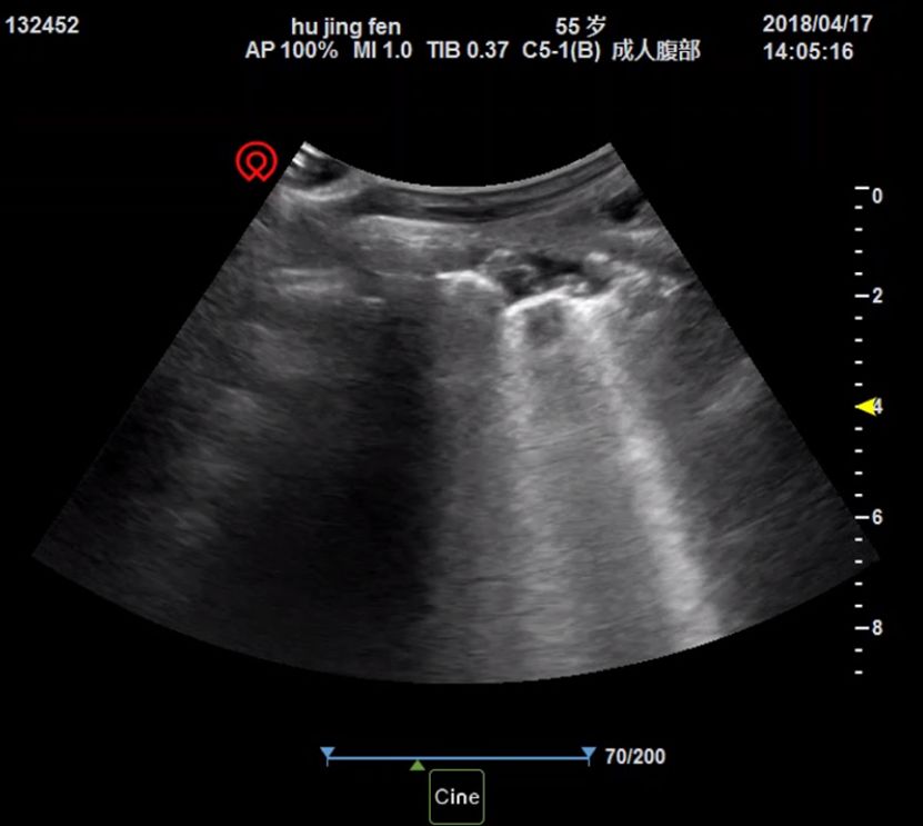
(4) High repeatability. US allows for repeated proce-           common cause of death in the UK after heart disease and
    dures at the patient’s bedside to monitor the progress      cancer5 . After the start of COVID-19 pandemic in 2020,
    of treatment.                                               analysis and diagnosis of lung disease became even more
(5) Real-time. US equipment can be used for dinamic             crucial. A rising demand for timely and correct diagno-
    imaging and hence boost doctor’s diagnostic effi-           sis, as well as an increasing need for patient monitoring,
    ciency.                                                     have been spurred6 .
(6) Hygiene. US equipment is easy to clean and disin-               LUS has been increasingly used to diagnose lung
    fect, particularly in the case of hand-held devices3 .      diseases.   This is not only because of the advantages
                                                                mentioned above, but LUS also helps in assessing the
    Despite all these advantages, it is worth noting that
                                                                fluid status of patients in intensive care as well as in de-
US remains an auxiliary tool after all since it has limita-
                                                                ciding management strategies for a range of conditions.
tions that cannot be easily overcome:
                                                                The common feature in all clinical conditions, both local
(1) US is operator-dependent, and the image quality may         to the lungs (e.g. pneumonia, chronic obstructive pul-
    vary due to the clinicians’ skills and the precision of     monary disease (COPD)) and those manifesting them-
    the device. Moreover, the interpretation of US im-          selves in the lungs (e.g. kidney disease, COVID-19) is the
    agery is subjective, in that the individual observa-        presence in LUS of a variety of artefacts, known as A-, B-
    tion, the movement of the probe, and the parameter          , and Z-lines. These carry important information about
    settings of the device effect image analysis.               the severity of diseases. Thence, the majority of research
(2) The operating environment also restricts US exam-           conducted in this area starts from detecting and quanti-
    ination. For example, critically ill patients need a        fying linear features in LUS images. A-lines are a kind
    variety of equipment (such as ventilators, blood pu-        of artifact caused by multiple reflection of sound waves
    rification tubes, chest drainage tubes, etc.) for life      due to the difference of acoustic impedance between the
    maintenance, which negatively affects US diagnosis          pleura and the lung. Their presence are indicative of a
    accuracy.                                                   healthy lung, whereas B-lines can indicate many diseases,
(3) Specific patient conditions also influence the US ex-       and characteristics such as their quantity or thickness are
    amination performance, in particular obesity, tho-          directly related to the severity of the disease.7 Therefore,
    racic deformity, skin lesions etc. For example, in          the quantification of B-lines can help with assessing the
    obese patients, thick subcutaneous fat may lead to          particular disorder8 . Z-lines are a type of artifact similar
    serious distortion, resulting in errors in US examina-      to B-line. The main difference consists in that Z-lines are
    tion. All these might influence US imagery character-       stationary and present brightness attenuation. Occasion-
    istics, and affect the results of clinical examination4 .   ally, they can be seen in pneumothorax. The position of
                                                                an artifact relative to the pleural line (which is the echo
    In this paper, we specifically review Lung US (LUS),
                                                                reflection formed by the surface of visceral and parietal
since lung disease is one of the most severe health prob-
                                                                pleura) is also important in determining whether the de-
lems causing the death of more than 100 thousand people
                                                                tected artifact is a B-line or merely air or another foreign
in the UK every year5 . According to the British Lung
                                                                body9 . Therefore, in some studies, the pleural line is used
Foundation, somebody dies due to lung disease in the
                                                                as reference for the positioning of other line artefacts10,11 .
UK every five minutes . Lung disease is in fact the third
                       5

2    24 March 2021                                                     Current Advances in Computational LUS Imaging: A Review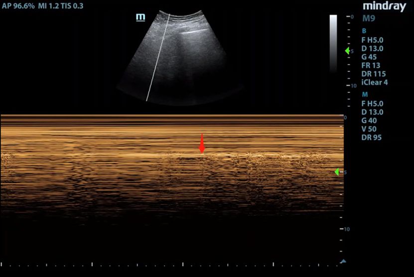
Considering the aforementioned importance of line arte-       the development of yet more accurate and efficient com-
facts, the literature spans various works focused on some     putational LUS imaging approaches.
standardized approaches of the assessment process in-
volving LUS. For instance, the first published attempt        II. ULTRASOUND IMAGING

at computerized B-lines scoring employed the 4-SCORE
                                                                  This section is intended to provide an overview of
rule . This principle is however not reliable in practice,
    12
                                                              medical ultrasound imaging, mainly in terms of instru-
so clinical diagnosis requires more accurate evaluation
                                                              mentation involved. While the discussion is for its more
standards13 .
                                                              part generic, we attempt to offer clear pointers to what
    In the computational LUS imaging literature, tradi-
                                                              is relevant to LUS in particular.
tional model-based methods are widely studied. Typi-
cally, they involve solving an inverse problem where a set    A. From sound to images
of unknown deterministic parameters which are observed
                                                                  Medical US is an imaging modality that uses the
through a linear transformation and corrupted by ad-
                                                              difference of physical characteristics of ultrasonics and
ditive noise are estimated14 . Despite their capability to
                                                              acoustic properties of human organs and tissues to dis-
produce results by just utilising a single corrupted image,
                                                              play and record the information in the form of wave-
the main disadvantages of such traditional approaches
                                                              form, curve or image. Different human organs and tis-
are that the prior distributions are handcrafted, and the
                                                              sues have their specific acoustic impedance and attenu-
generalization ability of the models is limited. In addi-
                                                              ation characteristics. When sound waves are transferred
tion, compared to other commonly used imaging modal-
                                                              into the body, their reflection and attenuation will vary
ities such as CT, X-ray and MRI, the resolution and
                                                              from organ to organ. According to their intensity, the
visual quality of US images are lower. Recently, data-
                                                              received echoes can be envelope detetcted and displayed,
driven methods which mainly exploit machine learning
                                                              and hence cross-sectional US image of human body can
(ML) and deep learning (DL) in particular can effectively
                                                              be formed15 . It took a long time before US has been
improve the quality of US images, so they have gained
                                                              adopted for imaging the lungs, as ribs, sternum and pul-
increased attention.
                                                              monary parenchyma filled with air are structures that
    This paper aims to provide a complete review of
                                                              reject ultrasonic waves16 .
the current advances on computational LUS imaging
                                                                  A widely accepted linear model of US image forma-
methodologies. We discuss modern equipment for med-
                                                              tion was proposed in Ng et al. 17 based on the physical
ical US image acquisition in Section II, and then de-
                                                              propagation characteristics of sound waves. The received
scribe the basic features and clinical applications of LUS
                                                              signal in the time domain is expressed as
imagery in Section III. In Section IV, processing meth-
ods for medical LUS images are presented in two sub-
                                                                            r(r0 , t) = h(r, t) ~ fm (r)|r=r0             (1)
categories, namely the model-based and the data-driven
methods. The discussions in Section V serve the purpose
                                                              where r0 represents the surface of the receiver, r refers to
of summarising the whole contribution. Since current
                                                              the location of the imaged tissue, fm (r) is the tissue re-
state of LUS development has not yet reached maturity,
                                                              flectively function (TRF), and h(r, t) refers to the system
this review aims to provide inspiration and references for
                                                              point spread function (PSF).

24 March 2021                                                   Current Advances in Computational LUS Imaging: A Review     3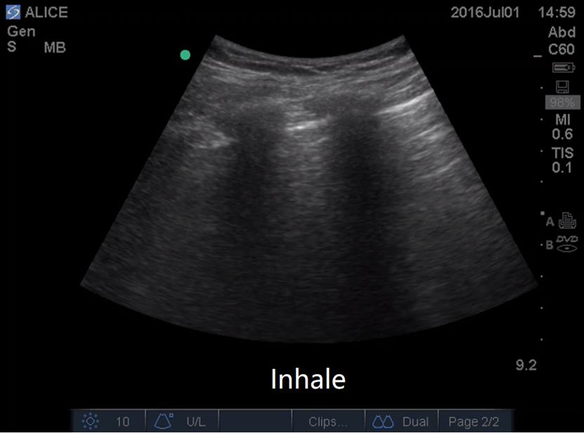
B. Instrumentation image is perpendicular to the direction of the sound
beam, which is similar to X-ray tomography. F-mode
Depending on clinical application, working princi-
imagers are a type of C-mode imagers, which are able
ples, tasks and operating systems, medical US images
to reconstruct three-dimensional images.
can be acquired using various modes as follows18 .
(1) A-mode: When the signal returns from the tissue
Among all these possibilities, for LUS, M-mode and
interface, its amplitude is directly displayed on the
B-mode are the two most commonly used for diagnosis.
screen. The abscissa of the screen represents the re-
Various types of probes are used in conjunction with dif-
flected US intensity. The propagation time corre-
ferent devices to receive and transmit ultrasonic signals.
sponds to the detection depth, and the ordinate rep-
Probes are the actual ultrasonic transducer, which em-
resents the amplitude of the returned echo - this is
ploy the piezoelectric effect to perform the bidirectional
called A-mode (amplitude mode) ultrasound.
conversion of mechanical waves into electrical signals and
(2) M-mode: This acquisition mode adds the echo in-
vice versa. A non-exhaustive list of most commonly used
formation obtained by the A-mode method to the
clinical probes is given in the following20
Cathode-ray tube (CRT) through brightness modu-
lation, and expands it on the time axis to obtain the
trajectory of the motions. This is especially suitable (1) Electronic convex array probes: These are com-
for the examination of the heart and other dynamic monly used for abdominal examinations, obstetrics
organs. and gynecology examinations. Their typical work-
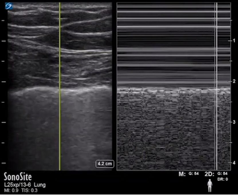
(3) B-mode: This is the most widely used US acquisi- ing frequencies are 3 ˜ 5MHz whereas, 5 ˜ 10MHz
tion mode and the one relevant to LUS. It uses the are recommended for children examinations at depths
amplitude of the echo pulse to modulate the bright- of more than 30cm. Small convex array probes are
ness of the display. The abscissa and ordinate of the used for transcranial scanning, whith 1.5 ˜ 3.5MHz
display correspond to the position of the sound ve- for adults, 3.5 ˜ 7.5MHz for infants and newborns
locity scan, thus forming an ultrasonic cross-sectional under the age of 3.
image modulated by the brightness. It is therefore (2) Electronic linear array probes: Their working
called B-mode (brightness mode). Different types of frequencies are over 7.5MHz, being often used for
B-mode scanners exist, including i. manual scanners, shallow investigations such as peripheral blood ves-
ii. real-time scanners, iii. mechanical scanners, iv. sels and superficial organs.
electronic scanners, with both linear-array scanners (3) Electronic phased array probes: They can be
and phased-array scanners19 . used for adult and pediatric heart examinations un-
(4) D-mode: This uses the Doppler effect to detect der working frequencies of 2 ˜ 5Hz.
the motion information of the human tissue. The (4) Transluminal probes: (including transesophageal,
Doppler detection method includes continuous wave transcatheter, and endoscopic probes, etc.) They
Doppler (CW) and pulse Doppler (PW). work at high frequency and can be used directly on
(5) C-mode and F-mode ultrasonic imagers: The the surface of organs or close to organs, clearly dis-
probe movement in C-mode and the synchronized playing the tissue structure and avoiding the inter-
scanning form a “Z” shape, and the displayed sound ference due to depth or other organs.
4 24 March 2021 Current Advances in Computational LUS Imaging: A Review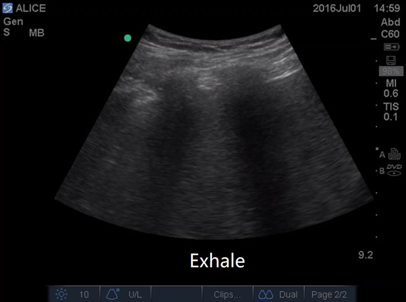
Probe selection is decided by the desired tissue            • Bat sign: If the probe is perpendicular to the ribs,
penetration as shown by .    2
                                  For the observation of             the upper and lower adjacent ribs, the shadow of the
in-depth lesions,low-frequency convex array probes or                ribs, and the pleural line together form a peculiar ap-
low-frequency phased array probes are usually selected,              pearance called the bat sign.
whereas high-frequency linear array probes are used for            • Pleural line and pleural sliding: In B-mode, if the
superficial pleural or sub-pleural detection. Image reso-            probe is placed perpendicular to the body surface, the
lution increases with frequency, at the cost of penetration          linear high echo below the ribs constituting the pleural
depth21 .                                                            line can be seen. The relative motion of the parietal
       Ultrasound scanners adapted to lung investigations            pleura and the visceral pleura forms a reciprocating
have been developed by a number of vendors, including                movement with breathing, which shows that the lungs
the Canon Xario Series22 , Aplio Series (by GE)23 or      24
                                                               .     are sliding relative to the chest wall when breathing.
In order to satisfy eventual point-of-care (PoC) needs,            • A-line: In B-mode, A-lines are a series of hyperechoic
many of these vendors have been developing portable de-              lines that are equally spaced and parallel to the pleural
vices. One such additional example is the Butterfly iQ+,             line and occur below the pleural line.
which is able to perform whole body imaging with one               • Seashore sign and sand beach sign: In the case of
small probe as well as provide real-time in-app monitor-             lung movement with breathing, in M-mode, a uniform
ing.                                                                 granular shape is formed below the pleural line, similar
                                                                     to a beach, called the “sand beach sign”, while the chest
III. CLINICAL APPLICATIONS                                           wall determines parallel lines due to its static state.
                                                                   • B-line or the comet tail sign: The B-line or comet
       This section describes clinical applications of LUS
                                                                     tail sign is a vertical artifact with clear boundaries and
imaging by reviewing the basic principles, signs, and con-
                                                                     moving synchronously with lung sliding. The appear-
ditions related to human lungs.
                                                                     ance of several B-lines in the image is also called the

A. Lung Signs                                                        “rocket sign” 25 .
                                                                   • Lung consolidation and atelectasis: The echogenic
       The lungs are the main air-containing organs of the
                                                                     US appearance of the lungs is similar to that of the liver
human body. In a pathological condition, the air-liquid
                                                                     or spleen.
ratio in the lungs changes, and abnormal artifacts may
                                                                   • Bronchial inflation sign: In uneven tissue-like con-
appear during a US examination. The analysis of LUS
                                                                     solidation, bronchial-like linear hyperechoic signals can
images is based on these signs, which are basically classi-
                                                                     often be found, indicating that there is residual air in
fied under 5 different categories, namely (i) normal lung
                                                                     the consolidated or atelectic lung. This type of appear-
signs (bat sign, pleural sliding, A-line), (ii) pleural effu-
                                                                     ance is called “Bronchial inflation sign”.
sion signs (quadrilateral sign, sine wave sign), (iii) lung
                                                                   • Lung points: A lung point is a dividing point seen in
consolidation signs (Fragment sign, tissue-like sign), (iv)
                                                                     real-time ultrasound where the lung sliding exists and
interstitial syndrome (pulmonary rocket sign) and (v)
                                                                     disappears alternately with breathing movement.
pneumothorax (stratospheric sign, lung point)1 . Some
                                                                   • Pleural effusion: When effusion occurs in the pleural
example LUS images showing these signs are shown in
                                                                     cavity, the space between the visceral pleura and the
Figures 1 and 2.

24 March 2021                                                        Current Advances in Computational LUS Imaging: A Review   5
parietal pleura is filled with fluid, thereby forming a        sion of B-lines and pleural effusion. Pneumonia can
    hypoechoic sign.                                               be characterized by B-lines, the quantity and char-
• Curtain sign: As air-filled lung moves up and down               acteristics of which vary according to the severity of
    with the breathing motion and other abdominal organs           the disease26 . A previous work by Chavez et. al.27
    are covered periodically. This is when “curtain sign”          shows that LUS had a high sensitivity (94%) and
    occurs.                                                        specificity (96%) for the diagnosis of pneumonia in
                                                                   adults, demonstrating that LUS is a reliable tool in
                                                                   diagnosing pneumonia.
B. Lung conditions
                                                                   Nowadays, the world is exposed to the COVID-19, a
      LUS provides a high amount of useful real-time infor-        condition which sometimes induces pneumonia and
mation on changes of lung morphology. Indirect signs in            has high infection rates. Recent studies6,28,29 sum-
LUS, such as the occurrence of abnormal pleural line, dis-         marize the symptoms of COVID-19 patients into sev-
appearance of A-lines, unexpected increase on the num-             eral points all of which can be identified with LUS.
ber of B-lines, or lung consolidations, can reveal the             These are: i. B-lines begin to increase in number and
severity of lung disease. In the following we include a            distribution. ii. The pleural line begins to become
non-exhaustive list of lung diseases, observable with LUS,         irregular. iii. Areas with B-lines are adjacent to nor-
along with their peculiar abnormalities.                           mal areas of lung sliding and A-lines. iv. There are
                                                                   ‘skip lesions’ or ‘spared areas’. v. Small consolida-
(1) Pleural effusion: The use of (M-mode) LUS in the
                                                                   tions around the lung.
     diagnosis of pleural effusions dates back to 196720 . In
                                                                (4) Chronic     obstructive       pulmonary         disease
     the evaluation of pleural effusion, LUS is highly sen-
                                                                   (COPD): COPD is listed as the fourth major cause
     sitive to small amounts of fluid. When the acoustic
                                                                   of human death by the World Health Organization
     impedance between the media is greater than 0.1%,
                                                                   (WHO). The ultrasonographic features of COPD
     US echo reflections appear. The quadrilateral sign is
                                                                   include pulmonary A-line, pulmonary sliding sign
     another important characteristic manifestation of M-
                                                                   and no right ventricular overload20 .
     mode LUS in the diagnosis of pleural effusion, with
                                                                (5) Acute respiratory distress syndrome (ARDS):
     a sensitivity of 93% and a specificity of 97%1 .
                                                                   It has been shown in a previous study20 that ARDS
(2) Pneumothorax: The initial sign of a pneumothorax
                                                                   accounts for 10.4% of ICU patients, and the mortal-
     is the disappearance of the pulmonary sliding sign.
                                                                   ity rate due to severe ARDS is as high as 40%. The
     Instead of the beach sign, the M-mode LUS shows a
                                                                   symptoms of ARDS mainly include lung consolida-
     superposition of parallel lines that lack motion char-
                                                                   tion with bronchial inflation sign, abnormal pleural
     acteristics. M-mode imaging can clearly identify lung
                                                                   line, diffuse pulmonary edema, and disappearance of
     signs, with the diagnostic specificity of 100% and the
                                                                   A-lines. Different from the diagnostic criteria defined
     sensitivity of 66%. For occult pneumothorax the sen-
                                                                   in Berlin for ARDS30 , a new study pointed out that
     sitivity values would reach up to 79% . 1
                                                                   EVLW (extravascular lung water) is more reliable as
(3) Pneumonia & COVID-19: The main manifesta-
                                                                   a new indicator to classify the severity of ARDS31 .
     tions of pneumonia are varying degrees of lung con-
     solidation and abnormal pleural lines, some with fu-

6     24 March 2021                                                   Current Advances in Computational LUS Imaging: A Review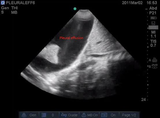
(a)                                       (b)                                        (c)

                   (d)                                       (e)                                        (f)

FIG. 1. Example images for various types of Lung signs.20 (a) Pleural line, (b) Bat sign, (c) Seashore sign, (d) A-line, (e)
B-line, (f) Atelectasis.

(6) Lung cancer: In the case of a peripheral lung tu-              (7) Congenital diaphragmatic hernia (CDH): A re-
    mor, LUS can clearly show the chest wall with the                 cent study32 has found that the LUS patterns for
    solid lung tissue as the “sound window”.       US can             CDH diagnosis include i. partial absence of the hy-
    clearly captures the chest wall, pleura and peripheral            perechoic line representing the normal diaphragmatic
    lung lesions, and can show the morphology, boundary               profile, ii. partial absence of the pleural line in the
    and blood flow of the lesions and the anatomical rela-            affected hemithorax, iii. absence of A lines in the
    tionship between fine structures and the surrounding              affected area, iv. presence of multi-layered area with
    tissue, which provides basic information for clinical             hyperechoic contents in motion (normal gut), and v.
    diagnosis.20 Through their US morphologies, one can               possible presence of parenchymatous organs inside
    distinguish benign and malignant lung tumors.                     the thorax (i.e., liver or spleen).

24 March 2021                                                       Current Advances in Computational LUS Imaging: A Review   7
(a)                                        (b)                                        (c)

                                 (d)                                                           (e)

FIG. 2. Example images for various types of Lung signs.20 (a) Bronchial inflation, (b) Lung point, (c) Pleural effusion, (d) and
(e) Curtain sign.

      In addition to the above conditions, LUS is also in-               62% of patients may develop pleural effusion during
valuable for the analysis and evaluation of lung related                 ICU hospitalization33 . Around 19% of mechanically
children diseases and in intensive care unit (ICU) appli-                ventilated patients are diagnosed with ARDS and an
cations.                                                                 increase in extravascular lung water is observed. The
                                                                         diagnosis and monitoring of these diseases can be done
• ICU uses of LUS: A study from 20161 confirmed that
                                                                         with LUS, thereby evaluating the respiratory system
    the diagnostic efficacy of LUS in some lung and pleural
                                                                         and circulatory system in real time.
    diseases for critically ill or mechanically ventilated pa-
                                                                       • Paediatrics: As a radiation-free imaging modality, ul-
    tients is equivalent to that of chest CT and better than
                                                                         trasonography is especially suitable for sensitive people
    that of X-ray. For example, LUS can effectively de-
                                                                         such as children, especially for newborns whose mus-
    tect lung consolidations with a thickness greater than
                                                                         culoskeletal system is not fully developed and whose
    20 mm in ICU patients, reaching an overall sensitiv-
                                                                         lung air content is low. LUS has high diagnostic sen-
    ity of 90% and a specificity of 98%. Another factor is
                                                                         sitivity and specificity for pediatric lung diseases such
    that the incidence of pleural effusion in ICU patients
                                                                         as: Neonatal respiratory distress syndrome (NRDS),
    is very high. About 41% of patients suffer from pleu-
                                                                         meconium aspiration syndrome, acute pneumothorax,
    ral effusion when they are transferred to the ICU, and

8     24 March 2021                                                           Current Advances in Computational LUS Imaging: A Review
occult atelectasis, etc. It has already replaced X-ray      the noise component (comprising multiplicative speckle
  diagnosis in neonatal intensive care units as the first     and additive measurement noise).
  choice for the diagnosis of lung diseases.
                                                                 f (x, y) = g(x, y)n(x, y) + w(x, y),    (x, y) ∈ Z 2     (2)
IV. LUS IMAGE PROCESSING AND ANALYSIS
                                                              where n(x, y) and w(x, y) represent the multiplicative
    This section presents the latest advances in image
                                                              and additive noise components, respectively.              (x, y)
processing methods for LUS. The methods are grouped
                                                              are the two-dimensional spatial coordinates; g(x, y) and
into two broad categories, specifically into the more stan-
                                                              f (x, y) represent the speckle free and the observed sig-
dard model-based approaches and the data-driven (or
                                                              nals, respectively. Since the influence of the additive
learning-based) methods.
                                                              noise is far less obvious than that of the multiplicative
                                                              noise, the image formation model (2) can usually be ap-
A. Model-Based Methods
                                                              proximated by
    In order to accurately identify the various signs of
disease in LUS images, the reconstruction of high quality           f (x, y) = g(x, y)n(x, y),   where (x, y) ∈ Z 2 .     (3)
images through observations and the extraction of infor-
mation therein are necessary. This is commonly posed          The term n(x, y) corresponds to speckle noise, which is

as an inverse problem. The solution however may not           an inherent phenomenon in US images, as well as in other

be unique, or it may be highly sensitive to changes in        coherent imaging modalities such as the synthetic aper-

the data, which illustrates the ill-posedness of the prob-    ture radar (SAR) or laser imaging. Speckle noise has

lem. The traditional methods convert this into a well-        been shown to be correlated with the tissue structure, so

posed one by using regularization/penalty functions or        its statistical description generally depends on the type of

prior information. Normally, the regularization term is       tissue and the imaging system. It exhibits granular pat-

related to the prior information of the parameter to be       terns, which obscure fine anatomical details, and thereby

estimated15 . In the relevant literature, there are three     reduce the diagnostic accuracy. Speckle noise is hence

main categories of approaches, which include (i) statisti-    regarded as an undesirable phenomenon in most clin-

cal methods, (ii) regularized geometric modeling methods      ical applications. There are situations however, when

and (iii) methods based on sparse representations34 or,       speckle noise can constitute useful information, such as

sometimes, a combination thereof. In the following, we        when used for speckle tracking (i.e. motion estimation)

discuss the most widely studied inverse problems in con-      and tissue characterization15 . To mitigate speckle, a log-

junction with medical US image processing, which are          arithmic transformation is usually employed to convert

also relevant to LUS. Approaches peculiar to LUS specif-      the multiplicative characteristics into an additive model.

ically are discussed in IV A 4.                                   In the computational US imaging literature, sev-
                                                              eral techniques have been developed for reducing speckle
1. Despeckling                                                noise. Achim et al. 35 use the heavy-tailed α-stable family

    Generally, the final envelope detected US image is        of distributions to describe US image subbands in a trans-

composed of two elements, the useful signal component         form domain, to capture the significant non-Gaussian

(corresponding to structure inside the human body) and        behaviour.    Based on this statistical characterisation,

24 March 2021                                                   Current Advances in Computational LUS Imaging: A Review     9
they develop a blind speckle-suppression processor, whith      Therefore, the multiplicative noise in the SRAD com-
non-linear characteristics related to the degree of non-       posite image is converted into additive noise through a
Gaussianity of the data.                                       homomorphic transformation. Following the application
     Duarte-Salazar et al. 36 thoroughly describe 27 tech-     of two-level DWT decomposition, in order to suppress
niques that can be utilized to smooth or eliminate speckle     the residual noise of an SRAD filtered image, GDGIF
noise in medical US images. These technologies can be          and WGIF are exploited to reduce noise from seven high-
divided into five categories: (1) dynamic analysis, (2)        frequency sub-band images and one low-frequency sub-
time-frequency analysis, (3) modern technology, (4) hy-        band image, respectively. Finally, a speckle-free image is
brid technology, and (5) machine learning. The majority        attained through inverse DWT and an exponential trans-
of the algorithms can eliminate speckle noise but neglect      formation.
the preservation of details. Therefore, in order to si-            Chen et al. 40 suggest a tehnique for removing
multaneously fulfill the need for excellent speckle noise      speckle noise, where the energy minimization model is
removal and edge information preservation, Garg and            solved by the alternating direction multiplier (ADMM)
Khandelwal   37
                  propose a method based on the model-         algorithm41 . The convexity, existence and uniqueness of
ing of the shear wave coefficients of the ultrasonic image     the solution of the new energy minimization model are
detail band after logarithmic transformation. In each          furthermore proved. It effectively reduces the speckle
detail shearlet subband, the coefficients corresponding to     and achieves considerable performance in terms of visual
the signal and speckle noise are modeled as normal in-         evaluation and quantitative measurements.
verse Gaussian and Gaussian priors, respectively. Based
on the local statistics of the image, these coefficients are   2. Deconvolution

categorized into homogeneous regions, heterogeneous re-            Another important type of model-based US methods
gions and strongly heterogeneous regions. Then, with           is image deconvolution, whereby US images are modeled
the prior distribution, maximum a posterior estimation         as a convolution between a blurring kernel or PSF, and
(MAP) is performed on all regions of the detail bands,         the tissue reflectivity function. The linear image forma-
except the strongly heterogeneous regions. An adaptive         tion model can be rewritten as
weight function is also used in the MAP expression to
reduce the loss of feature information.                                     y(r) = h(r) ~ x(r) + n(r),    r ∈ R,         (4)
     Choi and Jeong 38 employ various approaches such
as the speckle reduction anisotropic diffusion (SRAD)          where y(r) is the image pixel observed at position r, x(r)

filter39 , discrete wavelet transform (DWT) using sym-         is the TRF to be estimated, h(r) is the system PSF, n(r)

metric characteristics, gradient domain guided image           is the additive measurement noise. R refers to the image

filtering (GDGIF) and weighted guided image filter-            domain15 .

ing (WGIF). Under the multiplicative noise assumption,             Deconvolution in medical imaging is commonly em-

SRAD filtering can be used directly to suppress speckle        ployed to improve visual quality for and achieve better

noise, because it does not require log-compression. More-      contrast. This translates in easier interpretation for the

over, eliminating the additive noise (additive white Gaus-     physicians. Standard deconvolution schemes exploit sim-

sian noise) in the wavelet domain is straightforward.          plified models for the tissue reflectivity, prevalently Gaus-

10    24 March 2021                                                   Current Advances in Computational LUS Imaging: A Review
sian or Laplacian, which are typically chosen to produce on the complex plane, and iii. more advanced mod-
the most visually pleasing images. The statistics of the els than white Gaussian can be assumed for the tissue
restored solutions are hence badly biased by these prior reflectivity42 . Although commonly employed, MAP de-
assumptions, and are not always well suited for further convolution brings an increased computational cost, even
information extraction . 42
if simple schemes are employed such as based on Wiener
Among the completely blind deconvolution strate- filtering or `1 -norm optimization. Another problem is
gies, predictive deconvolution 43
represents an important that the PSF estimation is still tedious. In particular
solution, but very few authors spent considerable efforts this is due to the need for phase unwrapping procedures
in this direction. Their drawback relate to the simplis- where non-minimum phase PSFs are considered.
tic assumptions made in order to fit various real com- Due to the inherent bandwidth limitations of US
plex scenarios42 . The most common strategy is instead scanners and the adverse effects of measurement noise,
to exploit different approaches, relying upon statistical image deconvolution is very sensitive to errors occur-
estimation and convex optimization theory. ring in the PSF estimation. Even slight errors in the
Zhao et al. 44
study a blind deconvolution algorithm PSF estimates can lead to obvious artefacts that ren-
for ultrasound images in a predictive deconvolution man- der the reconstructed images worthless. To address this,
ner, where they model the ultrasonic PSF based on a Michailovich et al. 46 propose a “hybrid” deconvolution
Gaussian function modulated by a sinusoid. Given this technique. Their method is based only on partial infor-
parametric model, the estimation of the power spectrum mation about the PSF, especially its power spectrum to
requires estimation of its parameters. In addition, an `p - estimate tissue reflectivity. While directly estimating the
norm (0 ≤ p ≤ 2) regularization term of the US TRF reflectance of the tissue from the relevant radio-frequency
is employed, based on the assumption that the US im- (RF) data, the proposed approach simultaneously elimi-
ages follow a generalized Gaussian (GG) distribution. nates errors caused by inaccuracies in PSF estimation.
An alternate optimization method based on the block- Pham et al. 47 propose a novel algorithm for the re-
coordinate descent framework45 is proposed to estimate trieval of blood flow from a sequence of ultrafast US
US PSF and TRF. Due to the parametric nature of the images, based on the combination of two different tech-
PSF model, instead of estimating all the PSF pixels, only niques: deconvolution robust principal component anal-
a few parameters need to be estimated. This reduces the ysis (DRPCA) and blind deconvolution (BD). The pro-
computational load and estimation complexity. posed method overcomes the main limitation of the for-
Unlike the predictive deconvolution, MAP-based de- mer related to the requirementfor PSF estimation, whilst
convolution tackles the problem using a two-step scheme: providing similar performances. Thehe PSF is however
the PSF is estimated first and subsequently image assumed to be spatial-temporally invariant, and the al-
restoration is performed. These techniques represent the gorithm appears computational complex.
most common strategies for medical US image restora-
tion. Their main advantages are i. two dimensional or 3. Segmentation
even three dimensional PSFs can be accounted for, ii. no Classical approaches for image segmentation are for-
assumption is made on the PSF, neither on the number mulated in either a Bayesian or a variational frame-
of zeros or poles, and nor on the position of the zeros work. The literature spans three widely adopted types
24 March 2021 Current Advances in Computational LUS Imaging: A Review 11of segmentation approaches, which include (1) thresh- artefacts. For instance, Ramin et al. 8 aim to automati-
olding methods, (2) edge-based methods and (3) region- cally detect B-lines. They characterize the distribution of
based methods. Since US images suffer from low signal B-lines in conjunction with their ability to discriminate
quality caused by the attenuation, speckle, and shad- between healthy volunteers and patients with pulmonary
ows, many methods often utilize specific constraints edema. They use a random walk method to delineate
or priors. Some widely used constraints are intensity- the pleural line, and then exclude the upper pleural re-
based priors, geometric priors, and statistical priors. gion before identifying B-lines. This is achieved by an
The various US segmentation methods embed these con- alternate sequential filtration, and subsequently they ap-
straints into Bayesian methods, active contours, active ply the top-hat filter to ensure that B-lines are laterally
appearance models, level-sets, clustering, or graph-based detached. Finally, a Gaussian model is fitted to each
frameworks15 . detected B-line, and the peak point of the fitted Gaus-
Talebi et al. 48
propose a segmentation method com- sian models corresponding to the B-lines are calculated
bining genetic algorithms and active contours. This has and used to accurately determine the position of B-lines.
been provedn to overcome some limitations of classical B-lines are then overlaid on the B-mode images.
active contours, such as contour initialization and the Brusasco et al. 50 endeavor to develop a fully au-
entrapment of the contour in local minima caused by the tomatic method of quantifying B-lines and thereby de-
existence of speckle noise. But the limitation of Talebi termining extra-vascular lung water(EVLW). B-lines are
et al. 48 method is that it can only correctly segment cir- selected from the input gray-scale LUS images and con-
cular tissue in US images. firmed when white pixels take up at least 50% along the
Luo et al. 49 combine region and edge information image columns and over 70% of the total vertical length
into a robust graph-based segmentation method. The of the image. B-lines quantification is realized by per-
corresponding optimization problem is formulated as a forming a statistical regression on the segmented LUS
multi-objective problem. They maximize the difference images. Although this study provides a reliable operator-
between the target and the background, improve the uni- independent assessment of EVLW, this method may fail
formity in the target area, and consider the edge gradi- when multiple B-lines coalesce into a single white line
ent. The algorithm has several prerequisites. First, the rather than close-by B-lines.
operator should be well experienced. Second, the tumor Anantrasirichai et al. 51 propose an innovative way
centered image (TCI) should be carefully delineated to of detecting line artefacts in LUS images by solving an
capture the full lesion region with partial surrounding inverse problem. The method combines the Radon trans-
tissues. Third, the region of interest (ROI) around the form with the PSF of the US acquisition system in a sin-
lesion must be centrally located and the user’s participa- gle equation thereby achieving line detection and decon-
tion is necessary. Besides, the algorithm is computation- volution simultaneously. This inverse problem is solved
ally expensive. using the ADMM algortihm, offering a fast convergence
rate. The scheme firstly detects the pleural line in or-
4. Model-based LUS Image Processing der to locate the lung space. Then, the local peaks of
Most ultrasound image processing approaches specif- the Radon transform are detected and line-type classifi-
ically developed for LUS focus on the identification of line cation is done following clinical definitions, in the spatial
12 24 March 2021 Current Advances in Computational LUS Imaging: A Reviewimage domain. B-lines, A-lines and Z-lines are hence interpretation to image formation and reconstruction55 .
successfully identified. To enhance line detection per- The key to image reconstruction is to find a good sparse
formance and the visualization of restored lines, they representation of the image to be reconstructed (which
include an additional convolution factor in the Radon is also true for model-based approaches). DL methods
transform domain with an unknown blurring kernel. The assume that the representation is provided by deep neu-
penalty function employed is `p -norm with low values of ral networks, so it is nonlinear. The model parameters of
(0 ≤ p ≤ 1) in order to promote sparsity in the Radon the representation must be learned from a large amount
space. of data. Supervised learning is the most popular method
Extending the above work in the context of eval- since it can lead to highly accurate solutions. The de-
uating COVID-19 patients, Karakus et al. 52 further sign of the network is flexible but generally has a rela-
improve the line detection performance by regularizing tively high complexity. Stochastic optimization strate-
the solution using the Cauchy proximal splitting (CPS) gies, can effectively extract data features from low-level
algorithm53 . The key benefit of this type of work is that to semantic level. Representative methods include deep
it offers an unsupervised framework for the detection of image prior (DIP) and generative adversarial networks
B-lines in situations where annotated data are not freely (GAN)56,57 . Due to these advantages, data driven meth-
available. These methods converge rapidly, enabling thus ods have shown a great potential in various automated
the processing of LUS image sequences in a relatively US image analysis tasks.
short amount of time with high detection accuracy. A review by Liu et al. 58 describes several popular DL
Table I summarizes the algorithms presented in this architectures, including i. supervised deep networks or
section and their effectiveness in various applications. deep discriminative models, ii. unsupervised deep net-
works or deep generative models, and iii. hybrid deep
B. Data-Driven Methods networks. The authors point out that the current basic
models used in medical US analysis are mainly convo-
Compared to conventional signal processing meth-
lutional neural networks (CNNs), recurrent neural net-
ods, ML based methods can provide improved perfor-
works (RNNs), deep belief networks (DBNs), autoen-
mance in medical imaging, because they are able to cap-
coders (AEs), and variants thereof. They summarized
ture more complex patterns in the data. ML is referred
and discussed the application of these models in detail
to as being data-driven as large amounts of data are
for various specific tasks in US image analysis. These
generally required for training and testing. One of the
include i) traditional diagnostic tasks, such as classifi-
branches of ML is deep learning, a class of techniques
cation, segmentation, detection, registration, biometrics,
that have characteristic three important properties54 : i.
quality assessment, and ii) emerging tasks like image-
features are learned from the data instead of being hand-
guided interventions and treatments. They state that
crafted, ii. features are abstracted in a hierarchical way
the main problem is the small sample datasets gener-
from lower level to higher level, and iii. the number of
ally available. The common workaround is to perform
non-linear feature transformation layers is at least two.
transfer learning, which can be either cross-domain or
Over the past few years, DL techniques have rapidly
cross-modal. The former is the most common way to ac-
gained attention in the field of medical imaging applica-
complish a variety of tasks in medical US analysis, but
tions, their applications ranging from image analysis and
24 March 2021 Current Advances in Computational LUS Imaging: A Review 13TABLE I. Summary of Model-Based Methods
Application Algorithm Details Data Effectiveness Reference
Multiscale Bayesian The wavelet transform based on 44 abdominal US The method has a good adapta- Achim et al. 35
speckle suppressing subband representation function; images from the tion ability for biomedical images
algorithm and a Bayesian denoising algo- same patient and denoising when the noise can
De-speckling rithm based on an alpha-stable DICOM image be modeled as additive Gaussian
prior for the signal files donated by and signal-independent.
various vendors
GDGIF and WGIF GDGIF and WGIF are applied to 6 standard images Higher PSNR and better fea- Choi and Jeong 38
the discrete wavelet transformed and 6 real US ture conservation ability than
data that comes from SRAD fil- images of aligrant conventional methods. Low
tered images breast lesions computational cost in stan-
dard images(9.9913%) and US
images(2.7186%)
ADMM Minimization of a convex energy synthetic, natural, Highest PSNR and SSIM of the Chen et al. 40
model - an adaptive total varia- and real medical restored images by the different
tion model ultrasound images. model and visually fewer tex-
tures of residual images.
DST-MAP MAP estimation on the modeled Synthetic US im- Best performance at high noise Garg and Khan-
shear wave coefficient based on ages and 60 real variance in synthetic US images. delwal 37
DST of log-transformed images US images Highest mean to variance ratio
and equivalent number of looks
in all marked regions in real US
images.
Alternating opti- Parameter estimation of the US Simulated and in- Visually and quantitatively bet- Zhao et al. 44
mization algorithm PSF modeled by Gaussian func- vivo US images ter than the non-blind method in
Deconvolution tion modulated by a sinusoidal simulated images. Competitive
function performance but lower computa-
tional load compared with non-
blind deconvolution algorithm.
Alternating Proximal gradient In-vivo RF images Reasonable computational effi- Michailovich
minimization method(PGM) to estimate acquired from ciency. Noticeable increase in et al. 46
the tissue reflectivity function as healthy people spatial image resolution and im-
well as the minimization of the proved contrast. No “ringing”
DFT of the PSF effects or residual blur around
sharp image details.
BD-RPCA Iterative estimation of PSF and Simulated and in- Comparable NRMSE and PSNR Pham et al. 47
the blood component based on vivo US data with DRPCA in both simulated
Casorati matrix, using BD and and in-vivo US images but higher
DRPCA respectively. computational cost.
Genetic algorithm Genetic active contour with an US images with Satisfactory segmentation re- Talebi et al. 48
energy minimization procedure. circular tissue sults but time consuming.
Segmentation structure
Multi-objectively A multiobjective optimization 100 clinical breast Low averaged radial error(ARE) Luo et al. 49
optimized ro- function which combines region- US images and at 10.77% and good overall per-
bust graph-based based and edge-based informa- 18 clinical mus- formance in TPVF(volume frac-
(MOORGB) seg- tion is designed in the PSO to culoskeletal US tion) , FPVF and FNVF.
mentation method optimize the RGB. images
and Partial swarm
optimization (PSO)
algorithm
Alternate sequential After removing the pleural line US images from 4 Successfully differentiate be- Ramin et al. 8
filtering and top-hat detected by a random walk healthy subjects tween the healthy and unhealthy
filtering method, B-lines are identified us- and four patients class with a p-value of 0.015.
Detection ing ASF and white top hat fil- with pulmonary Average number of B-lines of
tering, and finally enhanced by edema. patients and healthy group
Gaussian model. are 0.28±0.06 and 0.03±0.06
respectively.
simple linear regres-B-lines are selected through con- 12 patients with Provides a reliable operator- Brusasco et al. 50
sion (SLR) and ro- trast adjustment, K-means clas- acute respiratory independent assessment of
bust linear regres- sification, ASF, and confirma- distress and each EVLW in ARDS, which is com-
sion (RLR) tion. Statistical regression is provided with 12 parable or superior to previous
used for quantification. LUS frames. scores.
ADMM Radon transform based non- 50 simulated im- Improves the performance of B- Anantrasirichai
convex optimization with the ages and in-vivo line detection approaches by up et al. 51
combination of the PSF function. US B-mode images to 50%.
Cauchy Proximal Radon transform based non- 12-segment LUS Up to 88% detection accuracy Karakus et al. 52
Splitting (CPS) convex optimization with for 9 COVID-19
Cauchy-based penalty function patients
its source and destination are not within the same do- considered to enhance performance. In this sense, cross-
main. However, the solution to train models on natu- modal transfer learning may be a wiser choice.
ral images might not be the optimal one and therefore Razzak et al. 59 also discuss the application of ML
domain-specific models in medical imaging are normally and DL in US, outlining the main challenges and solu-
tions. They highlight the importance of future ML sys-
14 24 March 2021 Current Advances in Computational LUS Imaging: A Reviewtems to provide real-time feedback to the sonographer inverse problems. They observe that in optimization al-
during image acquisition. It will also be important to re- gorithms for linear inverse problems, signal priors usu-
place manual ROI selection with automatic ROI selection ally appear in the form of proximal operators. Thus, the
or at least with a hybrid solution involving the human in proposed network acts as a proximal operator for an op-
the loop. timization algorithm and projects similar image signals
onto the set of natural images defined by the decision
1. Deep learning and inverse problems boundary of a classifier. The learned projection operator
A review on the use of DL methods to solve in- combines the high flexibility of deep neural nets with the
verse problems in imaging in general is provided in Lucas wide applicability of traditional signal priors. This has
et al. 60 . Some popular neural network architectures used the potential to lowers significantly the costs involved in
for imaging tasks are recalled, such as the classic mul- the design of specialized hardware and medical US is a
tilayer perceptron, autoencoder-based architectures and clear potential beneficiary.
U-Net with skip connections. The questions as to why The training of deep networks usually requires a large
and how these DL tools can solve specific inverse prob- collection of source-observation pairs, which can be pro-
lem are answered. Although not specifically focused on hibitively expensive and impossible to ensure in medical
medical ultrasound, most of the finding reported in this imaging. To cope with this problem, Tamir et al.64 pro-
review are relevant and can inspire the development of pose to jointly learn the prior and reconstruct images
DL techniques in LUS. using known noise statistics without access to ground-
There are obvious theoretical connections between truth data. They take advantage of (known) noise statis-
DL networks and traditional iterative algorithms. Jin tics for each training example and formulate the problem
et al. 61 explore the relationship between CNNs and iter- as an extension of basis pursuit denoising with a deep
ative optimization methods for a special type of inverse CNN prior in place of image sparsity. When it comes to
problem. Observing that the operator involved in the for- unsupervised learning however, reconstruction errors in-
ward model corresponds to a convolution, they propose crease, which highlights the importance of a large number
a method that combines a fast, approximate solver with of training datasets to offset the missing ground-truth in-
CNNs to solve inverse problems. The proposed method, formation. The choice of measurement loss function and
which is called FBPConvNet, combines filtered back pro- data signal-to-noise ratio also have a significant impact
jection (FBP) with a multiresolution CNN. The struc- on the quality of reconstruction.
ture of the CNN is based on U-Net62 , with the addi- Similarly, without the requirement of any training
tion of residual learning. This approach is motivated by data, deep image priors methods65 have been proposed
the convolutional structure of several inverse problems in and showed good performance in various image process-
biomedical imaging, including CT, MRI, and diffraction ing tasks, by capturing low-level image statistics with a
tomography (DT). generator-like network architecture. Sun and Bouman 66
Despite their superior performance, these specially- proposed a variational inference approach called Deep
trained solvers require retraining to solve other problems. Probabilistic Imaging (DPI), which uses an invertible
Chang et al. 63 propose a general framework to train a flow-based generative model to represent the posterior
single deep neural network that solves arbitrary linear probability distribution. In both convex and non-convex
24 March 2021 Current Advances in Computational LUS Imaging: A Review 15inverse imaging problems, this approach enables uncer- data is difficult to obtain, which reduces the applicability
tainty quantification and multi-modal solution character- of the method. The method proposed by Feng et al. 71
ization. was shown to retain all relevant anatomical and patholog-
GANs are also used for image reconstruction67 by ical information in the restored images. They introduce
utilizing pre-trained generative models to approximate a new CNN architecture, called US-Net, and propose a
the observed data Y . A GAN that models P (X) can nat- new hybrid loss that contains two parts. One is designed
urally act as a good prior for the true source X, thus elim- for speckle noise removal, the other consists of error sum-
inating the need to choose a prior that can regularize the mation of the structural similarity index with respect to
problem while also being tractable for optimization. Us- the denoised image and original image.
ing a pre-trained GAN in the space of source signals, one Li et al. 72 extended a DL algorithm for ultrasonic
can reliably recover solutions to under-determined prob- speckle reduction from 2D to 3D. For the first time, a 3D
lems in a ‘blind’ fashion, i.e., without knowledge of the dense U-net model was proposed to process 3D US B-
measurement process. For example, Marinescu et al. 68 mode data from clinical US systems. Apart from achiev-
leverage StyleGAN2, for building powerful image priors. ing a suppression and mean preservation index similar to
Bayesian reconstruction through generative models com- that of traditional denoising, the running time was also
bined with different forward operator models are applied reduced by two orders of magnitude.
in image super-resolution and in-painting tasks, using a
single pre-trained generator. However, although these 3. Deconvolution
methods were shown to be successful in generic image Deep neural networks have also found applications in
processing, their practical applicability to LUS is yet to ultrasound image restoration. Perdios et al. 73 propose to
be verified. exploit stacked denoising autoencoders (SDA), and suc-
cessfully applied them to the recovery of structured sig-
2. Despeckling
nals, including US images. They explore both a linear
Deep learning methods can play a compelling role in measurement case where a known Gaussian random ma-
improving the quality of US images, in terms of speckle trix is used as the measurement matrix (SDA–CNL) and
mitigation in particular. Such data-driven systems can a non-linear measurement case where the weight matrices
indeed be leveraged across the US imaging domain . 69
and bias vectors are learned (SDA–CL). It is shown that
Vedula et al. 70 proved the applicability of CNNs as a a 4-layer SDA–CL outperforms a state-of-the-art com-
method to quickly and accurately perform image restora- pressed sensing algorithm without the need to tune any
tion and produced very good results via advanced de- hyper-parameter. While increasing the quality of the re-
speckling algorithms. A multi-resolution fully convolu- constructed image is the main objective, reducing the
tional neural network (FCN) is used to approximate an calculation time is also an important requirement in ap-
ultrasound image of “CT quality”. This end-to-end ultra- plications. Yoon and Ye 74 propose a novel DL approach
sonic image de-speckling CNN structure effectively im- that interpolates the missing RF data by utilizing the
proves the image resolution and contrast, and at the same sparsity of the RF data in the Fourier domain. The algo-
time has a fast computation speed, making it applicable rithm effectively reduces the data rate without sacrific-
in real-time settings. However, in practice, CT-US paired ing the image quality and is also widely applicable. The
16 24 March 2021 Current Advances in Computational LUS Imaging: A ReviewCNN can be trained using a RF data measured by the used to optimize CovNet parameters. The network learns
linear array transducer for a particular organ, and can be a universal representation that can segment images not
extended for other types of transducers and/or different seen previously, based on their intensity statistics. In the
body parts. presence of ground truth data, the available segmentation
labels are utilized to incorporate structural information
4. Segmentation during network training. Wang et al. 85 provides a com-
Current US image segmentation methods often ex- parison of medical US image segmentation results using
hibit the following problems: i) most segmentation al- major DL-based segmentation algorithms.
gorithms cannot automatically segment the target area
5. Data-Driven LUS Image Processing
completely and accurately, and manual assistance is re-
quired to complete the image segmentation. ii) the cur- The data-driven algorithms described in the previ-
rent algorithms cannot make good use of the inherent ous sections work effectively in medical ultrasound im-
characteristics of US images. Due to the complex struc- age processing, and they have already started to make
ture of the human body, the use of some background an impact in LUS image analysis in particular. McDer-
medical knowledge is necessary to help segmentation of mott et al. 86 has demonstrated the applicability of LUS
the target contour. iii. standard US segmentation meth- imaging to COVID-19 diagnosis.
ods cannot accurately segment the target area, whilst the As a first example, Correa et al. 11 combine image
segmentation speed is slow75 . processing and vector classifiers for the diagnosis of con-
Lei et al. 76 focus on the applications of supervised solidations of pediatric pneumonia. The pleural line is
learning methods and weakly supervised learning meth- first identified, and skin/soft tissue removal is then car-
ods in medical image segmentation. Network architec- ried out. Based on brightness profiles, features can be
tures based on supervised learning are reported, includ- extracted. A standard neural network is trained to im-
ing U-Net, 3D Net, RNN, Skip connection and cas- plement the above process and is able to correctly iden-
cade 2D and 3D models. Although supervised learn- tify pneumonia infiltrates. However, only severe and ra-
ing shows strong adaptability to medical image segmen- diographically evident cases are analyzed in this study;
tation, the segmentation results rely heavily on high- this may imply reduced performance of the algorithm in
quality labels. To tackle this problem, weakly super- less severe cases, where a lower sensitivity is expected.
vised learning is studied, and the main methods in- Besides, vectors distributed in the training/testing set,
clude data augmentation (traditional augmentation77 corresponded to a single patient so the model could be
and synthetic augmentation78 ), transfer learning (pre- overfit, and the algorithm requires validation for individ-
trained model79 and domain adaptation80 ) as well as in- ual ultra-sonographers.
teractive segmentation (DeepIGeoS , BIFSeg
81 82
and GM Different network architectures were explored in the
Interactin83 ). An unsupervised method is proposed by literature. The approach introduced by Kulhare et al. 87
Chen and Frey 84 . The described ConvNet model min- automatically segment LUS features in simulated animal
imizes an active contour without edges (ACWE)-based models with single shot neural nets. However, the success
energy function, which depends only on the intensity of application to human LUS images has not been inves-
statistics of the given image. An image training set is tigated. Due to the reduced availability of LUS image
24 March 2021 Current Advances in Computational LUS Imaging: A Review 17You can also read