DETECTION AND TYPING OF THE ZOONOTIC HEV - Istituto Superiore di Sanità Ilaria Di Bartolo - VISAVET
←
→
Page content transcription
If your browser does not render page correctly, please read the page content below
DETECTION AND TYPING OF THE
         ZOONOTIC HEV

                         Ilaria Di Bartolo
                  Istituto Superiore di Sanità

Department of Food Safety, Nutrition and Veterinary Public Health
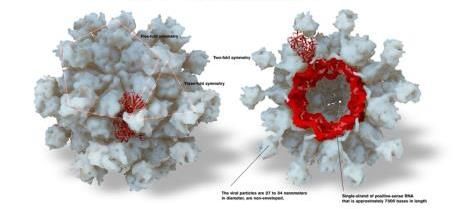
HEV GENOME
RNA quasi-envelope, ss+RNA
Capsid 27-34 nm
5’ UTR 3’
AAAAA
ORF1: non structural protein; ORF2: capsid protein; ORF3: multifunctional protein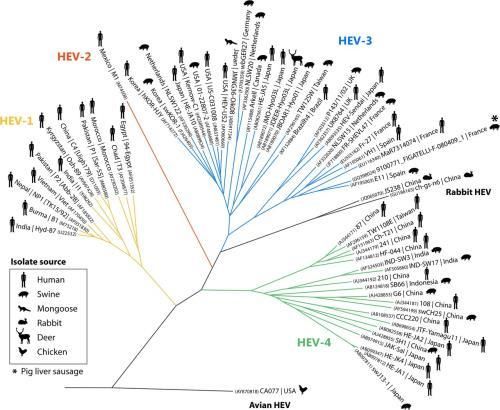
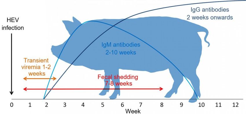
PATHOGENESIS IN PIGS
The incubation period varies from 3 to 8 weeks
Duration of viremia (as well as shedding in feces) is variable
The virus can be shed at high level in feces and bile (liver)
HEV shedded in feces for 7 to 8 weeks
Modified by https://www.pig333.com/public-health/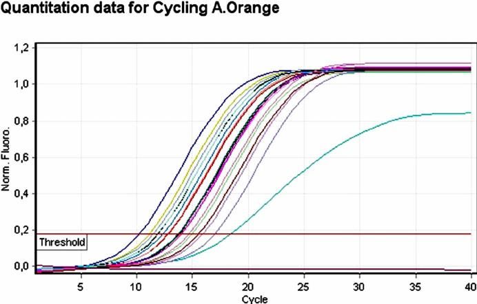
DETECTION OF HEV

• The techniques aimed at detecting different things
•     serological evidence of prior infection (IgM)
    • detection of virions (ELISA detects antigen)     Diagnosis
    • detection of virus nucleic acids
    • detection of virus infectivity
    Cell culture
    Animal model (living animals)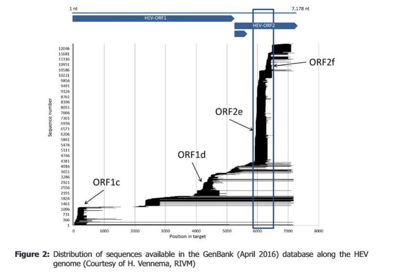
ANIMAL MODEL

   • Cynomolgus and rhesus monkeys HEV1 to 4. Model for infection in
     humans

Genotype        Origin of strain                       Infection

HEV3 and HEV4 pigs                                     +
              Human                                    +
HEV ra        rabbit                                   +
HEV           rat                                      -
              Food                    Thermal treat
HEV3          Pig liver               56°C for 1 hr    +           Feagins et al. 2008
HEV3          Patè (liver+fat+spices) 71°C for 5min    +           Barnaud et al. 2012
                                      71°C for 20min   -
NOVEL ANIMAL MODELS

• Animal model not only to evaluate infectivity of HEV but also to
  study replicative cycle and pathogenesis….

• Infections (evaluated by HEV RNA in the stool and by viremia) with
  human (HEV3f) and chimpanzee (HEV1, Sar-55) stool suspensions
  succeeded (Allweiss et al., 2016; Geldof et al., 2016)
DETECTION OF HEV

• The techniques can be arranged in four categories:
 • detection of virions (EM; ELISA; western blotting)
 • detection of virus nucleic acids
 • detection of virus infectivity
 • Cell culture
 • Animal model (living animals)

CELL CULTURE

                          First paper on cell cultivation Huang et al; 1992; 1995

human feces                          HEV4     A549 (human lung carcinoma cells)             (Wei et al., 2000)
human feces                          HEV3     human hepatoma cell lines (PLC/PRF/5, A549) (Tanaka et al., 2007)

rabbit liver homogenate              HEV3 ra human hepatoma cell lines (PLC/PRF/5, A549) (Jirintai et al., 2012)

12 swine and boar liver, feces, or   HEV      A549 and PLC/PRF/5 cells                       (Takahashi et al., 2012)
serum
swine feces                          HEV3     porcine stem cell line (PICM-19) porcine       (Rogee et al., 2013;
                                              hepatoma cell lines (HepaRG,)                 Talbot et al., 2013)

swine feces                          HEV3      primary cultured human hepatocytes (PHCs)    (Oshiro et al., 2014)
                                     HEV4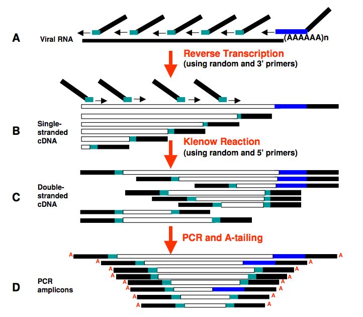
ISSUES

• initial inoculum (high viral load or MOI dependent)
• strain dependent (?)
• origin of inoculum (liver, feces)
• time of growing
• final titer
• visualization of growing (replication of HEV did not
  cause any cytopathic changes in the cell line)
                                                           Okamoto et al., 2011 HEV3 humans

        CELL CULTURE IS THE MOST PROMISING TEST
             TO EVALUATE INFECTIVITY (FOOD)
DETECTION OF HEV
• The techniques aimed at detecting different things
• serological evidence of prior infection (IgM)
• detection of virions (EM; ELISA detects antigen)
• detection of virus nucleic acids
• detection of virus infectivity
Cell culture
Animal model (living animals)DETECTION OF VIRAL NUCLEIC ACID
Humans: from plasma or faeces
Animals: liver, faeces, blood, bile
HEV genome
detection
RNA extractionRNA EXTRACTION • Food of animal and non animal origin No ISO available for RNA extraction from food either AO or NAO. Berries and shellfish (ISO/TS 15216-1:2013) e.g. Mesquita et al., 2016 >10%
RNA EXTRACTION FROM FOOD • Starting materials (30-100mg liver; 5gr meat, salami, 2 gr for liver sausages (Szabo et al., 2015; Moor et al., 2018 ) • Sample preparation: homogenize samples by Stomacher, Tissue lyser (zyrconia beads); lysis buffer (Vital project; Di Bartolo et al., 2012; Rose at al., 2011), water (Martin-Latil et al., 2014) or Trizol (Szabo et al., 2015) additional step with PEG (Martin-Latil et al., 2014) • Magnetic beads with silica • LOD 5.3 x 10⁴ GE / 2 g; 1.56x 10³ GE / g (liver sausage); • 2.9 x 10³ GE / 5 g (salami), 1.56x 10² GE / g (raw meat sausage) (Szabo et al., 2015; Moor et al., 2017)
HEV RNA DETECTION

• Real-time RT-PCR protocol by Jothikumar et al., 2006 Cited 304
• HEV1-4
• TaqMan® chemistry (4 copies for reaction)
• Performed using one or two steps (c-DNA followed by Real-time PCR),
  different kits, including IC
• WHO international standard for HEV RNA
Quantification in GE does not correspond to viable virus (1 up to 4 log difference with virus units)
RNA detection

                          RNA extraction                   End-point RT-PCR

                Phylogenetic analyses   Sequencing

      Subtype
Strain identification
TYPING OF HEV
Only 1 serotype has been described
Typing by molecular method: sequencing and comparisons
From Ricci et al., 2017 EFSA
Opinion on the public
health risks
associated with hepatitis E
virus (HEV) as a food-borne
pathogen. EFSA Journal
2017;15(7):4886,
Full genome alignment and p-distance calculationFULL GENOME SEQUENCING
• NGS Sample preparation: removal of bacteria; treatment with
Benzonase (removal of RNA and DNA);
Sequence-Independent, Single-Primer Amplification (SISPA)
Ion Torrent PGM
Matrices Subtype GE Reads HEV contings
Wild boar liver HEV3i 4.4 x 10⁶ 1,596,275 Full genome
Wild boar liver HEV3NA 1.7 x 10⁸ 1,243,513 6,900
Swine feces L75 HEV3l 2.8 × 10⁵ 1,206,199 2 contigs in ORF1 and 2 in ORF2 (300-900 bp)
Swine feces R13 HEV3l 7.1 × 10⁵ 4,884,086 No contig
Different approach: use of specific HEV-primers (limit the use of PCR amplification)ORTHOHEPEVIRUS A
• Orthohepevirus A: 7 genotypes (8 recently
proposed)
• p-distance threshold between genotypes of
0.088 for amino acid distances of
concatenated ORF1 and ORF2 (lacking
hypervariable regions) (Smith et al., 2014)
Lisa J. Krain et al. 2014TYPING OF ZOONOTIC HEV
a
b
c
h
• Genotypes are classified in subtypes (letters)
• Smith et al., 2016: set of reference sequences for each subtype
• HEV4: 10 reference strains; 9 subtypes and 1 strain not assigned
• HEV3: 20 reference strains; 2 major clades with six subtypes (3a, e
f
3b, 3c, 3h, 3i and 3j) and with the three subtypes (3e-3g). Two g
recently proposed novel subtypes
• HEVra not assigned, separate clade
Smith et al., 2016NOVEL SUBTYPE
FR-SHEV3c-like strain
• No threshold established for subtype
• By Smith et al. 2016, if at least three complete genome
sequences not related to any of the subtypes described and
that were epidemiologically unrelated (strains from different
studies or localities)NOVEL SUBTYPE
• 2 Italian swine HEV3 not epidemiologically correlated
• Full genome sequences
• Alignment using the reference set
The two Italian strains cluster together with the FR-SHEV3c-like strain
(JQ953664) (not assigned)
p-distance value of 0.129 with
the major clade represented by -3c, -3i, -3h prototype strains and some
not classified strains
Proposed novel subtype HEV3-l(De Sabato et al., 2018)NOVEL SUBTYPE
WHAT IS THE MEANING OF SUBTYPES? • silent mutations • some subtypes are more frequently observed • geographical distribution (pigs movement) Pigs experimental infections with subtypes 3c, 3e, and 3f (Rogee et al., 2015) the replication efficacy of the three different strains was similar 61 proteins differentially expressed during hepatitis E virus infection.
• The sequences databases: web-based typing platform
• strains from humans animals, food and environment
GRAZIE!

 Dr. Luca De Sabato
Dr.ssa Marina Monini
 Dr. Giovanni Ianiro
  Edoardo Vignolo

 Dr. Gabriele Vaccari

Dr. Franco Maria Ruggeri
 Dr.ssa Giorgia Angeloni
Dr.ssa Eleonora Ponterio

University of Bologna
Prof. Fabio Ostanello
You can also read