Development of prefrontal cortex - REVIEW ARTICLE - Nature
←
→
Page content transcription
If your browser does not render page correctly, please read the page content below
www.nature.com/npp

REVIEW ARTICLE                       OPEN

Development of prefrontal cortex
                    1✉                        ✉
Sharon M. Kolk           and Pasko Rakic2

© The Author(s) 2021

    During evolution, the cerebral cortex advances by increasing in surface and the introduction of new cytoarchitectonic areas among
    which the prefrontal cortex (PFC) is considered to be the substrate of highest cognitive functions. Although neurons of the PFC are
    generated before birth, the differentiation of its neurons and development of synaptic connections in humans extend to the 3rd
    decade of life. During this period, synapses as well as neurotransmitter systems including their receptors and transporters, are
    initially overproduced followed by selective elimination. Advanced methods applied to human and animal models, enable
    investigation of the cellular mechanisms and role of specific genes, non-coding regulatory elements and signaling molecules in
    control of prefrontal neuronal production and phenotypic fate, as well as neuronal migration to establish layering of the PFC.
    Likewise, various genetic approaches in combination with functional assays and immunohistochemical and imaging methods
    reveal roles of neurotransmitter systems during maturation of the PFC. Disruption, or even a slight slowing of the rate of neuronal
    production, migration and synaptogenesis by genetic or environmental factors, can induce gross as well as subtle changes that
    eventually can lead to cognitive impairment. An understanding of the development and evolution of the PFC provide insight into
    the pathogenesis and treatment of congenital neuropsychiatric diseases as well as idiopathic developmental disorders that cause
    intellectual disabilities.

    Neuropsychopharmacology (2022) 47:41–57; https://doi.org/10.1038/s41386-021-01137-9

PFC DEFINITION                                                                    new cell types and cytoarchitectonic areas. Species-specific
There is little disagreement that the human cerebral cortex is the                adaptations of prefrontal areas, steered by the environmental
organ that enabled abstract thinking and the creation of civilization,            demands, can explain the differences in size of frontal areas over
including architecture, science and all types of art. Using a wide                time. Among the most recent additions are several association
variety of methodologies, the size and cytoarchitecture of the frontal            areas, particularly the PFC, which has expanded enormously in
lobe, and more specifically the PFC, has been extensively studied                  primates culminating in humans [14]. In humans, the PFC occupies
over the years in various species. The PFC in humans and nonhuman                 as much as about 30% of its surface. Although still debated, the
primates can be divided into a collection of structurally and                     human frontal lobe seems to have evolved three times larger than
functionally different subdomains positioned anterior to the motor                that of our closest living relatives, the great apes. In fact, it has
cortex; the medial (mPFC), lateral prefrontal cortex (lPFC) and                   been argued that the human brain possesses prefrontal regions
orbitofrontal cortex (oFC). The lPFC is mostly involved in language               that are both qualitatively and functionally exclusive [15]. It is,
and executive processing, while the oFC and mPFC are known to                     nevertheless, remarkable that we use the rodent model for most
contribute to cognitive functioning and emotional control [1–4]. The              of the cellular and molecular neuroscientific studies, despite its
mPFC can be further subdivided into the infralimbic (IL), the                     lissencephalic brain which is clearly much simpler in both
prelimbic (PL) and anterior cingulate cortex (ACC). The most ventral              cytoarchitecture as well as function. A valid question still remains:
subdomain of the mPFC is the infralimbic cortex (IL) and is involved              Do rodents have a prefrontal cortex? [16, 17]. And if we were to focus
in coping with chronic stress eventually leading to structural changes            more on the evolutionary aspects of prefrontal development in terms
and prefrontal dysfunction [5–11]. Interestingly, the PFC of rodent               of structural organization and function, should we not include
models such as mice is limited in size, containing medial,                        longitudinal neurodevelopmental studies on more species [18, 19]?
orbitofrontal and cingulate areas, but probably lacking the equivalent            Although the basic principles of cortical development may be
of the primate dorsolateral PFC. In humans, the PFC can be                        similar in all mammals, the modifications of developmental events
considered to have evolved disproportionally large and it is thought              during millennia of primate evolution produce not only quanti-
to be the last region of the brain to gain full maturity [12, 13].                tative but also qualitative changes of its cellular structure and
                                                                                  synaptic circuitry [13, 20]. The origin of species-specific distinc-
                                                                                  tions can be traced either to the new or phylogenetically
EVOLUTIONARY VIEW ON PFC DEVELOPMENT                                              conserved genes that act at the time of the neural stem cell’s
During mammalian evolution, the cerebral cortex not only                          exit from the mitotic cycle and generate a different outcome,
increased in neuronal numbers and surface area but also acquired                  depending on the evolutionary context by interacting with a

1
Department of Molecular Neurobiology, Donders Institute for Brain, Cognition and Behaviour and Faculty of Science, Radboud University, Nijmegen, The Netherlands.
Department of Neuroscience and Kavli Institute for Neuroscience, Yale University, New Haven, Connecticut, USA. ✉email: s.kolk@donders.ru.nl; pasko.rakic@yale.edu
2

Received: 9 March 2021 Revised: 15 July 2021 Accepted: 22 July 2021
Published online: 13 October 2021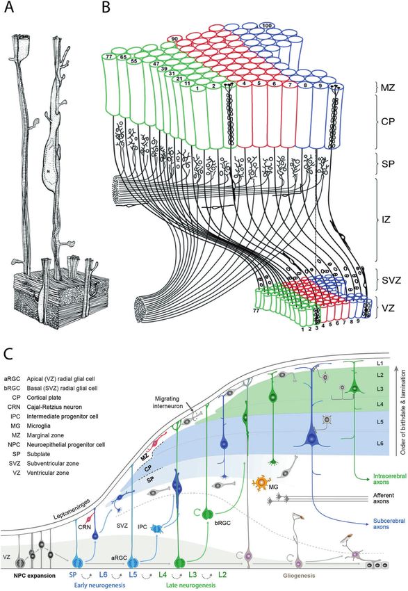
S.M. Kolk and P. Rakic
                  42
                       postmitotic neuron. Thus, the PFC as well as the Broca and                (E40) and E100, during the 165-day-long gestational period in this
                       Wernicke association areas in humans, which are formed in the             species [48]. Genesis of neurons destined for the PFC is completed
                       frontal and temporal lobes, display a temporarily enriched gene           by E90, before completion of neurogenesis in the primary visual
                       expression pattern that is distinct from the mice or macaque              cortex at E90 (Fig. 1 and [49]). Through close interplay between
                       cerebrum at the comparable prenatal stages (e.g., [21, 22]). More         cell-autonomous events and local as well as external cues, neurons
                       on evolution of the prefrontal cortex can be found in this volume,        generated close to the ventricle start migrating in radial columns
                       part I, chapter 1.                                                        [45, 46, 50, 51]. Gray matter continues to increase well into
                                                                                                 adolescence [52]. Astrocytes being the most abundant type of
                                                                                                 glial cells within cortical areas, are generated from the radial glial
                       THE EARLY STAGES OF PFC DEVELOPMENT                                       cells in the VZ and from the intermediate progenitors in the SVZ
                       Genetic determination of the PFC                                          after the peak of neurogenesis [53]. The oligodendrocyte
                       Still inside the womb, the generation of neural tissue (human,            precursor cells, or OPCs, are generated within the medial
                       third gestational week) begins with the induction of ectoderm into        ganglionic eminence and the anterior entopeduncular area and
                       neuroectoderm after which the neural tube will form through a             migrate toward the frontal cortical regions [54]. In the final stage
                       process called neurulation [23]. The detailed analysis of a series of     of OPC production, this generation occurs in the cortical regions
                       embryonic and fetal human postmortem brain tissue, as well as             themselves. Microglia cells, on the other hand, are of mesodermal
                       the evidence from experiments on animal models that range from            origin and migrate throughout the brain [55].
                       rodents to nonhuman primates, showed that specific genes and                  After the last cell division, postmitotic neurons migrate an
                       regulatory elements are involved in evolutionary elaboration of           increasingly long distance across the embryonic and fetal cerebral
                       the cranial part of the neural tube. More specifically, it is well         wall to their final positions in the cortex that develops below the
                       documented that differential gene expression and the gradients            pial surface [33, 56]. Although similar DNA labeling is not possible
                       of signaling molecules across the embryonic brain generate                to perform in humans, examination of histological and Golgi silver
                       prospective subdivisions of the neocortex [24–29]. Work of Cholfin         impregnation methods of the embryonic and fetal human
                       and Rubenstein in mice provide experimental evidence that the             cerebrum indicate the existence of similar timing and sequence
                       PFC can expand differentially and independently of the growth             of these developmental events [37, 57, 58]. The pyramidal
1234567890();,:

                       rate of the other areas [30, 31] and that its size can be regulated at    excitatory neurons born in the VZ and SVZ of the prefrontal
                       early stages by the change of expression of specific growth factors        subdomains, similar to other cortical areas, start to migrate radially
                       before they receive the afferent axonal input [32]. Through               toward the proper position in the CP under the influence of Fgfs
                       regional specification in which the Fgf family plays a significant          [50, 59, 60]. Migrating neurons are guided over an increasingly
                       role, the (pre)frontal cortical area starts to expand [32]. The           long and curvilinear pathway by the elongated radial glial cell
                       formation of the cytoarchitectonic map during evolution and               fibers that span the entire developing cerebral wall [61–63]. The
                       individual development can be explained by the Protomap                   radial glial processes that extend to the pial surface serve as a
                       Hypothesis (PMH) of cortical parcellation [33]. This hypothesis           scaffold for the migrating neurons, which will settle themselves in
                       postulates that intersecting gradients of molecules are expressed         an inside-out manner with the earlier-born neurons in the deeper
                       across the embryonic cerebral wall that guide and attract specific         layers and later born neurons in the more superficial ones
                       afferent systems to the appropriate position in the cortex where          [56, 64, 65]. Born in the ganglionic eminences, GABAergic
                       they can interact with a responsive set of cells [34]. The prefix          interneurons migrate tangentially to the proper place within the
                       “proto” indicates the malleable character of this primordial map,         prefrontal subdomains [66, 67]. Some recent findings in human
                       as opposed to the concept of equipotential cortical plate                 and primates, such as the role of outer radial glia cells (oRGCs) and
                       consisting of the undifferentiated cells that is eventually shaped        truncated glial cells, the diversity and complexity of cortical
                       and subdivided entirely by the instructions from those afferents          progenitors, the role of the subplate and the high specificity in
                       [35, 36]. The PMH is at present universally accepted even by its          axonal guidance events, again underline the complexity and
                       initial opponents (e.g., [29]).                                           evolution of cortical areas [68–75]. We now know from recent
                                                                                                 studies that it is the birth and migration of neurons derived from
                       Prefrontal expansion and lamination                                       oRGCs that play a role in the development of the primary sulci
                       The structural development of the various subdomains of the PFC           (superior frontal, inferior frontal and precentral) in week 25–26 of
                       is a meticulous process starting with a massive expansion of the          gestation [23, 76]. After the process of migration is completed,
                       most proximal part of the developing neural tube. The first step in        RGCs retract their apical process and generate astrocytes and
                       the expansion of the cortical surface during development starts           oligodendrocytes. In nonhuman primates and human, glial cells
                       with an increase in the number of symmetrical divisions of neural         seem to somewhat outnumber neurons in the PFC, albeit with
                       stem cells in the ventricular zone (VZ) before the onset of               regional variation which is likely to contribute to the formation of
                       neurogenesis and the formation of the subventricular (SVZ),               secondary and tertiary gyri [77–81].
                       intermediate (IZ) and subplate (SPZ) zones and cortical plate (CP)           Contrary to some initial concepts and theories [35, 36],
                       below the marginal zone (MZ) [33, 37–39], for review see [34]. This       embryonic VZ and CP are not uniform and equipotential. The
                       initial cortical expansion is also supported by experimental studies      enlargement and introduction of the new cytoarchitectonic areas
                       in mice [40–43] and provides an explanation for the mas-                  has been explained by the Radial Unit Hypothesis (RUH).
                       sive increase in cortical surface area during both individual             According to this hypothesis, increasing the size and proliferative
                       development and evolution.                                                capacity of the neuronal stem cells in the proliferative zone
                          By the time the apical radial glial progenitors within the             enables initial enlargement of the cortex as well as formation of
                       prefrontal subdomains start dividing asymmetrically, the number           the distinct anatomical and functional cytoarchitecture areas in
                       of neurons will increase rapidly and peaks between week 13 and            the mammalian evolution [33]. According to the RUH, tangential
                       16 of gestation in human (E10-E15 rodents/E43-E50 primates),              (horizontal) positions of cortical neurons are determined by the
                       specifically in the dorsal telencephalon [44–47]. The labeling of          positions of their precursor cells, now called stem cells in the VZ,
                       dividing cells by the DNA replication markers tritiated thymidine         while their radial (vertical) position in the overlying cortex is
                       (TdR) and bromodeoxyuridine (BrdU) showed that in nonhuman                determined by the time of their origin (Fig. 2). Therefore, the
                       primate rhesus macaque, most cortical neurons, including those            addition of the number of the radial columns increases the size of
                       destined for the PFC, originate in the proliferative VZ near the          the cortical surface, whereas the number of cells within the
                       cavity of the cerebral ventricle, between the 40th embryonic day          columns determines its thickness.

                                                                                                                            Neuropsychopharmacology (2022) 47:41 – 57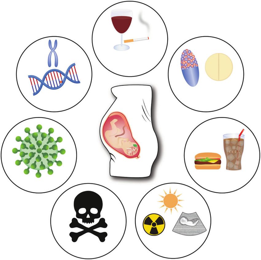
S.M. Kolk and P. Rakic
                                                                                                                                                         43
                            A                          B
                                                                                                                                   juvenile
                                                                                                                                    stage
                                                                E0   E34 E40 E43 E45 E50 E54 E62 E70   E80    E90   E100   Birth
                                                         (3H)dT
                                                        injection                                                                    sac

                            C
                                   I
                                   II

                                  III

                                  IV

                                  V
                                  VI

                                 WM

                                        E0       E40       E50         E60         E70        E80            E90     E100            E165
                                                                                                                                     TERM
Fig. 1 Prefrontal birthdating experiment in nonhuman primate. A Pen drawing of a macaque brain (side view) with the PFC indicated in
pink. B Schematic overview of the time line for which [3H] thymidine ([3H]dT) injections were given at particular embryonic (E) time points
indicated by green arrowheads. sac, sacrifice. C Relationship of time of origin and the final position of neurons destined for the PFC in
macaque monkeys based on autoradiographic labeling of DNA replication by [3H] thymidine at various days of gestation (for details of the
approach and methodology see Rakic [48]. Embryonic days are represented on the horizontal axis, vertical lines indicate the embryonic day
on which an animal received a pulse of [3H]dT and the horizontal markers on the vertical lines represent the positions of heavily labeled
neurons in the PFC. A schematic representation of the approximate position of layers I–VI and the white matter (WM) is indicated on the left
(green rectangle). The data show that all neurons in PFC are generated between embryonic (E) day 40 and E90 within the 165-day-long
gestational period in this primate species.

Differentiation and synaptogenesis                                                 spines compared to the adult PFC. The period of overproduction
After neurons assume their final position, they begin to                            of synapses is followed by a protracted plateau stage that lasts
differentiate further and form synaptic connections. In humans,                    from 2 months to 3 years of age when synaptic density remains
between 17 and 50 weeks of gestation (first to fourth postnatal                     relatively constant. In humans, the PFC synaptic density spikes
week in rodents), the pyramidal and interneurons in the various                    around 3.5 years of age (~4th postnatal week in rodents), which is
cortical layers of the PFC will further mature and differentiate                   relatively late compared to other cortical areas and almost double
[82, 83]. The basal and apical dendritic length will increase, the                 the net density of the adult PFC [82, 93, 94]. Examination of the
spines will further develop, specifically in layer III and V, and their             course of synaptogenesis in the macaque PFC, by detailed
axons will extend to other cortical and subcortical targets [82, 84].              quantitative electron microscopic analysis, showed that the
This is also the case for the inhibitory network where the                         number of synaptic contacts is initially grossly overproduced
interneurons mature extensively with a sharp increase in the                       before declining to the normal adult level (Fig. 3 and [49].
dendritic spine formation but also in terms of their intrinsic as well             Likewise, the axons of the corpus callosum, as well as other large
as their network properties as was shown in mice [85, 86].                         axonal tracts in the macaque cortex, including PFC, are grossly
Prefrontal synaptogenesis starts prenatally and peaks postnatally                  overproduced before decreasing to the adult level [95–97]. A
followed by a process called pruning or refinement of synaptic                      subpopulation of GABAergic neurons in the subplate zone also
connections, the removal of unused synaptic contacts [87]. When                    form transient synapses that are eventually eliminated [98, 99].
neurites to and from the PFC reach their final target position, an                  The period of synaptic decline in human PFC, which starts during
immature synapse is generated under the influence of, among                         childhood, is initially dramatic and continues during adolescence
others, cell adhesion molecules and reelin [88, 89]. Epigenetic                    and extends at a slower, but statistically significant rate into the
regulatory factors such as microRNAs (miRNAs) play an important                    3rd decade of life (Fig. 4 and [12]).
role in this process by modulating dendritic and synaptic                             The finding that synaptic density in the cerebral cortex is
maturation [90, 91]. The tempo and kinetics of synapse formation                   relatively stable from early adolescence through puberty (the
in the primate PFC closely resemble those described for other                      plateau period) is indicative that in primates the final synaptic
areas [92]. In young primate embryos, a precortical phase (E47-                    pattern is the result of selection and refinement of their higher
E78) is described when synapses are found only above and below,                    number during the formative years when learning experiences are
but not within, the CP. Following that, there is an early cortical                 most intense. These discoveries led to the proposal that the
phase, from E78 to E104, during which synapses accumulate                          Selective Elimination Hypothesis is a mechanism for tuning
within the cortical plate, initially exclusively on dendritic shafts.              synaptic connections by interaction with the environment during
The next rapid phase of synaptogenesis begins at 2 months before                   the period of most intense learning [92]. These days, selective
birth and ends approximately at 2 months after birth, culminating                  elimination or stabilization is commonly called “pruning”, and this
with a mean density of 750 million synapses per cubic micrometer.                  refinement of the differentiating cortical network via pruning of
This accumulation is largely accounted for by a selective increase                 dendritic branches, and/or efferent/afferent projections, is an
in axospine synapses in the supragranular layers. Therefore, the                   important process to fine-tune the meticulous intricate prefrontal
early childhood PFC contains a 2–3 fold higher density of dendritic                network [100, 101]. Within the rodent and primate PFC, this

Neuropsychopharmacology (2022) 47:41 – 57
S.M. Kolk and P. Rakic
44
Fig. 2 The evolution of corticogenesis. A Three-dimensional reconstruction of postmitotic neurons migrating along radial glial fibers, based
on electron micrographs of semi-serial sections of the monkey fetal cerebral cortex with permission from Rakic [56]. B Representation of the
radial unit hypothesis based on Rakic [33] with permission from Silver et al. [396]. C Illustration of the dynamics of major developmental
events and diversity of progenitors involved in the development of primate cerebral neocortex based on studies of Rakic, with permission
from Silver et al. [396].
process of synaptic pruning, which is most dramatic in layer III, zone (SPZ) which is thicker in the PFC compared to other cortical
continues well into adolescence leading to a long-lasting decline areas. A major change in development, which likely signals roots
in synaptic density across PFC subdomains [82, 102, 103]. It was in the evolution of the cortex, is in the specificity in neuro-
furthermore discovered that major neurotransmitter receptors are transmitter systems alongside a boost in receptor type hetero-
also initially overproduced in all eight primate prefrontal regions geneity in primates and human [108–112]. In humans the
examined [104, 105]. Moreover, during childhood the PFC thickening of the PFC subplate has evolved tremendously,
myelination process starts (white matter volume increase) which suggesting playing a role in the extensive prefrontal circuitry
continues into adulthood [106, 107]. [71, 102]. Vice versa, the multitude of pyramidal neurons in the
various layers and PFC subdomains will connect to other cortical
Getting connected and subcortical targets by extending their axons, once they have
The prolonged maturation of the PFC depends largely on the reached their final position in the PFC (human: birth till end of first
coordinated action of various external factors. Most neurotrans- year/rodent first 2 postnatal weeks). The intricate timely integra-
mitter projections arrive in the prefrontal subdomains in two tion of all these neurotransmitter systems is essential for
streams: within the marginal zone (MZ) and within the subplate prefrontal functioning. In this way, a unique and higher-order
Neuropsychopharmacology (2022) 47:41 – 57S.M. Kolk and P. Rakic
45
A B 8
Months after birth 2 6 12 24 60 240
Total number of synaptic contacts (*103/100 µm)
I
II
SP 6
III
4
SP IV
V
2
VI
WM
10 100 1000 10000
500 µm
birth puberty
Fig. 3 Primate synaptogenesis in the PFC assessed by quantitative electron microscopy. A Schematic representation of the site of the
block dissection from the depth of the Sulcus Principalis (SP). On the right: The section of the cortex in the SP showing the vertical (radial)
probes across layers I–VI, which were examined by electron microscopy. B The total number of synaptic contacts in each vertical probe as
represented by the green dots. The semi-log plot in abscissa represents the number of days after conception. Adapted from Bourgeois et al.
[49].
functional network capable of emotional processing and complex PFC showed that the α2-adrenoceptor and muscarinic M1
cognitive abilities is established. receptor modulate working memory via KCNQ potassium channel
[148–151]. Reciprocal direct connections from the mPFC to the LC
Developing PFC connections - from the neurotransmitter perspective mature over time, and this system is involved in a variety of
Serotonin: The brain matures from the brainstem to the more behaviors such as memory formation, attention, arousal, vigilance
frontal cortical regions, and it is therefore not surprising that and coping with stress [152–154].
serotonergic projections from both the dorsal as well as the
medial Raphe nuclei (DRN, MRN respectively) are among the first Dopamine: A subset of the medial part of the ventral tegmental
to emerge and are set towards cortical regions where they arrive area (VTA) starts to project to prefrontal subdomains around E15/
in the PFC around E16/E17 in rodent and week postnatal 10–13 in E16 (rodent) and week 10–13 in human [155–158]. Steering
humans [113–116]. Most of the work on molecular and cellular dopaminergic projections from the VTA via the medial forebrain
underpinnings of serotonin functioning and guidance during early bundle toward forebrain regions mostly depend on a coordinated
development have been investigated in rodents, although it is action of the guidance molecules Dcc and Netrin-1 mediated by
clear that the specificity of serotonergic prefrontal connectivity in microRNA miR-218 control of Dcc expression in the VTA [159–161],
primates and human increase tremendously in regional specificity while Semaphorin3F is orchestrating their fasciculation, rostral
[109]. In mice, the serotonergic projections toward the forebrain growth and targeting within the various mPFC subdomains [156].
are predominantly guided by the Epha5/ephrina5 interaction of The dopaminergic innervation of the mPFC in rodent surges
guidance cues [117]. Of note, early in the development of during adolescence hallmarked by massive changes in the
serotonergic signaling, molecules such as receptors and transpor- organization, shape and density of the dopaminergic fibers
ters are already expressed in the forebrain and an exogenous [162–164]. A similar surge in regional-specific dopaminergic
placental source of 5-HT has been considered to direct cortical connectivity to the PFC can be observed in primates, including
development even before raphe-derived projections have reached human modulating local microcircuits [165–169]. Of note here is
the forebrain [118–121]. Once the serotonergic projections have that some of these dopaminergic neurons projecting to the
arrived within cortical areas, they are able to make contacts with various PFC subdomains are capable of co-releasing glutamate as
Cajal Retzius cells within the MZ, thereby raising the possibility of well and have an exclusive excitatory effect on the GABAergic
playing a role in neuronal migration [122–124]. It has become interneurons in the various layers of the PFC [170–173]. Eventually,
widely accepted that serotonin exerts a significant trophic and the mature mesoprefrontal system is involved in attention,
modulatory function in neurodevelopmental processes such as behavioral flexibility, action planning, sustainability of motiva-
proliferation, migration and differentiation in cortical areas, tional and affective states, working memory and memory
including the PFC [119, 123, 125–129]. consolidation which is mediated in parallel by catecholaminergic
pathways [169, 174–178]. In many neurodevelopmental disorders
Noradrenalin: The Locus Coeruleus (LC) in the brainstem sends (NDDs) the developing dopamine system is affected playing a role
out its noradrenergic axonal projections to the PFC as early as E16/ in the diverse symptoms of these disorders [179].
17 (rodent) and week 10–13 in human [130–132]. It appears to be
a heterogeneous set of neurons innervating all aspects of the PFC GABA: Most of the GABAergic interneurons are born in the
subdomains [133–135]. Noradrenergic projections arrive in cortical ganglionic eminences of the ventral telencephalon and migrate
areas before all cortical neurons have finished migrating and have tangentially to the proper cortical areas and layers to form a
adopted their final appearance [136]. During the embryological network with the radially migrated pyramidal neurons [180–182].
development of prefrontal areas specifically, noradrenaline plays a Initially being excitatory through the GABAA receptors expressed
role in cell division, neuronal migration, differentiation as well as on radial glia cells and migrating interneurons, GABA plays a role
synaptogenesis [137–141]. Like serotonin, noradrenergic axons in proliferation, migration and synaptogenesis [183–186]. It has
make contact with the Cajal Retzius cells in the marginal zone, furthermore been shown that dopamine and GABA interactions
suggesting a role in the laminar formation of cortical regions can influence these processes [187, 188]. Around the second
[132, 142, 143]. In addition, noradrenalin seems to have an effect postnatal week in rodents (~first postnatal week in human), the
on the development of dopaminergic projections in the PFC by depolarizing effect slowly transitions into an inhibitory net effect
providing a dopamine reuptake mechanism through the nora- depending on place and time [189, 190]. A remarkable feature in
drenalin transporter [144, 145] as well as on GABAergic signaling GABA signaling from an evolutionary perspective is that in
in the PFC [146, 147]. Recent studies of rat and primate nonhuman primates and human there seems to be a cell-type
Neuropsychopharmacology (2022) 47:41 – 57S.M. Kolk and P. Rakic
46
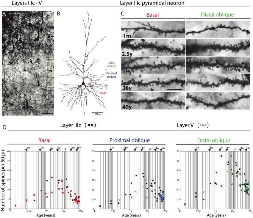
Fig. 4 Development of dendritic spines on layer IIIC and layer V pyramidal neurons in the human PFC. A Low-magnification photograph of
the rapid Golgi-impregnated layer IIIc and V pyramidal cells in the dorsolateral PFC of a 16-year-old subject. B Neurolucida reconstruction of
layer IIIc pyramidal neuron of a 49-year-old subject showing distal oblique (green), proximal oblique (blue) and basal dendrites (red). C
Representative high-power magnification images of rapid Golgi-impregnated layer IIIc pyramidal neurons in a 1 month old infant, 2.5-year-old
child, and 16-, 28-, and 49-year-old subjects. D Graphs representing number of dendritic spines per 50-μm dendrite segment on basal
dendrites after the first bifurcation (red); apical proximal oblique dendrites originating within 100 μm from the apical main shaft (blue); and
apical distal oblique dendrites originating within the second 100-μm segment from the apical main shaft (green) of layer IIIc (filled symbols)
and layer V (open symbols) pyramidal cells in the human dorsolateral PFC. Squares represent males; circles represent females. The age in
postnatal years is shown on a logarithmic scale. From Petanjek et al. [12].
specific expression of the GABA transporter GAT-1 in early from the PFC itself such as the various thalamic regions. Recently it
childhood [191, 192]. Furthermore, it appears that nonhuman was found that retinoic acid (RA) plays a critical role in PFC
primates and humans have distinct populations of GABAergic development and specifically in this thalamus-prefrontal connectivity
neurons which originate in proliferative zones of the dorsal [203]. The PFC furthermore sends out glutamatergic afferents to the
telencephalon [193, 194]. VTA as well as to the nucleus accumbens modulating dopaminergic
signaling [204–206]. In addition, DRN serotonergic neurons are
Glutamate: There are various sources of glutamatergic input controlled by glutamatergic projections from the PFC [152, 207–209].
projections including a subset of (non-)dopaminergic VTA neurons
to GABAergic interneurons in the PFC [170–173]. The most prominent Acetylcholine: Around birth, the numerous cholinergic projections
monosynaptic inputs of the PFC are derived from hippocampus, arising from the basal forebrain nuclei innervate the primary cortical
mediodorsal (MD) thalamus and amygdala [195–201]. In fact, the regions where they influence cortical ultrastructure [210–213].
medial pulvinar part of the medial thalamus or PM, which Acetylcholine modulates primarily the prelimbic subdomain of the
evolutionary expanded alongside the association cortex in nonhuman PFC during development targeting GABAergic interneurons [210]. But
primates and human, is characterized by a distinct prefrontal even before the cholinergic projections arrive in the cortical areas, the
glutamatergic connectivity that seems to play a significant role in nicotinic and muscarinic receptors are expressed on neural progeni-
NDDs [202]. A multitude of cortical and subcortical targets are tors playing a role in proliferation/differentiation and axonal guidance
progressively innervated by developing glutamatergic projections events [214–217]. In the PFC of nonhuman primates, the muscarinic
Neuropsychopharmacology (2022) 47:41 – 57S.M. Kolk and P. Rakic
47
M1 receptors modulate working memory via KCNQ potassium peaking [245]. Higher order cognitive functions, in which PFC
channels [151]. Alongside, a transient expression of the enzyme plays a prominent role, such as language and intelligence,
acetylcholinesterase seems to play a role in the thalamocortical circuit continue to develop into adulthood [254, 255]. More on the role
formation [218–220]. Within the PFC, the cholinergic innervation of the PFC in cognitive control and executive functioning can be
initially terminates in layers III and IV slowly losing laminar preference found in this volume in the reviews by Robbins and Friedman (I.6)
over time [221–223]. Key developing PFC circuitry is shaped by and Menon and D’Esposito (I.7).
acetylcholine, and PFC pyramidal neurons depend on its proper
signaling in terms of dendritic branching, spine formation and
synaptogenesis [224–226]. PFC DEVELOPMENT AND MENTAL ILLNESS
Although stress-induced structural changes in the PFC are equally
Convergence of developing transmitter systems within the PFC. important in their contribution to the pathophysiology of
There is, furthermore, ample evidence now that during embryonic neuropsychiatric conditions [256–262] (see also part III of this
development there is convergence of the various neurotransmit- volume), we focus in this paragraph on particular risk factors
ter signaling pathways and influence each other’s development involved in the onset of NDDs) in which PFC functioning is
and functioning [153, 227–231]. These neurotransmitter systems affected. It has been speculated that, as the PFC takes so long to
can act as neurotrophic factors steering various neurodevelop- fully mature, it also has the largest critical window of all
mental events in their target areas. Serotonergic and dopaminer- developing brain areas. The various risk factors, either genetic or
gic markers are jointly present in their developmental origins, environmental, can hamper the intricate developmental events
guidepost areas, as well as within the subdomains of the PFC, and pose a risk in developing NDDs (Fig. 5).
which is important for their intricate interaction later in life to
establish higher cognitive functions [209, 232–234]. The same From a genetic point of view
holds true for noradrenergic and dopaminergic projections The group of patients having a NDD is enormously heteroge-
towards forebrain regions as well as dopaminergic-glutamatergic neous. The genetic causes underlying NDDs are diverse ranging
and dopaminergic-cholinergic interactions controlling PFC from single gene mutations, copy number variations to whole‐
maturation and functioning [227, 228, 231]. These neurotransmit- chromosome aberrations [263]. Even with monogenic causes, the
ter projections initially innervate prefrontal regions via two parallel severity and comorbidity of the symptoms can vary tremendously
paths; one via the subplate and one via the marginal zone where and neurological/neuropsychiatric symptoms are often accompa-
the Cajal Retzius cells reside [235]. Being in close proximity of the nied by additional clinical features such as maldevelopment of
CR cells, it is likely that volume transmission is used to release the organ systems. But there are also some clear examples of
neurotransmitter. Receptors, transporters as well as synthesizing environmental risk factors that specifically hamper PFC develop-
enzymes are already expressed (~E10 rodent and week 4–5 ment resulting in behavioral and cognitive deficits. Below we list
human) even when the actual axonal projections have not yet specifically those NDDs where the structural development of the
arrived in the PFC [236–240]. In fact, neurotransmitter receptors PFC is clearly affected.
are found to be expressed by progenitor cells throughout
development [231]. External neurotransmitter sources, such as Monogenic causes. Many of the Mendelian monogenic NDDs are
the placenta, can play a role in this early shaping of cortical areas characterized by intellectual disability and behavioral problems
[114, 118, 241, 242]. All this is especially important in light of (anti-
depressant) use of pharmacological drugs during pregnancy as
they can interfere with these early signaling pathways and
hamper the structural development of brain areas including
the PFC.
Smoking
Drinking
PFC COGNITIVE DEVELOPMENT
The PFC, as the seat of our higher-order cognitive functions,
continues to develop into adulthood [52, 243]. It is among the Genetic Drugs
latest brain regions to fully mature in humans as well as rodents causes
[106, 159, 244, 245]. The primary somatosensory cortex, as well as
the primary motor cortex, mature earlier, however the dendritic
trees and the density of spines within the subdomains of the PFC
seem to be more complex [246–249]. Cognitive abilities are
shaped by experience over time and seem to be in synchrony with
PFC structural changes such as synaptogenesis and pruning [250]. Nutrients
Following the ‘use it or lose it’ principle, the developing PFC Virus
dynamically rearranges incoming and outgoing wiring depending
on usage and need [12]. Specific for the PFC, the non-coding
microRNAs mir-128b and mir-30a-5p have shown to be involved in
prefrontal-dependent cognitive maturation by affecting epige-
netic mechanisms [251, 252]. The constantly developing cognitive Physical
Toxins factors
and executive capabilities occur parallel to the neurophysiological
changes within the PFC and its connected areas and seem to
reach a plateau in teenagers (around 12 years in human, around Fig. 5 Risk factors in PFC development. Schematic overview of
genetic and environmental risk factors during pregnancy to be the
P50 in rodents) [253]. Adolescence is typically characterized by possible cause of NDDs (Clockwise: Genetic causes, Smoking,
changes in social interactions and cognitive abilities in order to Drinking, prescription or recreational Drugs, certain combination
gain independence and adult skills and competences [245]. In of Nutrients, Physical factors such as UV, ultrasound or various
nonhuman primates this is characterized by risk-taking, novelty radiations, Toxins, Virus infections). Possible causes for NDDs include
seeking, and increased vigilance; whereas in rodents by play specific genetic or environmental factors as well as a combination
behavior, increased exploratory activity and impulsivity are of both.
Neuropsychopharmacology (2022) 47:41 – 57S.M. Kolk and P. Rakic
48
due to, in part, an altered prefrontal functionality. Fragile X condition known as Alcohol-related Neurodevelopmental Disorder
syndrome (FX) is a NDD where the causative gene, Fragile X Mental (ARND). There is a clear correlation of children with FAS and prefrontal
Retardation Protein (FMRP), is completely absent causing a executive functioning [316]. MRI studies showed reductions in
plethora of developmental abnormalities [264, 265]. It is clear that brainstem as well as cerebellum volume in a primate FAS model
in FX the many behavioral and cognitive deficits can be attributed, and a sex-dependent change in functional connectivity and
at least in part, to prefrontal dysfunction. Some of these aspects metabolism in prefrontal areas in a rat FAS model [317, 318].
could be rescued in an animal model where FMRP production was Structurally, the prefrontal cortical thickness is affected after prenatal
initiated in the mutant PFC [266]. In Rett syndrome, a severe NDD alcohol exposure and it matures with a smaller number of excitatory
with specific cognitive and behavioral features, there is a strong neurons and more GABAergic ones disrupting the excitatory/
PFC hypofunction with structural abnormalities [267–269]. Restor- inhibitory balance severely [319–321]. Similar structural and beha-
ing Mecp2 levels within the PFC in mice via state-of-the-art vioral defects of the PFC can be observed in kids with prenatal
techniques such as CRISPR-Cas9 or DREADDS can restore some of exposure to opioids, cocaine, amphetamines and other drugs-of-
the endophenotypes such as social recognition deficits or long- abuse [322–330]. Similarly, we can find lead and other pollutants to
term retrieval of auditory conditioned fear [267, 270, 271]. Other be damaging to the developing brain and PFC [331–334]. Another
monogenic syndromes like Kleefstra, KBG, WitKoS, Angelman, field of recent study is the perinatal exposure to pharmaceuticals
Coffin-Sirris, Rubinstein-Taybi, Phelan-McDermid, Smith–Magenis given to treat the pregnant mother. Perinatal HIV infections can alter
Syndrome and Kabuki syndrome also have a clear prefrontal the course of brain development (see below), on the other hand
component in their behavioral and cognitive phenotype [272–280]. perinatal exposure to antiretroviral drugs such as Efavirenz (EFV) to
For some of these syndromes it has recently been shown that treat HIV leads to an altered prefrontal cytoarchitecture [335, 336].
deficits in the structural development of the PFC underlie these Although maternal stress itself can be detrimental to brain
problems [281–287]. development in general and the developing PFC in particular (for
review see [125]), treatments against maternal depression such as
Chromosomal abnormalities. In all human chromosomal aberra- SSRIs can cause substantial structural damage to prefrontal
tion syndromes, including trisomies, monosomies (e.g., Turner subdomains [125, 128].
syndrome, monosomy 1p36), polyploidies, disomies and imprinting
errors or sex chromosome anomalies, structural abnormalities of Viral infections. Traditionally, pregnant women were warned for
(pre)frontal as well as many other areas are common [61, 288–292]. TORCH (TOxoplasmosis, Rubella, Cytomegalovirus, and Herpes
Trisomy of chromosome 21 or Down syndrome can be considered a simplex viruses type 1 and 2) infections especially during the first
NDD with significant developmental deficits. Cognitive abilities are two trimesters of pregnancy as they were shown to cause severe
affected due to a developmental delay including the maturation of congenital abnormalities [337]. Later, the O was referring to Other
brain areas such as the PFC [293]. Particular neurodevelopmental infections such as syphilis, varicella-zoster, and parvovirus. Zika
events are delayed in forebrain regions such as neurogenesis, viral infection during pregnancy can cause microcephaly including
migration and synaptogenesis eventually resulting in altered severe structural damage to the prefrontal areas [338–340]. The
prefrontal circuitry [294–296]. Williams (WBS or WS) syndrome is a Zika virus is able to infect neuroepithelial stem cells and cortical
rare NDD with a deletion of approximately 25 genes on chromo- radial glial cells and to a lesser extent postmigratory neurons
some 7 and characterized by an unusual sociability and cognitive causing structural disorganization in these cells eventually leading
deficits [297]. The structural organization of prefrontal pyramidals, to cell death [341–343]. Even a postnatal viral infection can lead to
specifically their density and dendritic arboring, is severely affected postnatal meningitis and neurodevelopmental problems due to
[298, 299]. Prader-Willi syndrome (PWS) is a disorder in which structural and functional damage of frontal areas [344]. In the
imprinted genes on chromosome 15 are affected and is character- recent SARS-CoV-2 or COVID-19 viral outbreak, similar structural
ized by increased volume of prefrontal subdomains important in the damage of frontal cortical areas could be observed, most likely
reward circuitry [300, 301]. In the 22q11.2 deletion syndrome (or due to an inflammatory response in which parenchymal cells and
DiGeorge/Velo-Cardio-Facial syndrome), individuals are character- the choroid plexus are involved [345–347]. Little is known
ized by loss of executive function and working memory alongside however on the short- and long-term effects of a COVID-19
other cognitive problems and MRI studies showed a clear loss of infection during pregnancy and the possible neurodevelopmental
volume of the various PFC subdomains [302–307] changes it can make during corticogenesis leading to NDDs [348].
From an environmental point of view Other perinatal causes. Multiple fetuses per pregnancy, intrau-
Food/drugs. One of the most studied risks during pregnancy is the terine growth restriction (IUGR, due to placental failure other than
composition of our diet. Many food-derived molecules can reach the by causes described above), X-ray, UV, nuclear or cosmic radiation,
unborn baby in one form or another, and therefore could directly or (ultra)sound as well as high temperature, preterm birth or hypoxia
indirectly influence brain development when crossing the immature whether or not by traumatic causes can pose serious threats to
blood-brain barrier and potentially affect PFC development [308]. It is proper corticogenesis as well [349–356]. Recently it has become
therefore important to realize that with the change of our diet clear that IUGR is associated with an increase in impulsive
through the ages, having become more processed and high-fat and behavior due to an altered dopamine signaling in the PFC
high-sugar in contents, this can have a dramatic effect on the [357, 358]. Perinatal hypoxia can furthermore change the
development and functioning of the PFC. Particular consumption of expression of cytokine and ceramide metabolism genes in the
high-fat and/or high-sugar during pregnancy, childhood and PFC and hampers cognitive functioning in later life [359, 360]. In
adolescence can result in structural changes in the PFC and deficits preterm birth, changes in white and gray matter including
in executive functioning [308–311]. Maternal metabolic disorders reductions in cortical surface area and cortical thickness of the
including diabetes and obesity can pose another threat to the unborn PFC are described [361–364]. Disruptions in PFC network activity
child as placental dysfunctioning alters the prenatal exposure to often further aggravates the already compromised neurocognitive
nutrients and toxins [312–314]. In the early ‘70s it was found that development in these children [365, 366].
women who abused alcohol during pregnancy may deliver children
with severe developmental delays, smaller brains and cognitive The multifactorial view
problems called Fetal Alcohol Syndrome (FAS) [315]. These children It is now generally accepted that the etiology of many NDDs is
often have various conditions that are collectively known as Fetal considered to be multifactorial. Often, comorbidity of two or more
Alcohol Spectrum Disorder (FASD), which includes FAS and a NDDs is observed. Variable environmental exposure to risk factors
Neuropsychopharmacology (2022) 47:41 – 57S.M. Kolk and P. Rakic
49
combined with variable genetic background makes it hard to of corticogenesis can be recapitulated reliably; however the later
pinpoint possible causes. Yet, as the PFC takes the longest to fully stages in development still need to be optimized.
mature, we can argue that it is most vulnerable to any risk factor It has become clear that most neurotransmitter systems play
when presented early enough. We will here review three of the neurotrophic roles during neurodevelopment as well. More
‘classical’ NDDs that are considered to be multifactorial in their holistic studies into the extrasynaptic neurotrophic functions of
onset with affected PFC development. Intellectual disability (ID) is neurotransmitters during prefrontal development might also
an umbrella term and is a comorbidity of many of the described provide more understanding of their potential roles in the
NDDs and will therefore not be discussed separately. etiology of NDDs and eventually will enable us to design critical
Autism spectrum disorder (ASD) is an example heterogeneous developmental windows in which we may be able to intervene. In
NDD characterized by impaired communication and social the future, more longitudinal as well as interspecies studies will
interaction accompanied with repetitive behavior and stereotyped be needed to corroborate our understanding of prefrontal
interests. It has been described that the PFC of individuals with development.
ASD show structural and functional changes, specifically in the Abnormal PFC development may lead to a variety of behavioral
ACC, oFC and lPFC [367, 368]. The number of neurons (but also and cognitive problems inherent to psychiatric disorders including
their size), specifically the chandelier cells, basket cells and other NDDs. In order to create tailored interventions targeted to the
parvalbumin-expressing interneurons, is decreased in the PFC specific genetic syndromes, there is a strong need for research
[369–371]. There are also indications that serotonergic signaling is into the specific developmental and behavioral aspects accom-
affected during PFC development [372]. In the first couple of years panying these syndromes. A better understanding of the under-
of life there is a prefrontal hyperconnectivity in children with ASD lying neurodevelopmental and biological mechanisms will open
followed by a hypoconnectivity resulting in a ‘disconnection’ with doors to investigate the possibility of therapeutic (early/preven-
other cortical areas involved in higher-order associative proces- tive) interventions and subsequent improvement of care.
sing [373–376]. The short- and long-range prefrontal axons,
particularly those from the ACC, are affected in their guidance
to subcortical targets and may underlie the network disruption REFERENCES
characteristic for ASD [377, 378]. A new and interesting finding is 1. Fuster JM. Prefrontal neurons in networks of executive memory. Brain Res Bull.
that there appears to be a major change in the levels of various 2000a;52:331–6.
metabolites in the PFC of autistic individuals [379]. 2. Fuster JM. Memory networks in the prefrontal cortex. Prog brain Res.
2000b;122:309–16.
Attention deficit/hyperactivity disorder (ADHD) is a NDD
3. Goldman-Rakic PS. Circuitry of the primate prefrontal cortex and the regulation
characterized by signs of inattention, impulsivity, and hyperactiv- of behavior by representational memory. Handbook of Physiology, The Nervous
ity [380, 381]. Control processes mediated by the PFC are System, Higher Functions of the Brain. In: Plum F, editor. Handbook of phy-
hampered. Imaging studies have shown the PFC to be thinner siology, the nervous system, higher functions of the brain. Bethesda: American
in ADHD individuals, thereby hampering proper maturation and Physiological Society; 1987. pp. 373–417.
prefrontal connectivity leading to attentional dysfunction 4. Li H, Yan G, Luo W, Liu T, Wang Y, Liu R, et al. Mapping fetal brain development
[382, 383]. Loss of catecholaminergic innervation underlies the based on automated segmentation and 4D brain atlasing. Brain Struct Funct.
most important ADHD symptoms [381]. 2021;226:1961–72.
Epilepsy, specifically childhood frontal lobe epilepsy (FLE) has 5. Flak JN, Solomon MB, Jankord R, Krause EG, Herman JP. Identification of chronic
stress-activated regions reveals a potential recruited circuit in rat brain. Eur J
various clinical outcomes but most often resulting in multi-
Neurosci. 2012;36:2547–55.
cognitive symptoms [384–387]. Elevated prefrontal oscillations 6. Izquierdo A, Wellman CL, Holmes A. Brief uncontrollable stress causes dendritic
and hippocampal-prefrontal theta coherence could be observed retraction in infralimbic cortex and resistance to fear extinction in mice. J
after FLE [388]. And eventually the FLE seizures can cause Neurosci. 2006;26:5733–8.
structural and functional damage of the prefrontal areas including 7. Moench KM, Maroun M, Kavushansky A, Wellman C. Alterations in neuronal
altered short-term plasticity [389, 390]. Important is also that the morphology in infralimbic cortex predict resistance to fear extinction following
anti-epileptic drugs, given at young age or during pregnancy, can acute stress. Neurobiol Stress. 2016;3:23–33.
have major neurodevelopmental implications as well. These drugs 8. Dias-Ferreira E, Sousa JC, Melo I, Morgado P, Mesquita AR, Cerqueira JJ, et al.
act upon the major neurotranmitter and second messenger Chronic stress causes frontostriatal reorganization and affects decision-making.
Science 2009;325:621–5.
systems including ion channels, thereby affecting neurodevelop-
9. Shansky RM, Hamo C, Hof PR, McEwen BS, Morrison JH. Stress-induced dendritic
mental events [391–393]. remodeling in the prefrontal cortex is circuit specific. Cereb Cortex.
2009;19:2479–84.
10. Radley JJ, Rocher AB, Miller M, Janssen WG, Liston C, Hof PR, et al. Repeated
FUTURE RESEARCH DIRECTIONS stress induces dendritic spine loss in the rat medial prefrontal cortex. Cereb
The multidisciplinary nature of the field of developmental Cortex. 2006;16:313–20.
neurobiology has made enormous progress in recent years. The 11. McKlveen JM, Morano RL, Fitzgerald M, Zoubovsky S, Cassella SN, Scheimann JR,
combination of classical (immuno)histological techniques with et al. Chronic stress increases prefrontal inhibition: a mechanism for stress-
physiological, behavioral and high power molecular approaches induced prefrontal dysfunction. Biol Psychiatry. 2016;80:754–64.
12. Petanjek Z, Judas M, Simic G, Rasin MR, Uylings HB, Rakic P, et al. Extraordinary
such as large-scale genome-wide (single-cell) transcriptome and
neoteny of synaptic spines in the human prefrontal cortex. Proc Natl Acad Sci
epigenome profiling studies have brought us an enormous USA. 2011;108:13281–6.
amount of insight and resolution into the development and 13. Donahue CJ, Glasser MF, Preuss TM, Rilling JK, Van, Essen DC. Quantitative
evolution of developing brain areas, specifically the PFC and their assessment of prefrontal cortex in humans relative to nonhuman primates. Proc
role in the onset of NDDs [21, 394]. Advances in molecular labeling Natl Acad Sci USA. 2018;115:E5183–E92.
and imaging techniques have added to this understanding 14. Povinelli DJ, Preuss TM. Theory of mind: evolutionary history of a cognitive
[4, 395]. But maybe the most exciting field is the rapid emergence specialization. Trends Neurosci. 1995;18:418–24.
of stem cell approaches such as the generation of brain organoids 15. Carlen M. What constitutes the prefrontal cortex? Science 2017;358:478–82.
that have led to some tremendous breakthroughs. A dazzling 16. Uylings HB, Groenewegen HJ, Kolb B. Do rats have a prefrontal cortex? Behav
Brain Res. 2003;146:3–17.
number of studies in gyrencephalic species have led to scientific
17. Preuss TM. Do rats have prefrontal cortex? The rose-woolsey-akert program
breakthroughs and the description of novel types of cortical reconsidered. J Cogn Neurosci. 1995;7:1–24.
progenitors, including the basal and outer RGCs, both of which 18. Wise SP. Forward frontal fields: phylogeny and fundamental function. Trends
have been linked to cortical expansion and folding. Early features Neurosci. 2008;31:599–608.
Neuropsychopharmacology (2022) 47:41 – 57S.M. Kolk and P. Rakic
50
19. Warren JM. Evolution, behavior and the prefrontal cortex. Acta Neurobiol Exp. 50. Noctor SC, Flint AC, Weissman TA, Dammerman RS, Kriegstein AR. Neurons
1972;32:581–93. derived from radial glial cells establish radial units in neocortex. Nature.
20. Llinares-Benadero C, Borrell V. Deconstructing cortical folding: genetic, cellular 2001;409:714–20.
and mechanical determinants. Nat Rev Neurosci. 2019;20:161–76. 51. Miyata T, Kawaguchi A, Okano H, Ogawa M. Asymmetric inheritance of radial
21. Johnson MB, Kawasawa YI, Mason CE, Krsnik Z, Coppola G, Bogdanovic D, et al. glial fibers by cortical neurons. Neuron. 2001;31:727–41.
Functional and evolutionary insights into human brain development through 52. Gogtay N, Giedd JN, Lusk L, Hayashi KM, Greenstein D, Vaituzis AC, et al.
global transcriptome analysis. Neuron. 2009;62:494–509. Dynamic mapping of human cortical development during childhood through
22. Pletikos M, Sousa AM, Sedmak G, Meyer KA, Zhu Y, Cheng F, et al. Temporal early adulthood. Proc Natl Acad Sci USA. 2004;101:8174–9.
specification and bilaterality of human neocortical topographic gene expres- 53. Ge WP, Miyawaki A, Gage FH, Jan YN, Jan LY. Local generation of glia is a major
sion. Neuron. 2014;81:321–32. astrocyte source in postnatal cortex. Nature. 2012;484:376–80.
23. Stiles J, Jernigan TL. The basics of brain development. Neuropsychol Rev. 54. Kessaris N, Fogarty M, Iannarelli P, Grist M, Wegner M, Richardson WD. Com-
2010;20:327–48. peting waves of oligodendrocytes in the forebrain and postnatal elimination of
24. Rakic P, Suner I, Williams RW. A novel cytoarchitectonic area induced experi- an embryonic lineage. Nat Neurosci. 2006;9:173–9.
mentally within the primate visual cortex. Proc Natl Acad Sci USA. 55. Muffat J, Li Y, Yuan B, Mitalipova M, Omer A, Corcoran S, et al. Efficient deri-
1991;88:2083–7. vation of microglia-like cells from human pluripotent stem cells. Nat Med.
25. Rubenstein JL, Rakic P. Genetic control of cortical development. Cereb Cortex. 2016;22:1358–67.
1999;9:521–3. 56. Rakic P. Mode of cell migration to the superficial layers of fetal monkey neo-
26. Elsen GE, Hodge RD, Bedogni F, Daza RA, Nelson BR, Shiba N, et al. The pro- cortex. J Comp Neurol. 1972;145:61–83.
tomap is propagated to cortical plate neurons through an Eomes-dependent 57. Sidman RL, Rakic P. Neuronal migration, with special reference to developing
intermediate map. Proc Natl Acad Sci USA. 2013;110:4081–6. human brain: a review. Brain Res. 1973;62:1–35.
27. Hevner RF, Haydar TF. The (not necessarily) convoluted role of basal radial glia in 58. Paredes MF, James D, Gil-Perotin S, Kim H, Cotter JA, Ng C, et al. Sidman RLRP.
cortical neurogenesis. Cereb Cortex. 2012;22:465–8. Development of the human central nervous system. Haymaker WARD, editor.
28. Miyashita-Lin EM, Hevner R, Wassarman KM, Martinez S, Rubenstein JL. Early Sprinfield, IL: Thomas; 1982.
neocortical regionalization in the absence of thalamic innervation. Science. 59. Nobrega-Pereira S, Marin O. Transcriptional control of neuronal migration in the
1999;285:906–9. developing mouse brain. Cereb Cortex. 2009;19:i107–13.
29. O’Leary DD, Stocker, AM, Zembrycki A. Area patterning of the mammalian 60. Paredes MF, James D, Gil-Perotin S, Kim H, Cotter JA, Ng C, et al. Extensive
cortex. In: Rubenstein JL, Rakic P editors. Patterning and Cell Type Specification migration of young neurons into the infant human frontal lobe. Science.
in the Developing CNS and Pns. San Diego: Elsevier. vol. 1, pp. 61–85. 2016;354:6308.
30. Cholfin JA, Rubenstein JL. Frontal cortex subdivision patterning is coordinately 61. Shiba N, Daza RA, Shaffer LG, Barkovich AJ, Dobyns WB, Hevner RF. Neuro-
regulated by Fgf8, Fgf17, and Emx2. J Comp Neurol. 2008;509:144–55. pathology of brain and spinal malformations in a case of monosomy 1p36. Acta
31. Borrell V, Reillo I. Emerging roles of neural stem cells in cerebral cortex devel- Neuropathol Commun. 2013;1:45.
opment and evolution. Dev Neurobiol. 2012;72:955–71. 62. Rakic P, Stensas LJ, Sayre E, Sidman RL. Computer-aided three-dimensional
32. Cholfin JA, Rubenstein JL. Patterning of frontal cortex subdivisions by Fgf17. reconstruction and quantitative analysis of cells from serial electron microscopic
Proc Natl Acad Sci USA. 2007;104:7652–7. montages of foetal monkey brain. Nature. 1974;250:31–4.
33. Rakic P. Specification of cerebral cortical areas. Science. 1988;241:170–6. 63. Schmechel DE, Rakic P. A Golgi study of radial glial cells in developing monkey
34. Geschwind DH, Rakic P. Cortical evolution: judge the brain by its cover. Neuron. telencephalon: morphogenesis and transformation into astrocytes. Anat
2013;80:633–47. Embryol. 1979;156:115–52.
35. O’Leary DD. Do cortical areas emerge from a protocortex? Trends Neurosci. 64. Rakic P. Radial versus tangential migration of neuronal clones in the developing
1989;12:400–6. cerebral cortex. Proc Natl Acad Sci USA. 1995;92:11323–7.
36. Creutzfeldt OD. Generality of the functional structure of the neocortex. Die 65. Ayala R, Shu T, Tsai LH. Trekking across the brain: the journey of neuronal
Naturwissenschaften. 1977;64:507–17. migration. Cell. 2007;128:29–43.
37. Bystron I, Blakemore C, Rakic P. Development of the human cerebral cortex: 66. Lim L, Pakan JMP, Selten MM, Marques-Smith A, Llorca A, Bae SE, et al. Opti-
Boulder Committee revisited. Nat Rev Neurosci. 2008;9:110–22. mization of interneuron function by direct coupling of cell migration and axonal
38. Stancik EK, Navarro-Quiroga I, Sellke R, Haydar TF. Heterogeneity in ventricular targeting. Nat Neurosci. 2018;21:920–31.
zone neural precursors contributes to neuronal fate diversity in the postnatal 67. Mi D, Li Z, Lim L, Li M, Moissidis M, Yang Y, et al. Early emergence of cortical
neocortex. J Neurosci. 2010;30:7028–36. interneuron diversity in the mouse embryo. Science. 2018;360:81–5.
39. Hilgetag CC, Barbas H. Developmental mechanics of the primate cerebral cortex. 68. Kosik KS, Nowakowski T. Evolution of new miRNAs and cerebro-cortical devel-
Anat Embryol. 2005;210:411–7. opment. Annu Rev Neurosci. 2018;41:119–37.
40. Chenn A, Walsh CA. Regulation of cerebral cortical size by control of cell cycle 69. Pollen AA, Nowakowski TJ, Chen J, Retallack H, Sandoval-Espinosa C, Nicholas
exit in neural precursors. Science. 2002;297:365–9. CR, et al. Molecular identity of human outer radial glia during cortical devel-
41. Kuida K, Haydar TF, Kuan CY, Gu Y, Taya C, Karasuyama H, et al. Reduced opment. Cell. 2015;163:55–67.
apoptosis and cytochrome c-mediated caspase activation in mice lacking cas- 70. Nowakowski TJ, Bhaduri A, Pollen AA, Alvarado B, Mostajo-Radji MA, Di Lullo E,
pase 9. Cell. 1998;94:325–37. et al. Spatiotemporal gene expression trajectories reveal developmental hier-
42. Huang C, Liu T, Wang Q, Hou W, Zhou C, Song Z, et al. Loss of PP2A disrupts the archies of the human cortex. Science. 2017;358:1318–23.
retention of radial glial progenitors in the telencephalic niche to impair the 71. Molnar Z, Clowry GJ, Sestan N, Alzu’bi A, Bakken T, Hevner RF, et al. New insights
generation for late-born neurons during cortical developmentdagger. Cereb into the development of the human cerebral cortex. J Anat. 2019;235:432–51.
Cortex. 2020;30:4183–96. 72. Molnar Z, Hoerder-Suabedissen A. Regional scattering of primate subplate. Proc
43. Grasby KL, Jahanshad N, Painter JN, Colodro-Conde L, Bralten J, Hibar DP, et al. Natl Acad Sci USA. 2016;113:9676–8.
The genetic architecture of the human cerebral cortex. Science. 2020;367:6484. 73. Molnar Z, Kaas JH, de Carlos JA, Hevner RF, Lein E, Nemec P. Evolution and
44. Samuelsen GB, Larsen KB, Bogdanovic N, Laursen H, Graem N, Larsen JF, et al. development of the mammalian cerebral cortex. Brain Behav Evol.
The changing number of cells in the human fetal forebrain and its subdivisions: 2014;83:126–39.
a stereological analysis. Cereb Cortex. 2003;13:115–22. 74. Molnar Z, Luhmann HJ, Kanold PO. Transient cortical circuits match sponta-
45. Oberst P, Fievre S, Baumann N, Concetti C, Bartolini G, Jabaudon D. Temporal neous and sensory-driven activity during development. Science. 2020;370:6514.
plasticity of apical progenitors in the developing mouse neocortex. Nature. 75. Molnar Z, Pollen A. How unique is the human neocortex? Development.
2019;573:370–4. 2014;141:11–6.
46. Telley L, Agirman G, Prados J, Amberg N, Fievre S, Oberst P, et al. Temporal 76. Hansen DV, Lui JH, Parker PR, Kriegstein AR. Neurogenic radial glia in the outer
patterning of apical progenitors and their daughter neurons in the developing subventricular zone of human neocortex. Nature. 2010;464:554–61.
neocortex. Science. 2019;364:6440. 77. Rash BG, Duque A, Morozov YM, Arellano JI, Micali N, Rakic P. Gliogenesis in the
47. Rice D, Barone S Jr. Critical periods of vulnerability for the developing nervous outer subventricular zone promotes enlargement and gyrification of the pri-
system: evidence from humans and animal models. Environ Health Perspect. mate cerebrum. Proc Natl Acad Sci USA. 2019;116:7089–94.
2000;108:511–33. 78. Sherwood CC, Stimpson CD, Raghanti MA, Wildman DE, Uddin M, Grossman LI,
48. Rakic P. Neurons in rhesus monkey visual cortex: systematic relation between et al. Evolution of increased glia-neuron ratios in the human frontal cortex. Proc
time of origin and eventual disposition. Science. 1974;183:425–7. Natl Acad Sci USA. 2006;103:13606–11.
49. Bourgeois JP, Goldman-Rakic PS, Rakic P. Synaptogenesis in the prefrontal 79. Dombrowski SM, Hilgetag CC, Barbas H. Quantitative architecture distinguishes
cortex of rhesus monkeys. Cereb Cortex. 1994;4:78–96. prefrontal cortical systems in the rhesus monkey. Cereb Cortex. 2001;11:975–88.
Neuropsychopharmacology (2022) 47:41 – 57You can also read


