Developments, Results, Impressions - European XFEL
←
→
Page content transcription
If your browser does not render page correctly, please read the page content below
s , I m p re s s io n s
              e n t s , Re s ul t
Deve   lo p m
Contents
                         Forewords                                                                 04

                         Highlights                                                                08

         e a n X F E L   Tracking the ultrafast dynamics of photoinduced spin state switching      10

  Euro p                 in metallogrid complexes
                         Probing nanoscale dynamics with MHz repetition rates                      12
                         Opening new vistas on nanoparticle structure                              14

                  20
Annual Repor t 20
                         Glimpsing the secrets of crystallization                                  16
                         Developing an ultrafast thermometer for matter at extremes                18
                         Visualizing spin–lattice coupling in iron–platinum nanoparticles          20
                         Observing optically driven 4f orbital transitions in rare-earth metals    22
                         Probing stimulated X-ray Raman scattering using photon recoil imaging     24
                         Mapping the electronic structure of transient atomic states               26
                         Realizing a two-colour pump–probe setup at the SASE3 undulator            28

                         News and Events                                                           30

                         Magazine: Remote Operations                                               40

                         Operations                                                                48

                         Facility Update                                                           54
                         Campus Development                                                        56
                         Facility Development                                                      58
                         Company Development                                                       66
                         Budget and Third-Party Funding                                            68
                         Quality Management in Safety and Administration                           70
                         International Collaboration                                               72
                         Contacts to Industry                                                      74
                         Outreach                                                                  76
                         Director’s Outlook                                                        78

                         Facts and Figures                                                         80
                         At a Glance                                                               82
                         Staff                                                                     84
                         Shareholders                                                              88
                         Management, Council, and Committees                                       89

                         Scientific Record                                                         96
                         Publications                                                              98
                         Workshops and Seminars                                                   104

                         Glossary                                                                 108

                         Imprint                                                                  110
4   Foreword                                                                                                                                                                                                Foreword by the Management Board Annual Report 2020   5

     Foreword 2020
    The year 2020 will go down in history as a very special       the accelerator into a safe shutdown mode to prevent
    year, also at European XFEL: It started out very well for     mechanical damage in case of an uncontrolled warmup.
    us, with a Users’ Meeting in January that again featured a    The period from mid-March to early May, before the
    record number of participants and excellent presentations     restart of the accelerator, was nevertheless very produc-
    of experiments done at the facility. It was really great to   tive. Many important tasks that had been postponed,
    see, for the first time, such a broad spectrum of research    due to the time pressure of operation, were performed,
    from all of our scientific instruments. The prospects for     including data analysis and writing of scientific and
    new scientific discoveries in 2020 were great and, in         technical papers as well as documentation, program-
    February, the proposal review panels assessed an              ming, risk assessment, and so on.
    exciting set of proposals for experiments.
                                                                  Fortunately, we were able to restart the facility relatively
    User experiments started as planned in March, but             quickly and could start user operation in the fall. How-
    shortly afterwards we were overrun by the pandemic in         ever, nearly all user experiments had to be postponed
    the second week of the month, and countries all over          and rescheduled, and, due to travel restrictions, most
    the world went into lockdown. We will never forget when       users were not able to come on site. In fact, during the
    some our users had to leave the facility in the middle of     fall, we registered only 16 users on site, mostly from the
    the night to catch the last plane home to the USA, while      local area. Also, important commissioning work had to
    others continued their measurements until the very end        be postponed, including the split-and-delay line at the
    of the beam delivery. This was the morning of Monday,         MID instrument, commissioning of the DIPOLE laser at
    16 March. In the management board, we had to take             the HED instrument, and testing the new beam shutter
    decisions that we had never imagined would be necessary.      systems that would allow more X-ray pulses in the                                             1: Robert Feidenhans’l, 2: Nicole Elleuche,
    An extraordinary staff meeting, the last in-person            experiment hall.                                                                              3: Serguei Molodtsov, 4: Sakura Pascarelli, 5: Thomas Tschentscher
    gathering for more than a year, was held on Friday,
    13 March, where staff members were informed that the          However, we also quickly realized our role in fighting
    company would go into a reduced mode of operation in          the pandemic. Our XBI biology laboratories for instance
    which nearly all staff had to work from home. The shift       could immediately help in this important task, and our         By August, most of us thought that the worst was over
    to home office functioned exceptionally well, due to          lab experts worked closely with scientists from the            and that we were on the way back to normality. Under
    fantastic efforts by our IT & Data Management (ITDM)          Hamburg area in the fight against the pandemic by              the circumstances, the fall went well, and a range of
    and Legal groups, with the help of many others.               identifying potential drug candidates using the excellent      exciting experiments was performed at all instruments,        Robert Feidenhans’l             Serguei Molodtsov
    From one day to another, everything was done online:          tools for mass spectrometry available in our labs.             the highlights of which you can read about in this report.
    seminars, routine staff meetings, interviews, Council
    and committee meetings, and social events. Extremely          It also became rapidly clear that the pandemic could only      As the winter of 2020–2021 showed, the world would not
    quickly, we all became Zoom experts, learning by doing        be conquered by developing vaccines, and that such a           get out of the grips of the pandemic as easily as we had      Nicole Elleuche                 Sakura Pascarelli
    how to mute and unmute our microphones, share our             development had to be based on science and techno-             hoped. With only a limited number of staff members on         Managing Directors

    screens, and so on. We also quickly introduced a new          logy, where our expertise and capabilities in bio-crystallo-   site, social activities and scientific interactions are at
    internal communication platform, on which all staff           graphy could be helpful. Hence, we issued a rapid call for     least partially missing. We miss the daily small talk
    members could share news and achievements from                COVID-19–related experiment proposals. The deadline            with staff members in which we get the newest and                                             Thomas Tschentscher
    their groups as well as social activities.                    for the call was in June, and, with Scientific Advisory        most interesting information about progress within the                                        Scientific Directors

                                                                  Committee (SAC) support and the help of the Peer Review        company. We are confident, that the pandemic situation
    Nearly the entire user programme—with many exciting           Panel (PRP), the proposals were quickly reviewed, so first     will improve by fall 2021 and that more life will return to
    experiments we had been looking forward to—had to             beamtime could be allocated for November, with the             campus soon.
    be shifted to the second half of 2020 or even to 2021.        remaining experiments to be performed in spring 2021.
    Together with the DESY Directorate, we decided to put
Foreword by the Council Chairperson Annual Report 2020   7

                                  Co u n ci l  C  hai r
                                  				    F   o rew  o rd
                                  Life is definitely not a walk in the park, and the COVID-19       In 2021, I am happy to welcome Sabine Carl from the
                                  pandemic has brought us its share of inconveniences.              German Federal Ministry of Education and Research
                                  But, as Albert Einstein said, “in the middle of difficulty lies   (BMBF) as the new Chair of the Administrative and
                                  opportunity”, and I believe that European XFEL has grown          Finance Committee (AFC). And I would like to thank Xavier
                                  stronger through this crisis.                                     Reymond for the tremendous work he has done in this
                                                                                                    position with professionalism and a constructive spirit.
                                  It has been an unprecedented year, starting with the
                                  lockdown in March, the shutdown of the accelerator to             There are still many uncertainties ahead of us, and the
                                  safe mode, and the implementation of massive home-                start of 2021 is still overshadowed by the pandemic and
                                  working. Unprecedented decisions had to be made, and              lockdown restrictions, impacting the restart of the facility
                                  the Council commended the European XFEL management                as well as the user programme after the winter shutdown.
                                  for their responsive and responsible approach. Despite all        However, European XFEL is prepared for further challeng-
                                  the burdens, the management and the staff have main-              es, so this shall not prevent us from welcoming the new
                                  tained the performance and progress of the company                year with prudent optimism and inspiration.
                                  in an impressive way with a successful, if restricted,
                                  user programme during the fall with most users online.            Again, I would like to thank the management, the staff,
                                                                                                    the governance bodies and the scientific community for
                                  The Council has again been very active this year, taking          their deep involvement and strong commitment to the
                                  important decisions for the future, giving its green light        success of European XFEL—today and in the future.
                                  for the HiBEF user consortium Agreement and for the               Thank you for keeping us enlightened!
                                  building of a third instrument at the SASE3 beamline.
                         n
Maria Faury – Chairperso
                                  Many delegates are actively involved in a working group
                          uncil
of the European XFEL Co
                                  dealing with potential actions to mitigate the impact of
                                  the transition from share- to usage-based cost repartition.
                                  Following their recommendation, the Council decided
                                  to postpone the transition from 2023 to 2024 in order
                                  to get more robust statistics. In November, the Council           Maria Faury
                                  expressed its strong support for the development of a
                                  strategy for the next decade and beyond.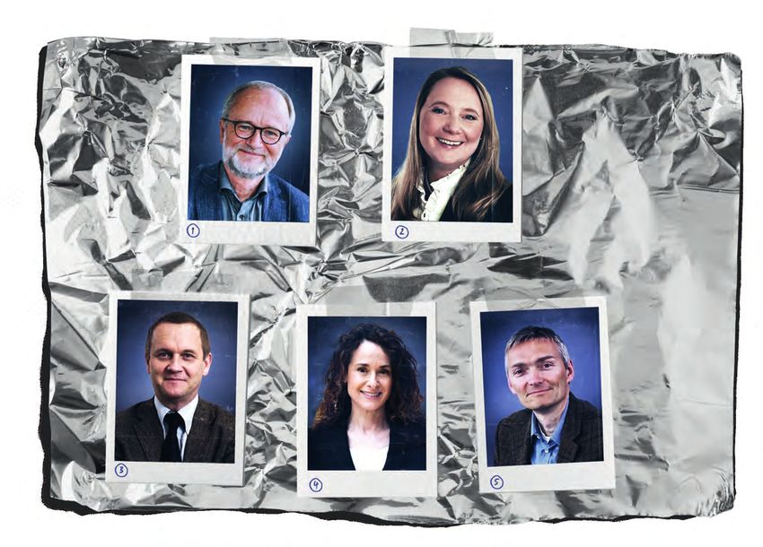
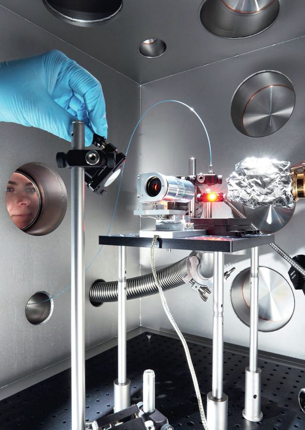
The REMI endstation at SQS
being prepared for experiments
10 FXE Highlight                                                                                                                                                                                                                                                                Highlights Annual Report 2020       11

     Tracking the ultrafast dynamics of
     photoinduced spin state switching in
     metallogrid complexes
          Maria A. Naumova, Deutsches Elektronen-Synchrotron, Germany
          Sophie E. Canton, ELI Attosecond Light Pulse Source, Hungary

     Polynuclear complexes incorporating several transition        dissipation, which can all be influenced by the nuclearity
     metal ions are intensively investigated for their pro-        of the assembly. Identifying the numerous deactivation
     spective photoconversion applications. Their rational         pathways is a prerequisite for maximizing the yield of the
     design crucially depends on charting detailed maps of         metastable state and extending its lifetime into the regime
     the energy and charge flows following photoabsorp-            of diffusive processes in order to ensure its subsequent
                                                                                                                                     Figure 1: XES measurements of Fe metallogrid complexes at the FXE instrument. (a) Molecular structures of Fe3 and Fe4 from density-functional theory
     tion. In contrast to mononuclear complexes, which             reactivity. To gain this understanding, the coupled dynam-        (DFT). (b) Location of the LS (blue) and HS (red) Fe atoms in the ground and excited states of Fe3 and Fe4 in acetonitrile. (c) Red dots: transient Kα1,2
                                                                                                                                     signals at Δt = 1 ps for Fe3 after 515 nm excitation (top); transient Kβ spectrum at Δt = 1 ps after 400 nm excitation (bottom). Black lines: reference
     involve only one metal ion, the photoinduced dynamics         ics of the spin, electronic, and nuclear degrees of freedom       traces constructed by subtracting the static line shapes for LS and HS mononuclear complexes. The transient spectra are scaled to match the references.
                                                                                                                                     (d) For Fe3: area under the transient Kα1 emission line (6400.2–6409.4 eV) as a function of the time delay between the laser and X-ray pulses; fit with a
     in polynuclear complexes remain scarcely studied to           need to be exhaustively mapped on an ultrafast time               Gaussian-broadened step function. (e) Photocycle for Fe3 and Fe4. The inset shows the vibrational coherence observed in the optical spectra.
     date due to the intricacies associated with the nume-         scale. Owing to its intrinsic elemental and spin sensitivity,
     rous competing channels. In particular, the intrinsic         femtosecond X-ray emission spectroscopy (XES) is
     rates of spin state transition cannot be accessed with        uniquely suited for monitoring the dynamical changes in          background removal and normalization. Figure 1c                                            will focus on hetero-metallogrids where the spin state
     transient absorption spectroscopies in the ultraviolet,       spin multiplicity across the full manifold of excited states.    shows the ΔXES spectra of the Kα1,2 and Kβ lines for                                       transition is coupled to intramolecular electron transfer
     visible, and infrared range. In an experiment at the FXE                                                                       Fe3 at Δt = 1 ps, along with the respective differences                                    in order to realize synergistic energy, charge, and spin
     instrument of the European XFEL, we employed femto-           Building on the pioneering work on mononuclear iron (Fe)         of reference lineshapes. The profile characteristic for the                                manipulation.
     second X-ray emission spectroscopy to directly track          complexes performed over the last decade at X-ray FEL            formation of an HS state appears quasi-instantaneously,
     the ultrafast dynamics of photoinduced spin state             sources worldwide, we applied this technique to track for        considering the time resolution that was achievable in the                                 Authors
     switching within two metallogrid complexes containing         the first time the ultrafast dynamics of photoinduced spin       experiment (~330 fs) [3]. The kinetics of the photoinduced                                 M.A. Naumova, A. Kalinko, J.W.L. Wong, M. Abdellah, H. Geng,
                                                                                                                                                                                                                               S.A. Gutierrez, J. Meng, M. Liang, P.-A. Mante, W. Lin, P. Zalden, A. Galler,
     three and four iron ions, respectively. In combination        state switching in two homometallic grid-like complexes          process can be captured by following the temporal
                                                                                                                                                                                                                               F. Lima, K. Kubicek, M. Biednov, S. Checchia, E. Domenichini, A. Britz,
     with complementary spectroscopic observations, it             containing three and four Fe(II) centres, respectively [1, 2].   evolution of the integrated areas of the ΔXES profiles.                                    V. Kabanova, M. Wulff, J. Zimara, D. Schwarzer, S. Demeshko, V. Murzin,
     was possible to unequivocally reveal the definite im-         The structures of the complexes, denoted Fe3 and Fe4,            Fitting with a Gaussian-broadened step function yields the                                 D. Gosztola, M. Jarenmark, J. Zhang, M. Bauer, L.M. Lawson Daku,
     pact of nuclearity—that is, the number of metal ions—         are displayed in Figure 1a. In the solution phase, Fe3           time constant of the spin-switching process (Figure 1d).                                   W. Gawelda, D. Khakhulin, C. Bressler, F. Meyer, K. Zheng, S.E. Canton
     on the photoinduced dynamics. More generally, our             primarily contains two Fe centres in the low-spin (LS)
     study paves the way to the systematic development of          state (blue dots) and one Fe centre in the high-spin (HS)        Combining these observations with the results from
                                                                                                                                                                                                                               References
     novel polynuclear complexes optimized for photocon-           state (red dots), denoted 2LS-1HS. Fe4 consists of 39%           complementary femtosecond absorption measurements
                                                                                                                                                                                                                               [1] M. Steinert et al.: “A trinuclear defect-grid iron(II) spin crossover complex
     version with energy-rich excited states.                      in the 2LS-2HS state and 61% in the 1LS-3HS state                in the ultraviolet, visible, and infrared ranges allows us to                                  with a large hysteresis loop that is readily silenced by solvent vapor”,
                                                                   (Figure 1b).                                                     propose the reversible photocycle displayed in Figure 1e.                                      Angew. Chem. Int. Ed. 53, 6135−6139 (2014),
     Transition metal complexes (TMCs) are primary building                                                                         Photoabsorption by an Fe centre in the LS state yields a                                       doi:10.1002/anie.201403068
     blocks ubiquitous in chemistry and materials science.         The XES measurements of the Kα1,2 and Kβ emission                vibrationally hot HS state that exhibits coherent behaviour                                [2] B. Schneider et al.: “A double-switching multistable Fe4 grid complex
                                                                                                                                                                                                                                   with stepwise spin-crossover and redox transitions,” Angew. Chem. Int.
     Linking several TMCs into organized assemblies is an          lines were performed at the FXE instrument of the                over the first 1–2 ps. Vibrational cooling to the thermalized
                                                                                                                                                                                                                                   Ed. 49, 9274–9277 (2010), doi:10.1002/anie.201001536
     efficient synthesis strategy frequently adopted to augment    European XFEL [3]. Solutions of Fe3 and Fe4 acetonitrile         HS state takes place within tens of picoseconds.                                           [3] D. Khakhulin et al.: “Ultrafast X-ray photochemistry at European XFEL:
     the performance of photoconversion applications. For          were delivered as a thin liquid sheet into the interaction       The lifetimes of the metastable states are drastically                                         Capabilities of the Femtosecond X-ray Experiments (FXE) instrument”,
     example, compact arrays of TMCs tend to exhibit wider         region where the optical laser pump pulses (400 nm and           prolonged compared to closely related mononuclear                                              Appl. Sci. 10(3), 995 (2020), doi:10.3390/app10030995
     spectral coverage with higher extinction coefficient than     515 nm) and the X-ray probe pulses (9.3 keV) overlapped          complexes (123 ns for Fe3, 210 ns for Fe4).                                                [4] M. A. Naumova et al.: “Revealing hot and long-lived metastable spin
                                                                                                                                                                                                                                   states in the photoinduced switching of solvated metallogrid complexes
     single chromophores.                                          in both space and time. The signals were collected as a
                                                                                                                                                                                                                                   with femtosecond optical and X-ray spectroscopies”, J. Phys. Chem.
                                                                   function of the variable pump–probe delay Δt using an            These experimental findings demonstrate that the                                               Lett. 11, 2133−2141 (2020), doi:10.1021/acs.jpclett.9b03883
     Following efficient photoabsorption, the Franck–Condon        energy-dispersive von Hamos spectrometer and a 2D                nuclearity has a profound impact on the photoinduced                                       [5] M. A. Naumova et al.: “Exploring the light-induced dynamics in
     excited state decays to a thermalized metastable state        CCD Greateyes detector. The transient difference XES             dynamics. They also reveal the unique potential of Fe3,                                        solvated metallogrid complexes with femtosecond pulses across
     through a complex interplay between intersystem cross-        (ΔXES) spectra were obtained by subtracting the “laser           Fe4, and other Fe metallogrids as advanced photoreac-                                          the electromagnetic spectrum”, J. Chem. Phys. 152, 214301 (2020),
                                                                                                                                                                                                                                   doi:10.1063/1.5138641
     ing, charge transfer, structural rearrangements, and energy   off” XES signals from the “laser on” XES signals after           tants [4, 5]. Based on the present work, future studies
12 SPB/SFX Highlight 1 Highlights Annual Report 2020 13
Probing nanoscale dynamics
resulting in a stepwise increase in temperature and thus a
speeding up of the sample dynamics.
with MHz repetition rates
In order to quantify this process, we performed a
pulse-resolved XPCS analysis. For this purpose,
diffusion coefficients were extracted after each pulse,
giving access to a pulse-resolved effective temperature
(Figure 3). At a fluence of 56.8 mJ/mm2—while the sample
Felix Lehmkühler, was still in the liquid phase, as evidenced by the diffusive
Deutsches Elektronen-Synchrotron DESY, Germany
dynamics of the nanoparticles—the effective temperature
crossed the boiling point of water and rose to superheat-
ed states at about 445 K at the end of the pulse train.
This heating exceeded the heating of pure water (dashed
Dynamics and kinetics in soft-matter physics, 3.6 µm x 4.4 µm and a repetition rate of 1.128 MHz, corre- lines). It could be modelled taking into account the explicit
Figure 1: Sketch of the experiment at the SPB/SFX instrument. The X-ray pulse trains
biology, and nanoscience frequently occur on (sub-) sponding to a time interval of 886 ns between the pulses, time resolution of heat relaxation and convective heat scattered off the sample filled in quartz capillaries. The speckle patterns were recorded
in small-angle scattering geometry 5.5 m downstream of the sample by the AGIPD detector.
microsecond time scales, which are difficult to probe hit a fresh spot of the colloidal sample every 100 ms. The transfer between the nanoparticles and the water (solid The pulse trains contained 120 pulses each and were separated by 0.1 s. Within each pulse
experimentally. The European XFEL enables such coherent diffraction patterns—called speckle patterns— lines). However, at short times and large Teff, both models train, consecutive pulses were separated by 886 ns (1.128 MHz repetition rate).
experiments down to atomic length scales for the were measured using the SPB/SFX AGIPD detector. fail, suggesting that the nanoparticles move faster than
first time, thanks to its MHz repetition rate. We used what is expected from the Stokes–Einstein relation. This
microsecond X-ray photon correlation spectroscopy As a coherent X-ray scattering technique, XPCS relies on motivates further studies on heat transfer at the nano-
(XPCS) at the SPB/SFX instrument to observe the the coherence properties of the X-ray pulses. The sample scale to understand the emergence of non-equilibrium q (nm-1)
Correlation function (g2-1)βc
dynamics of nanoparticles dispersed in water. In the dynamics are obtained from intensity–intensity correlation dynamics in liquids and soft matter probed at MHz X-ray
experiment, series of speckle patterns were measured functions from successive X-ray pulses, which are FEL sources.
with sub-microsecond time resolution. We found modelled, among others, using the Stokes–Einstein
exceptional stability over the pulse train, suggesting relation for diffusion of spherical particles through a liquid. To conclude, our study demonstrated the application of
very weak—if any—shot-to-shot fluctuations of beam The pointing stability during the pulse train is of utmost XPCS at the European XFEL. Currently, XPCS is further
size, pointing, and coherence. By fine-tuning the importance because these correlations are calculated used in various studies at the MID instrument and will be
fluence of the European XFEL pulses, we were able from different X-ray pulses. Pulse-to-pulse SASE fluctua- extended to femtosecond and picosecond time scales
to observe different degrees of beam-induced heating. tions have been observed in previous XPCS experiments using the MID split-and-delay line.
At fluences above 50 mJ/mm2, superheated-water at other FEL sources [3, 4, 5]. The degree of such
states above 170°C were reached, which persisted fluctuations can be quantified by comparing the speckle Lag time τ (s)
at least for 100 µs. contrast from single-pulse speckle patterns with the Figure 2: Normalized correlation function g2 measured in real
time from a single train of 120 pulses at a fluence of 27.7 mJ/mm2.
contrast obtained from the intensity–intensity correlation Solid lines are fits to the data.
In the last decade, hard X-ray FELs have opened up new function from static samples. We found that both values Authors
t µs
research directions. These facilities hold special promise match, which shows that the European XFEL provides F. Lehmkühler, F. Dallari, A. Jain, M. Sikorski, J. Möller, L. Frenzel, I. Lokteva,
G. Mills, M. Walther, H. Sinn, F. Schulz, M. Dartsch, V. Markmann, R. Bean,
for the investigation of equilibrium and non-equilibrium unprecedented beam stability within a pulse train.
Y. Kim, P. Vagovic, A. Madsen, A.P. Mancuso, G. Grübel:
processes with XPCS [1]. Whereas at storage ring light “Emergence of anomalous dynamics in soft matter probed at the
sources, XPCS typically enables studies of dynamics In our XPCS experiment, we used silica nanoparticles with European XFEL”, Proc. Natl. Acad. Sci. USA 117, 24110–24116 (2020),
between milliseconds and hours in real time, the time a radius of 69 nm that were dispersed in water as model doi:10.1073/pnas.2003337117
T
scale accessible at FELs is defined by the pulse repetition samples for nanoscale materials. Different X-ray fluences
rate. The unique pulse scheme at the European XFEL between 1.3 and 56.8 mJ/mm2 per single pulse were
References
allows studies of (sub-)microsecond dynamics by applied. Some results from a single pulse train are shown
[1] G. Grübel, G.B. Stephenson, C. Gutt, H. Sinn, T. Tschentscher: “XPCS at
exploiting the MHz repetition rate within the pulse trains in Figure 2. The measured diffusion coefficients resemble the European X-ray free electron laser facility”, Nucl. Instrum. Meth. B
in such XPCS experiments. These temporal regimes the results from theory when low fluences are used. 262, 357–367 (2007), doi:10.1016/j.nimb.2007.05.015
correspond to the natural time scale of diffusion process- Increasing the fluence leads to a speeding up of the [2] A.P. Mancuso et al.: “The Single Particles, Clusters and Biomolecules
es of nanoparticles and biological macromolecules in their particle dynamics. and Serial Femtosecond Crystallography instrument of the European
XFEL: initial installation”, J. Synchrotron Radiat. 26, 660–676 (2019),
native aqueous environment. We explored this time scale
doi:10.1107/S1600577519003308
in the first experiment in which the MHz repetition rate of Due to the exceptionally high intensity of the European XFEL [3] J. Carnis et al.: “Demonstration of feasibility of X-ray free electron laser
the European XFEL was a crucial requirement. The study pulses, the exposed sample volume heats up instantane- studies of dynamics of nanoparticles in entangled polymer melts”, n
revealed the dynamics of colloidal nanoparticles as a ously. Subsequently, its temperature relaxes back. While Sci. Rep. 4, 6017 (2014), doi:10.1038/srep06017
Figure 3: Effective temperature Teff as a function of the pulse number
model system for nanoscale materials. this scenario has been observed at the LCLS and SACLA [4] F. Lehmkühler et al.: “Sequential single shot X-ray photon correlation in the pulse train (np) for different fluences. The black lines represent the
spectroscopy at the SACLA free electron laser”, Sci. Rep. 5, 17193 time-dependent heating model, the dashed lines the heating of water as
X-ray lasers in the USA and Japan, respectively [3, 4, 5], solvent only. The boiling temperature of water is given by the horizontal
(2015), doi:10.1038/srep17193 dashed-dotted line.
The scheme of the experiment carried out at the SPB/SFX it is not fully valid here because the temperature relaxation [5] F. Lehmkühler et al.: “Dynamics of soft nanoparticle suspensions at hard
instrument [2] is shown in Figure 1. Pulse trains of 120 time is longer than the pulse repetition rate [1]. Conse- X-ray FEL sources below the radiation damage threshold”, IUCrJ 5,
X-ray pulses at a photon energy of 9.3 keV, with a size of quently, the next pulse hits a non-equilibrated sample, 801–807 (2018), doi:10.1107/S2052252518013696
14 SPB/SFX Highlight 2 Highlights Annual Report 2020 15
Opening new vistas on
In 2019, our team of researchers from Germany, Sweden, Authors
Singapore, the USA, and Australia came together at the K. Ayyer, P.L. Xavier, J. Bielecki, Z. Shen, B.J. Daurer, A.K. Samanta,
S. Awel, R. Bean, A. Barty, M. Bergemann, T. Ekeberg, A.D. Estillore,
European XFEL to overcome the biggest hurdles in
H. Fangohr, K. Giewekemeyer, N.S. Hunter, M. Karnevskiy, R.A. Kirian,
nanoparticle structure
achieving the goal of high-resolution 3D structure H. Kirkwood, Y. Kim, J. Koliyadu, H. Lange, R. Letrun, J. Lübke, T. Michelat,
determination. These two challenges were the collection A.J. Morgan, N. Roth, T. Sato, M. Sikorski, F. Schulz, J.C.H. Spence,
of a sufficiently large number of high-quality diffraction P. Vagovic, T. Wollweber, L. Worbs, O. Yefanov, Y. Zhuang, F.R.N.C. Maia,
patterns and the robust decomposition of structural D.A. Horke, J. Küpper, N. Duane Loh, A.P. Mancuso, H.N. Chapman:
“3D diffractive imaging of nanoparticle ensembles using an X-ray laser”,
variability among the particles. The samples chosen were
Optica 8, 15–23 (2021), doi:10.1364/OPTICA.410851
cubic and octahedral gold nanoparticles, which are ideal
Kartik Ayyer, Max Planck Institute for the test samples since they scatter relatively strongly but also
Structure and Dynamics of Matter, Germany have natural structural variability, due to the way they are
synthesized. Fully capitalizing on the high repetition rate References
of the European XFEL, we collected 10 million diffraction [1] H.N. Chapman, K.A. Nugent, “Coherent lensless X-ray imaging”,
Nature Photon. 4, 833–839 (2010),
patterns over a 60-hour beamtime experiment at the
doi:10.1038/nphoton.2010.240
SPB/SFX instrument and reconstructed four structures [2] H.N. Chapman , A. Barty, M.J. Bogan, S. Boutet, M. Frank,
X-ray single-particle imaging (SPI) using X-ray FELs signal is generated before the particles get destroyed with sub-3 nm resolution. In the process, we also had to S.P. Hau-Riege et al., “Femtosecond diffractive imaging with a
holds great promise as a method to determine the by the radiation in a process termed “diffraction-before- develop algorithms to separate patterns from particles soft-X-ray free-electron laser”, Nature Phys. 2, 839–843 (2006),
structure of individual biomolecules at physiological destruction” [2]. But a single object can still be exposed with different structures; otherwise, the reconstruction doi:10.1038/nphys461
temperatures without crystallization. In order to realize only once, so in order to obtain 3D structures, data from a quality was noticeably worse.
its potential, one needs to collect enough high-quality large number of particles in random orientations must be
diffraction patterns and develop analytical tools to combined computationally. Not only do these orientations The results of the experiment are exciting for several
classify their structural variability. In this work, we have to be determined from the data, but any variations in reasons. First, we were able to demonstrate how the
report on the collection of 10 million patterns on gold the structure across the different particles must be European XFEL is a game changer in terms of being able
nanoparticle test samples in a single beamtime at the decomposed. When combined with the fact that the to collect a truly enormous amount of useful data, which
SPB/SFX instrument of the European XFEL, setting a scattering signal from sub-50 nm objects is quite weak is crucial for the eventual success of the SPI technique.
blueprint for a new class of SPI experiments. Since and sensitive to background, the technique has been Of course, this also requires a reliable sample delivery
nanoparticle synthesis is inherently heterogeneous, challenging to realize experimentally. system, and the steady developments by various groups
the data must be structurally classified to obtain useful have brought us to the point where we were collecting
oct30
3D structures. We show that the standard methods data for up to 10 hours in every 12-hour shift. Using the
used previously work relatively poorly and we develop template set by this work, we are now significantly closer
new algorithms to perform the 3D structural sorting to the long-term goal of visualizing the dynamics of
that is crucial to imaging such ensembles. In the biomolecules with atomic resolution.
process, we produce the highest-resolution structure
oct40
reconstructions using the SPI technique. The experi- The experiment also highlights the power of the serial
ment also paves the way towards the high-throughput imaging method for the 3D structure characterization of
3D characterization of nanoparticle ensembles. nanoparticle ensembles. Since each particle is exposed
individually, it enables us to identify and image rare events
cub42
Coherent diffractive imaging (CDI) is a technique in which (Figure 2), which would not be possible otherwise, as
objects, when exposed to coherent light, weakly perturb they would be washed out by the average in an ensemble
the light waves, leading to the creation of a diffraction imaging technique such as powder diffraction or be nearly
pattern on a detector far from the sample [1]. Just as in a impossible to find by techniques such as transmission or
textbook Young’s double-slit experiment, where the scanning electron microscopy. And, once you combine
cub17
pattern of fringes can be related to the distance between this with the fact that the short pulses also enable you to
the slits, the diffraction pattern can be used to computa- image ultrafast processes, the possibilities are endless.
tionally derive the structure of the objects in the beam.
Figure 2: 2D classification. Representative examples of reconstructed 2D
This is also termed “lensless imaging” since one obtains models shown on a logarithmic scale, with each row representing a different
sample. The numbers indicate how many patterns had that model as the most
images of the objects without the need for imaging likely one. The first two columns show models selected for further processing.
The third column shows diffraction from rounded/spherical particles, except in
lenses, which are used in conventional microscopy. the cub17 case, where there were no spherical particles and the model shows
Figure 1: Experiment setup at the SPB/SFX instrument. The European XFEL
X-ray SPI is a CDI technique in which coherent X-ray FEL pulses were focused by a series of Kirkpatrick–Baez mirrors into a 3 x 3 µm2
diffraction from a dimer instead. The fourth column shows some of the
low-contrast models generated by averaging patterns from a diverse set of
spot and scattered off particles in the aerosol stream to produce diffraction
beams are used to sequentially collect diffraction patterns patterns on the AGIPD detector. The lower inset shows the timing structure
particles. The resolution at the edge of the circle is 3.3 nm.
from nanoscale objects, such as nanoparticles or biomol- of the European XFEL pulses at the instrument, while the top inset shows
representative scanning electron microscope images of the cub42 and oct30
ecules (Figure 1). The X-ray beams from FELs like the samples; scale bars are 100 nm. The low-resolution part of the detector used
for the structural sorting is highlighted in green.
European XFEL are bright and short enough that the
atoms are effectively frozen during the exposure and the16 MID Highlight Highlights Annual Report 2020 17
Glimpsing the secrets
the continuous, self-replenishing nature of the liquid
jet allows us, in fact, to take full advantage of the MHz
repetition rate of the European XFEL X-ray pulses.
of crystallization Figure 2a shows averaged scattering patterns clearly
revealing the evolution from the liquid phase (bottom
panel) to the crystalline phase (top panel), evidencing
the characteristic (111) and (200) face-centred cubic (fcc)
diffraction rings. Such averaged diffraction patterns
resemble those that can be obtained at synchrotron
Robert E. Grisenti, GSI Helmholtzzentrum facilities, where long acquisition times make averaging
over large sample volumes inevitable. Figure 2b shows
für Schwerionenforschung, Germany
difference images obtained from the same data as in
Johannes Möller, European XFEL, Germany Figure 2a by exploiting the pulse-resolved nature of the
experiment, allowing for subtraction of the scattering Figure 1: Microscopic images of a krypton jet in the MID sample
chamber. (a) Shadow image of a solidified krypton jet ejected
contribution of the liquid fraction. These patterns clearly at a speed of 100 m/s from a 3.5 μm diameter capillary orifice.
(b) Image of the jet after exposure to the focused X-ray beam.
The process of crystallization by which a “super- that stresses this impasse is supercooled water, for corroborate the presence of a solidified fraction (bottom X-ray–induced fluorescence can be observed at the interaction
spot of the jet and the X-ray beam. The trigger of the laser-based
cooled” liquid—that is, a liquid at some temperature which experimental and numerical crystal nucleation panel) that was hidden in Figure 2a and thus demonstrate background illumination for the optical camera was set so as
to obtain an image of the jet shortly after the fifth X-ray pulse
below its melting point—transforms into a solid is one rates can diverge by up to 20 orders of magnitude [2]. the feasibility of directly capturing the early stages of of a train hitting the jet, resulting in four gaps in the jet below
of the most fundamental phase transitions occurring A further, highly debated aspect of crystal formation is crystallization with the intense X-ray pulses of the the interaction point where argon was blasted away by previous
exposures.
in nature. It substantially determines many physical the possibility, suggested by numerical simulations, European XFEL.
phenomena, from materials science to atmospheric of a crystal nucleation process driven by precursor forms
physics. Microscopic supercooled water droplets at with some local symmetry formed within the supercooled At each position along the jet, which, as mentioned earlier,
very low temperatures, for example, are naturally liquid, but in fact this scenario is not yet supported by corresponds to a specific temperature of the supercooled
present in upper layers of the Earth’s atmosphere, experimental evidence. liquid, we have recorded over 106 single-shot images
such as in aerosol-poor arctic stratiform clouds. with the MID AGIPD detector, each of them from a new
Understanding how and at which rates such droplets Investigations of crystallization in atomic and molecular sample delivered by the jet. The very high repetition
transform into ice is important because small differ- liquids are extremely challenging because of the inherent- rate of the X-ray pulses at the European XFEL has thus
ences in the ratio of water to ice have a huge impact ly stochastic nature of HCN, which greatly hinders access allowed us to obtain a valuable data set orders of magni-
on the radiative effects of clouds and thus on the to its details, as no a priori knowledge of the exact spatial tude larger than in any previous study of HCN, enabling a
development of reliable climate models. Experimen- and temporal coordinates of the spontaneous nucleation genuine statistical study of crystallization.
tally accessing such details in supercooled liquids is, event in a macroscopic liquid sample is available.
however, very difficult because of the spontaneous To address this challenge, we combine X-ray pulses This is important not only to obtain a reliable estimation
nature and the very short time scale of the crystalliza- from the European XFEL with microscopic liquid jets in of the crystal nucleation rate but also to address funda-
tion process. To address these challenges, we employ vacuum to investigate the early stages of crystallization. mental aspects related to the structural path during the
microscopic liquid jets in vacuum in combination with The vacuum-exposed jet rapidly cools below melting by crystallization process. For example, bulk rare-gas liquids Figure 2: Scattering patterns from a krypton jet. (a) Averaged scattering
signal on one of the modules of the MID AGIPD detector, with the distance
X-ray scattering at the MID instrument of the European surface evaporation until it undergoes a first-order phase crystallize into the fcc crystal structure, but numerical from the capillary orifice increasing from the bottom to the top. (b) Data set
XFEL. The unique features of this approach allow us to transition driven by the onset of HCN [3]. The whole studies also indicate the formation of a metastable obtained from that shown in (a) by displaying the maximum intensities after
subtracting the scattering contribution from the liquid.
investigate the early stages of crystallization. Finally, evolution of the crystallization process, from critical hexagonal close-packed (hcp) solid phase, suggesting
we have the opportunity to shed light on so-far unex- seed formation to crystal growth occurring along the jet that crystallization may proceed with the nucleation and
plored aspects of the liquid-to-solid phase transition. propagation direction, is effectively mapped onto the time growth of the least stable phase. However, the frequency
Authors
axis through the jet velocity. Information on the structure of the diverse transformation pathways is very difficult to
J. Möller, U. Bösenberg, F. Dallari, D.E. Galli, L. Gelisio, C. Goy, G. Grübel,
Crystallization is generally viewed as a two-step process and size of the rapidly growing crystals is then captured determine, further illustrating the complexity of the phase A. Kalinin, C. Kim, R. Kurta, D. Lapkin, F. Lehmkühler, A. Madsen, F. Mambretti,
in which thermal fluctuations in the metastable liquid in the single-shot diffraction patterns produced by the transition even in such simple liquids. A. Schottelius, R. Shayduk, F. Trinter, I. Vartaniants, R.E. Grisenti
phase trigger the spontaneous formation of a small, ultrashort, intense X-ray pulses, which are focused down
localized seed of the new ordered phase, which subse- to a 300 x 300 nm2 spot size, scattering off the jet. The data analysis is presently ongoing, but once accu-
References
quently grows to macroscopic dimensions [1]. However, rately determined, the resulting crystal nucleation rates
[1] K.A. Jackson: Kinetic Processes: Crystal Growth, Diffusion, and Phase
the details of this process, and particularly of the initial In our experiments at the MID instrument [4], we have and probability distributions for the transformation
Transitions in Materials (Wiley-VCH Verlag, Weinheim, 2004)
nucleus formation commonly known as homogeneous probed liquid jets of the atomic elements argon and pathways will offer a unique benchmark for theory and [2] A. Haji-Akbari, P.G. Debenedetti: “Direct calculation of ice homogeneous
crystal nucleation (HCN), are still very poorly understood. krypton, which are particularly attractive model systems computer simulations in which the particle interaction in nucleation rate for a molecular model of water”, PNAS 112, 10582 (2015),
For example, although the classical crystal nucleation due to their simplicity and because crystallization in these the simplest atomic liquids of our study can be reliably doi:10.1073/pnas.1509267112
theory provides a qualitatively valid description of HCN, liquids is comparable to that observed in pure metals. described by a Lennard–Jones potential. Ultimately, [3] R.E. Grisenti et al.: “Evaporating laminar microjets for studies of
rapidly evolving structural transformations in supercooled liquids”,
theoretical crystal nucleation rates often depart by orders A shadow image of a solidified krypton jet generated in our results will provide long-awaited experimental input
Adv. Phys. X 3, 1418183 (2018), doi:10.1080/23746149.2017.1418183
of magnitude from those observed experimentally, and the MID sample chamber is presented in Figure 1a. to help guide any future refinements of theories of crystal [4] A
. Madsen et al.: “Materials Imaging and Dynamics (MID) instrument at
the latter are also found to differ substantially from those Figure 1b shows the jet explosion during exposure to the nucleation and growth in supercooled liquids, thereby the European X-ray Free-Electron Laser Facility”, J. Synchrotron Radiat.
predicted by computer simulations. A prominent example focused, non-attenuated X-ray beam. We emphasize that advancing our understanding of crystallization. 28 (2) (2021), doi:10.1107/S160057752100130218 HED Highlight Highlights Annual Report 2020 19
Developing an ultrafast
thermometer for matter
at extremes Adrien Descamps and Emma E. McBride,
SLAC National Accelerator Laboratory, USA T
T
ΔE (meV)
Warm dense matter is an exotic state on Earth, yet The generated states of pressure and density are there-
ubiquitous throughout the universe, found in the fore, by nature, transient, with a large density of free Figure 1: Schematic of the experiment setup used to
perform millielectronvolt inelastic X-ray scattering at the
interiors of stars and giant planets such as Jupiter and electrons making them challenging to probe using optical HED instrument of the European XFEL. Incident X-ray
pulses at 7492 eV are first monochromatized using a
Saturn, or even at the centre of the Earth. Typically, techniques. The solution comes with the emergence of succession of two double-bounce silicon monochroma-
such states are generated in the laboratory using hard X-ray FEL sources, such as the European XFEL. tors. The subsequent X-ray pulses are then scattered
from a diamond sample and energy-dispersed by three
high-intensity pulsed lasers to drive either compres- These light sources provide extremely bright, time- diced silicon analysers, before being recorded on the
instrument’s ePIX100 detector. The inset shows example
sion waves or rapid heating on femtosecond to resolved X-rays with pulse lengths of just few tens of spectra for the diamond sample at room temperature
(blue) and heated to 500 K (red). The intensity ratio
nanosecond time scales. These transient states of femtoseconds, which can penetrate and probe extreme between the positive and negative energy transfer
is used to determine the temperature through the
extreme pressure and temperature are challenging to states. As such, the measurement of the structure and application of the principle of detailed balance.
probe using traditional optical techniques. Hard X-ray density of compressed matter has become almost
FELs, in contrast, provide the ultrabright, ultrashort routine, providing a wealth of information on the behav-
X-rays pulses necessary to allow these short-lived iour of these exotic states. However, despite being a vital
states to be captured and probed. While inferring thermodynamic parameter, temperature remains challeng-
density from X-ray diffraction data has become a ing to measure experimentally.
well-established technique, the measurement of
temperature remains a major challenge. Here, we In such experiments, temperature is often calculated three diced and spherically curved single-crystal silicon Authors
demonstrate a novel first-principles technique at the using a hydrodynamic model, which needs to be bench- analysers to obtain the phonon spectrum. A. Descamps, B.K. Ofori-Okai, K. Appel, V. Cerantola, A. Comley,
J.H. Eggert, L.B. Fletcher, D.O. Gericke, S. Göde, O. Humphries,
HED instrument of the European XFEL for measuring marked, or is sometimes inferred through the thermal
O. Karnbach, A. Lazicki, R. Loetzsch, D. McGonegle, C.A.J. Palmer,
temperature using high-resolution inelastic X-ray self-emission of a sufficiently hot sample. This latter Example phonon spectra from single-crystal diamond, C. Plueckthun, T.R. Preston, R. Redmer, D.G. Senesky, C. Strohm,
scattering. This method, combined with the high- technique assumes a priori knowledge of the material both at room temperature and resistively heated to 500 K, I. Uschmann, T.G. White, L. Wollenweber, G. Monaco, J.S. Wark,
repetition-rate lasers available at the HED instrument, properties, and one accesses only the surface tempera- are shown in the inset of Figure 1. We measured only the J.B. Hastings, U. Zastrau, G. Gregori, S.H. Glenzer, E.E. McBride
will allow new studies to greatly improve our under- ture rather than the bulk temperature. In our experiment inelastic Brillouin peaks corresponding to the longitudinal
standing of warm dense matter. at the HED instrument, we demonstrated the measure- phonon mode along the (100) crystallographic direction,
References
ment of bulk temperature in both room temperature and as the sample is a single crystal. The positions of the
[1] D. Kraus et al.: “Formation of diamonds in laser-compressed hydrocar-
Throughout the universe, matter exists at extremes of resistively heated single-crystal diamond using ultrafast peaks are related to the sound speed of the material, bons at planetary interior conditions”, Nat. Astron. 1, 606–611 (2017),
pressure and temperature, from the cold vacuum of X-ray pulses. The experiment resolved the diamond and the asymmetry of the Stokes and anti-Stokes peaks doi:10.1038/s41550-017-0219-9
interstellar space to the hot, dense centres of neutron phonon modes, whose intensities yield the temperature arises due to Bose–Einstein statistics. The ratio of these [2] B.I. Cho et al.: “Electronic structure of warm dense copper studied by
stars. Particularly intriguing is a state known as warm through the application of the principle of detailed peaks allows one to directly obtain the sample tempera- ultrafast X-ray absorption spectroscopy”, Phys. Rev. Lett. 106, 167601
(2011), doi:10.1103/PhysRevLett.106.167601
dense matter, found at the centre of stars and planets balance. The applicability of this principle was then ture. Here, we measured a temperature of 294 ± 25 K for
[3] L. Wollenweber et al.: “High-resolution inelastic X-ray scattering at the
as well as in inertial confinement fusion processes. While confirmed using the temperature reading from a thermo- the diamond at room temperature (blue) and 496 ± 26 K High Energy Density scientific instrument at the European X-Ray
understanding it is vital for comprehending pathways to couple attached to the sample. when heated to 500 K (red) [4]. Free-Electron Laser”, Rev. Sci. Instrum. 92, 013101 (2021),
fusion energy or creating accurate models of planetary doi:10.1063/5.0022886
and stellar interiors and their evolution, warm dense The experiment setup for high-resolution inelastic Our work demonstrates the feasibility of using inelastic [4] A. Descamps et al.: “An approach for the measurement of the bulk
temperature of single crystal diamond using an X-ray free electron
matter lies in a pressure–temperature region that is not scattering at the HED instrument is shown in Figure 1 X-ray scattering to measure the bulk temperature of a
laser”, Sci. Rep. 10, 14564 (2020), doi:10.1038/s41598-020-71350-x
well described by condensed matter physics or by and described in detail in Ref. [3]. While the self-amplified sample at the HED instrument of the European XFEL. [5] P. Mason et al.: “Development of a 100 J, 10 Hz laser for compression
plasma physics. Therefore, a direct experimental charac- spontaneous emission mode of the European XFEL has By combining this technique with the high-repetition-rate, experiments at the High Energy Density instrument at the European
terization of these states is essential to establish a robust a bandwidth of several tens of electronvolts, the X-ray high-intensity nanosecond and femtosecond lasers at XFEL”, High Power Laser Sci. Eng. 6, E65 (2018),
theoretical description. beam was further monochromatized to ~32 meV, suffi- HED [5], we will soon be able to extend the technique to doi:10.1017/hpl.2018.56
cient to resolve the characteristic energy transfer for directly measure the bulk temperature and the material
Extreme states are often created in the laboratory through phonon modes, which is typically in the range of tens sound speed of transient matter states at extreme
shock compression using nanosecond laser drivers or to hundreds of millielectronvolts. The narrow-band X-ray pressures and temperatures.
through ultrafast heating using femtosecond lasers [1, 2]. beam was then scattered from the diamond sample onto20 SCS Highlight 1 Highlights Annual Report 2020 21
dispersing with wave vector q as well as new modes that Authors
Visualizing spin–lattice
do not disperse with q. For example, in Figure 1c, the first D. Turenne, I. Vaskivkyi, M. Schneider, N. Zhou Hagström, A. Yaroslavtsev,
X. Wang, G. Mercurio, L. Le Guyarder, L. Mercadier, J. Brock, D. Mukkat-
oscillation corresponding to the acoustic phonon mode
tukavil, N. Agarwal, D. Lomidze, M. Turcato, A. Samartsev, M. Teichmann,
appears at a high q ≈ 0.9 nm-1 after a time delay of 1 ps N. Gerasimova, J. Schlappa, D. Potorochin, B. Van Kuiken, R. Gort,
coupling in iron–platinum
and drifts to a lower q ≈ 0.25 nm-1 after a time delay of R. Carley, V. Unni, Y. Takashi, S. Molodtsov, A. Scherz, E. Jal, E. Fullerton,
4 ps. Another feature shows up at 4 ps in a broad q range, S. Bonetti, S. Eisebitt, H.A. Dürr
being the most distinct at higher q ≈ 0.7–0.9 nm-1 and
nanoparticles
almost merging with the acoustic phonon mode in the
References
middle q range near 0.5–0.6 nm-1. Apparently, the non-
[1] J. Li et al.: “Observation of magnon polarons in a uniaxial
dispersive features are caused by the coupling between
Hermann Dürr,
antiferromagnetic insulator”, Phys. Rev. Lett. 125, 217201 (2020),
the lattice motion and the ferromagnetic resonance doi:10.1103/PhysRevLett.125.217201
Uppsala University, Sweden precession of the magnetization. A more detailed analysis [2] J. Holanda et al.: “Detecting the phonon spin in magnon–phonon
(not shown) results in a precession frequency reasonably conversion experiments”, Nat. Phys. 14, 500 (2018),
doi:10.1038/s41567-018-0079-y
close to the one observed previously [4].
[3] M. Weiler et al.: “Spin pumping with coherent elastic waves”, Phys. Rev.
Lett. 108, 176601 (2012), doi:10.1103/PhysRevLett.108.176601
Studying the spin dynamics initiated in magnetic In this experiment, we have studied the magneto-acoustic In conclusion, we have directly visualized, for the first [4] J. Becker et al.: “Laser induced spin precession in highly anisotropic
materials by a sub-picosecond laser pulse is an coupling of ferromagnetic resonance spin precession time, the magneto-acoustic coupling in ferromagnetic granular L10 FePt”, Appl. Phys. Lett. 104, 152412 (2014),
emerging and rapidly developing research field in modes and acoustic phonons in FePt nanoparticles with FePt nanoparticles. This experiment has become possible doi:10.1063/1.4871869
[5] A.H. Reid et al.: “Beyond a phenomenological description of
fundamental magnetism, which promises novel an average diameter of around 16 nm. FePt nanoparticles thanks to the improved performance of X-ray FELs as
magnetostriction”, Nat. Commun. 9, 388 (2018),
applications in information technology. The goal of are a promising material for future high-density magnetic powerful research tools, which now cover the tender doi:10.1038/s41467-017-02730-7
these studies is to understand and ultimately control data storage media, thanks to their extremely high X-ray range. The impact of our research is a better
the energy and angular-momentum transfer processes magnetic anisotropy, which makes them good candidates understanding of the basic physical processes in magnet-
occurring in the laser-excited non-equilibrium state. for pushing back the superparamagnetic limit—a funda- ic materials on the nanometre length scale and at THz
Since the advent of femtosecond optical lasers, mental restriction of the data storage density of hard disk frequencies, which is highly relevant for state-of-the-art
research has focused on exploring the electronic drives due to the minimum size of the particles that can spintronic applications.
and magnetic structure of the excited state. The be used. Figure 1: Illustration of the experiment. (a) The process under
investigation: Schematic illustration of how femtosecond
availability of FEL radiation in the tender and hard laser excitation generates spin precession (blue arrows) and
lattice expansion propagating via strain waves (red rings)
X-ray range has recently opened up the possibility Figure 1 describes the experiment. A laser pulse induces into the interior of the nanoparticle. The transmission electron
to study the coupling of spin excitations to the lattice. the propagation of a strain wave through the material from micrograph at the bottom shows the FePt nanoparticles
(brown) suspended in a carbon matrix (black). (b) Schematics
In this experiment at the SCS instrument of the the grain boundaries towards the centre, which results of the experiment setup: A femtosecond optical laser pulse
(50 mJ/cm2 fluence) arrives at the sample, followed after a
European XFEL, we used iron–platinum (FePt) in the expansion of the FePt lattice and grain as a whole certain time delay by a tender X-ray FEL pulse (2500 eV),
which scatters off the nanoparticles and is measured by the
nanoparticles—promising candidates for future (Figure 1a). At the same time, a ~0.2 THz ferromagnetic DSSC detector of the SCS instrument. (c) Experimental data:
Time traces for different q vectors showing the normalized
high-density magnetic data storage media—to demon- resonance precession of the FePt magnetization is scattering yield from the nanoparticles. At every q, a number
strate the coupling of a ferromagnetic resonance excited through ultrafast demagnetization with femto- of distinct frequencies are observed, which correspond to the
coupled phonon and spin oscillations.
spin precession mode to longitudinal acoustic pho- second optical laser pulses [4].
nons on a length scale corresponding to the nano-
particle size (~16 nm). We isolated the phonon response At the SCS instrument, we detected the laser-induced a b
using X-ray scattering, which allowed us to probe the coherent lattice expansion of the FePt nanoparticles [5]
ultrafast lattice expansion of the FePt nanoparticles by tender X-ray scattering (Figure 1b), using the instru-
through coherent phonon wave packets, and we ment’s DSSC detector to record the scattering patterns
detected new modes caused by the non-linear at a photon energy of 2500 eV. X-ray diffraction from the
coupling of acoustic phonons to large-angle spin FePt grains in the sample produced a broad scattering c
precessions. ring on the detector with a radius defined by the grain size
distribution. We varied the time delay between the laser
The magneto-acoustic coupling of phonon and magnon pump and X-ray FEL probe pulses to explore the temporal
modes offers novel functionalities for spintronic applica- behaviour of the system with a resolution down to a few
tions in information technology. Magnon polarons can tens of femtoseconds.
form by magneto-elastic coupling [1], circularly polarized
phonons can form and transport angular momentum [2], The change in intensity of the scattering ring after the
and phonons can be used to generate spin currents in arrival of the laser pump pulse depends on the wave
metallic contacts [3]. However, many of these phenomena vector and shows the dynamics on the sub-picosecond
have, so far, been demonstrated only at GHz frequencies time scale (Figure 1c). Such a wave-vector-dependent
and micrometre dimensions. X-ray FELs enable us to scattering yield is a direct probe of coherent acoustic
explore the potential of moving towards THz frequencies phonon propagation from the perimeter to the interior of
and nanometre dimensions, which are far more attractive the nanoparticles. Surprisingly, in the time domain, we find
for applications. phonon spectral features of the acoustic phonon modeYou can also read