DIAGNOSTIC VALUE OF MORPHOLOGICAL CHANGES IN GASTROESOPHAGEAL REFLUX DISEASE IN BIOPSY MATERIAL OF THE DISTAL ESOPHAGUS IN ADOLESCENTS SICKLY WITH ...
←
→
Page content transcription
If your browser does not render page correctly, please read the page content below
Original Research Article:                                                             (2019), «EUREKA: Health Sciences»
full paper                                                                                         Number 1

                       [17] Schröder, F. H., Hugosson, J., Roobol, M. J., Tammela, T. L. J., Ciatto, S., Nelen, V. et. al. (2009).
               Screening and Prostate-Cancer Mortality in a Randomized European Study. New England Journal of Medi-
               cine, 360 (13), 1320–1328. doi: http://doi.org/10.1056/nejmoa0810084
                       [18] Zhane, A. K. (2000). Ways to improve inpatient care during the period of health care reform.
               Saint Petersburg, 23.
                       [19] Pavlov, Yu. V. (2002). Medical and organizational bases of improvement of work of hospitals of
               intensive treatment in the conditions of reforming of inpatient care. Saint Petersburg, 40.

                   DIAGNOSTIC VALUE OF MORPHOLOGICAL CHANGES
                      IN GASTROESOPHAGEAL REFLUX DISEASE IN
                    BIOPSY MATERIAL OF THE DISTAL ESOPHAGUS IN
                   ADOLESCENTS SICKLY WITH ACUTE RESPIRATORY
                                     DISEASES

                                                           Olena Zhuravel
                                                    Department of Pediatrics No. 1
                                             А. А. Bogomolets National Medical University
                                             T. Shevchenko blvd., 13, Kyiv, Ukraine, 01601

                                                         Tetyana Pochinok
                                                    Department of Pediatrics No. 1
                                             А. А. Bogomolets National Medical University
                                             T. Shevchenko blvd., 13, Kyiv, Ukraine, 01601

                                                        Tamara Zadorozhna
                                                     Department of Pathological
                              Institute of Pediatrics, Obstetrics and Gynecology of NAMS of Ukraine
                                           8 Platona Mayborody str., Kyiv, Ukrainе, 04050

                                                         Tetyana Archakova
                                                     Department of Pathological
                              Institute of Pediatrics, Obstetrics and Gynecology of NAMS of Ukraine
                                           8 Platona Mayborody str., Kyiv, Ukrainе, 04050

                                                          Valentyna Zamula
                                          Children’s Clinical Hospital No. 9 Podolsk district
                                               1/7 Kopilivska str., Kyiv, Ukrainе, 04073

          Abstract
          The article dedicated to the problem of the diagnostic value of morphological changes in gastroesophageal reflux disease in
  the biopsy of the distal esophagus in pubertal children of childbearing age.
          Aim of the research is to investigate the diagnostic value of morphological changes in gastroesophageal reflux disease in
  esophageal biopsy material in adolescents sickly with acute respiratory diseases.
          Methodology. The objective of the study was achieved through examination of 90 adolescents (10 to 16 years old, average
  age 13.1±3.54 years) kept under observation at the Children’s Clinical Hospital No. 9 of Kyiv and on the basis of the Department of
  Pediatrics No. 1 Center of Primary Health Care No. 4 of the Desnianskyi district of Kyiv. All adolescents belonged to the group of
  sickly with a number of respiratory diseases averaging 6–8 times a year, lasting from 8 to 18 days (on average 12.8±5.41 days). All
  children have undergone endoscopic examination of the esophagus, stomach and duodenum with the esophagus mucosa biopsy using
  the OLYMPUS GIF-P3 flexible fiberscope.

                                                                           15

                                                                                                                      Medicine and Dentistry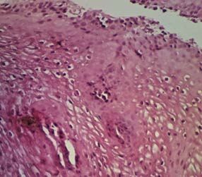
Original Research Article:                                                          (2019), «EUREKA: Health Sciences»
full paper                                                                                      Number 1

             Results. It was found that the least valuable diagnostic feature in the morphological examination of the mucous membrane of
    the distal esophagus in the pain-causing children with GERD was thickening of the epithelium with a sensitivity of 13,0 %, a specific-
    ity of 96.0 %, and total value of 65.0 %. It has been proved that hyperplasia of cells of the basal layer of the mucous membrane of the
    distal esophagus at the GERD in the infected children is 46.7 % (specificity – 93.3 %, the total value is 75.6 %). Increase in the num-
    ber of papillae and their prolongation in 33.3 % cases (sensitivity – 33.3 %, specificity – 93.3 %, overall diagnostic value – 70.8 %).
             Conclusion. The peculiarity of the morphological manifestations of GERD in childbearing children is dystrophic changes
    in keratocytes in the superficial parts of the multilayer squamous epithelium, which are detected at 100.0 % of patients (specificity
    is 93.3 %, total value is 96.8 %), with parakeratosis centers at 13.3 % of cases. It has been shown that a frequent and diagnostically
    valuable indication is inflammatory infiltration of the esophageal mucosa, which are verified in all cases (100.0 %, with dilatation
    and hyperemia in 46.7 % of patients (specificity – 40.0 %, total value – 81.3 %).
             Keywords: gastroesophageal reflux disease, childhood fever, distal esophagus, morphological changes.

                                                                                                  © Olena Zhuravel, Tetyana Pochinok,
    DOI: 10.21303/2504-5679.2019.00847                                         Tamara Zadorozhna, Tetyana Archakova, Valentyna Zamula

                1. Introduction
                         Gastroesophageal reflux disease (GERD) is a chronic relapsing disease with a spontaneous reg-
                ular discharge of the gastric and/or duodenal contents into the esophagus, leading to the lower esoph-
                agus affection [1, 2]. Incidence of the disease from 1 to 12 years and further increases reaching the
                maximum at the age of 16–17 [3, 4]. Although there are no large prospective population surveys in this
                area, it is anticipated that many children are diagnosed with GERD in adolescence and teen-age [5].
                         For the first time, the modern term GERD was proposed by M. Rossetti in 1966 [6]. The cur-
                rent general consensus determines the GERD in the occurrence of endoscopically indentifiable le-
                sions, cracks, ruptures of the esophagus mucosa immediately above the gastroesophageal junction.
                Detection of esophagitis is specific for the GERD in 90–95 %, but with a sensitivity of 50 % [7].
                         The endoscopic method is the main method of diagnostics of the GERD which allows con-
                firming the occurrence of reflux esophagitis, assessing its severity and make sampling for histodiag-
                nosis and bacterioscopy [8]. For the endoscopic classification of esophagitis, the Hetzel-Dent (2016)
                [9], Savary-Miller classifications [10, 11] in the Carisson [12] and Los Angeles (2011) [10, 13–14]
                modifications are used in the academic research of adults and children. Endoscopically normal esoph-
                agus mucosa, however, does not exclude the diagnosis of the GERD or esophagitises of other etiolo-
                gies [15]. The results of histology are affected by sampling faults, the heterogeneity of inflammatory
                changes, the lack of standardization of the biopsy site and the interpretation of morphological data.
                Therefore, these observations are important and still remain the focus of research [16].
                         There are also recent works dealing with the role of infectious agents, in particular persistent in-
                fection, in the development of chronic esophagitises and the GERD [17–19]. That is, the data of research
                conducted in recent years, testify to the etiological heterogeneity of chronic esophagitises, which may
                involve at least three factors in their development - pathological gastroesophageal reflux (GER), food
                and respiratory allergy, chronic infection. The above urges study of the esophagus mucosa biopsy data
                in adolescents sickly with acute respiratory diseases (ARD) against the background of the GERD.

                2. Aim of the research.
                       Research objective is to investigate the diagnostic value of morphological changes in gas-
                troesophageal reflux disease in esophageal biopsy material in adolescents sickly with acute respi-
                ratory diseases.

                3. Material and methods of the research
                        The study was conducted for the period 2015–2018. The objective of the study was achieved
                through examination of 90 adolescents (10 to 16 years old, average age 13.1±3.54 years) kept under
                observation at the Children’s Clinical Hospital No. 9 of Kyiv and on the basis of the Department of
                Pediatrics No. 1 Center of Primary Health Care No. 4 of the Desnianskyi district of Kyiv. All ado-
                lescents belonged to the group of sickly with a number of respiratory diseases averaging 6-8 times
                a year, lasting from 8 to 18 days (on average 12.8±5.41 days).

                                                                          16

 Medicine and Dentistry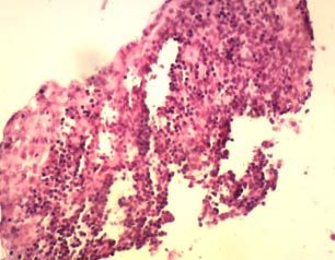
Original Research Article:                                                    (2019), «EUREKA: Health Sciences»
full paper                                                                                Number 1

                       All children have undergone endoscopic examination of the esophagus, stomach and duode-
               num with the esophagus mucosa biopsy using the OLYMPUS GIF-P3 flexible fiberscope.
                       In order to solve the tasks, the general histological method was used in the study. The ma-
               terial was treated through paraffin embedding, the sections were stained with hematoxylin & eo-
               sin (the technique gives a general idea of the organ structure, reveals well all cellular elements
               and some non-cellular structures) and picro-fuchsine following van Gieson method (the technique
               allows to detect the connective tissue; the collagen fibers of the connective tissue when stained
               with picro-fuchsine have a red colour, muscular and elastic tissues have brownish-yellow or yel-
               low-green colour, the nuclei have dark brown colour).
                       The statistical processing of the research results was made using the STATISTICA 10.0
               package (StatSoft Inc., USA). The objective parameters were used to describe the informativeness
               of the morphological study, which were determined as the operational characteristics of the tests.
               The most important operational characteristics of the diagnostic methods included sensitivity (Se),
               specificity (Sp), diagnostic accuracy (E, efficacy). The probability of the difference in frequency
               distribution was determined by Fisher’s criterion χ2.

               4. Results
                     Research findings and their consideration. In the study of morphological features of the
               esophagus mucosa among biopsy material of 60 children, the following criteria were evaluated:
                     1) occurrence of erosion (Fig. 1, 2);
                     2) thinning of the epithelium (Fig. 3);
                     3) thickening of the epithelium (Fig. 4);
                     4) dystrophy of the epithelium (Fig. 5);
                     5) papillae elongation (Fig. 6);
                     6) inflammatory infiltration (Fig. 7);
                     7) basal cell hyperplasia (Fig. 8);
                     8) dilatation and plethora of vessels.

                             Fig. 1. Esophagus mucosa of sickly children with the GERD. Mucosa erosion.
                                                   Hematoxylin & eosin (10×40)

                              Fig. 2. Esophagus mucosa of sickly children with the GERD. Deep erosion.
                                                   Hematoxylin & eosin (10×10)

                                                                    17

                                                                                                          Medicine and Dentistry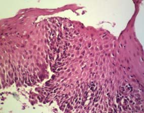
Original Research Article: (2019), «EUREKA: Health Sciences»
full paper Number 1
Fig. 3. Esophagus mucosa of sickly children with the GERD. Focal thinning of squamous
epithelium. Hematoxylin & eosin (10×10)
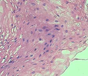
Fig. 4. Esophagus mucosa of sickly children with the GERD. Focal thickening of squamous
epithelium. Hematoxylin & eosin (10×10)
The activity of the GERD in sickly children with acute respiratory diseases (Fig. 1, 2) was
characterized by erosive defects of the esophagus mucosa of varying degrees of severity with the
death of a small number of superficial cells of the stratified squamous epithelium (Fig. 1), in some
cases extending to the basal layer of the esophagus mucosa – deep erosion (Fig. 2).
Morphological changes in the stratified squamous epithelium of the distal esophagus were
detected as focal thinning (Fig. 3), areas of thickening - moderate or pronounced (Fig. 4), alternat-
ing areas of thinning with thickening focuses with dystrophic changes (Fig. 5).
Fig. 5. Esophagus mucosa of sickly children with the GERD. Dystrophy of superficial cells of the
stratified squamous epithelium. Hematoxylin & eosin (10×10)
In all cases, signs of dystrophic changes in the epithelium and keratocytes were observed in
the biopsy material (Fig. 5). In some of them, balloon dystrophy of the epithelium (Fig. 9), micro-
18
Medicine and Dentistry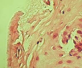
Original Research Article:                                                    (2019), «EUREKA: Health Sciences»
full paper                                                                                Number 1

               focuses of parakeratosis, hyperplasia of cells of varying degrees of severity (Fig. 8) with esophagus
               mucosa papilla elongation (Fig. 6) were observed.

                    Fig. 6. Esophagus mucosa of sickly children with the GERD. Esophagus mucosa papillae
                                          elongation. Hematoxylin & eosin (10×10)

                    Fig. 7. Esophagus mucosa of sickly children with the GERD. Inflammatory infiltration of
                                the stratified squamous epithelium. Hematoxylin & eosin (10×10)

                      Fig. 8. Esophagus mucosa of sickly children with the GERD. Basal cell hyperplasia of
                                     the stratified squamous epithelium. Van Gieson (10×10)

                       Along with the dystrophic changes in the cells of the stratified squamous epithelium, infil-
               tration of the esophagus mucosa by lymphocytes, plasma cells, occasionally by single eosinophil
               and neutrophil cells (Fig. 7) was identified in all observations. Inflammatory infiltration was ob-
               served in the depth of the epithelium, in the papillary area, in the submucous layer with dilatation
               and plethora of the mucosa vessels (Fig. 10).
                       The frequency of verification of morphological signs of the distal esophagus mucosa lesions
               in sickly adolescents is given in Table 1.

                                                                   19

                                                                                                         Medicine and Dentistry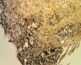
Original Research Article:                                                     (2019), «EUREKA: Health Sciences»
full paper                                                                                 Number 1

                 Fig. 9. Esophagus mucosa of sickly children with the GERD. Focuses of the balloon dystrophy of
                                    the stratified squamous epithelium. Van Gieson (10×10)

                 Fig. 10. Esophagus mucosa of sickly children with the GERD. Mucosa dilatation and plethora of
                                            vessels. Hematoxylin & eosin (10×20)

                Table 1
                Morphological signs of the distal esophagus mucosa lesions with diagnosed GERD in children sickly with
                acute respiratory disease

                                 Morphological signs                                   Abs.                       %

     Occurrence of erosions:                                                            30                       33.3
     – superficial                                                                      24                       26.7
     – deep                                                                              6                        6.6
     Thinning of epithelium                                                             54                       60.0
     Thickening of epithelium                                                           12                       13.3
     Alternation of the areas of thinning and thickening of the epithelium              24                       26.7
     Basal cell hyperplasia                                                             42                       46.7
     Papillae elongation                                                                30                       33.3
     Dystrophy of the epithelium                                                        90                       100.0
     Parakeratosis                                                                      12                       13.3
     Inflammatory infiltration                                                          90                       100.0
     Dilatation and plethora of vessels                                                 42                       46.7

                                                                         20

 Medicine and Dentistry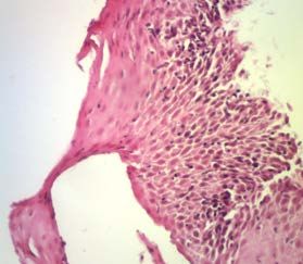
Original Research Article:                                                      (2019), «EUREKA: Health Sciences»
full paper                                                                                  Number 1

                      As we see, erosive reflux esophagitis (erosive form of the reflux disease) was detected in
               30 observations (33.3 %), of which superficial – in 24 (26.7 %), deep – in 6 (6.6 %) cases. Often, in-
               stead of erosion, there was a thinning of the epithelial layer (54 cases, 60 %) or its focal thickening
               (12 observations, 13.3 %).
                      In 24 case (26.7 %), there were areas of thinning of the esophagus mucosa, which alternated
               in some places with its thickening. Hyperplasia of the basal cells of the esophagus mucosa was
               observed in 42 observations (46.7 %), sometimes hyperplasia was accompanied by an increase in
               the number of papillae and their elongation (30 cases, 33.3 %).
                      Dystrophic changes in keratocytes in the superficial parts of the stratified squamous epithe-
               lium were found in all patients (90 people, 100.0 %), with focal parekeratosis in 13.3 % of patients
               (12 people). Infiltration of the esophagus mucosa was verified in all cases (90 patients, 100.0 %),
               with dilatation and plethora in 42 patients (46.7 %).
                      The diagnostic value of the morphological signs of the distal esophagus lesion in children
               sickly with acute respiratory diseases with diagnosed GERD is presented in Table 2.

               Table 2
               Diagnostic value of the morphological signs of the distal esophagus lesion in children sickly with acute
               respiratory diseases with diagnosed GERD, %
                    Signs                          Sensitivity                  Specificity                Diagnostic value
           Occurrence of erosions                     33.3                         96.0                          77.4
           Thinning of epithelium                     60.0                         93.3                          76.7
          Thickening of epithelium                    13.0                         96.0                          65.0
            Basal cell hyperplasia                    47.0                         93.3                          75.6
             Papillae elongation                      33.3                         93.3                          70.8
        Dystrophy of the epithelium                  100.0                         93.3                          96.8
          Inflammatory infiltration                  100.0                         40.0                          81.3
      Dilatation and plethora of vessels              47.0                          86.7                         66.7

                       Thus, in the morphological study of the biopsy material of children sickly with acute respi-
               ratory diseases with diagnosed GERD, the diagnostic sensitivity of the esophagus mucosa lesions
               in the form of erosions was 33.3 %, the specificity was 96.0 %, and the total diagnostic value was
               77.4 %. Similar results were obtained for such a morphological feature as the mucosa epithelium
               papillae elongation – sensitivity – 33.3 %, specificity – 93.3 %, total diagnostic value – 70.8 %.
                       Instead, the thinning of the epithelium in children sickly with acute respiratory diseases
               with diagnosed GERD had a greater diagnostic value (76.7 %) compared with signs of thickening
               of the epithelium (65.0 %) and epithelium basal cell hyperplasia (75.6 %), with a sensitivity of
               47 % and specificity of 93.3 %.
                       The highest coefficients of diagnostic value in sickly children with diagnosed GERD were
               obtained for such signs as dystrophy of the epithelium (sensitivity - 100.0 %, specificity – 93.3 %,
               total value – 96.8 %) and inflammatory infiltration (sensitivity – 100.0 %, specificity – 40.0 %, total
               value – 81.3 %).
                       Consequently, the findings are partly consistent with the generally accepted recommenda-
               tions for the evaluation of the reflux esophagitis, where, regardless of the endoscopic diagnosis for
               the morphological confirmation of the erosive esophagitis, the following criteria are required:
                       1) papillae enlargement of more than 2/3 of the thickness of the mucosa;
                       2) hyperplasia of the basal layer;
                       3) intraepithelial granulocytosis and even single neutrophil or eosinophil cells [6, 9].
                       For children sickly with acute respiratory diseases with diagnosed GERD, we have estab-
               lished a slightly different trend. Particularly, in this group, not the thickening of the epithelium,

                                                                     21

                                                                                                            Medicine and Dentistry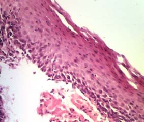
Original Research Article:                                                          (2019), «EUREKA: Health Sciences»
full paper                                                                                      Number 1

                hyperplasia of the basal layer and papillae elongation, but thinning (sensitivity – 60.0 %, spec-
                ificity – 93.3 %, total value – 76.7 %) and dystrophy of the epithelium (sensitivity – 100.0 %,
                specificity – 93.3 %, total value – 96.8 %) have the largest value.
                        According to the existing data in literature regarding morphological changes of the esoph-
                agus mucosa for diagnosing GERD is important: capillary dilation, intraepithelial lymphocytes,
                balloon dystrophy of squamous epithelium cells [13].
                        We also identified the above features in the examined group of children sickly with acute
                respiratory diseases with diagnosed GERD.
                        In the event that the endoscopic examination does not reveal any apparent defects in the
                mucosa, signs of inflammation may still be detected in the biopsy material. In children sickly with
                acute respiratory diseases with diagnosed GERD, the sensitivity of this sign is 100.0 %, and the
                overall diagnostic value is 81.4 %.

                5. Discussion
                        Before discussing the results of 60 adolescents (10 to 16 years old, average age 13.1±3.54 years)
                that were observed at the Children’s Clinical Hospital No. 9 of Kyiv and on the basis of the Depart-
                ment of Pediatrics No. 1 Primary Health Care Center No. 4 of the Desnianskyi district of Kyiv the
                following should be noted. At present, there is no, and probably, cannot be, the unified criteria for
                evaluating the results of the study of morphological changes in gastroesophageal reflux disease in the
                biopsy materials of the esophagus in adolescents sickly with acute respiratory diseases.
                        For the endoscopic classification of esophagitis in academic research in adults and children,
                the [9] Savary-Miller classifications in the Carisson and [10] modifications are used. However, the
                endoscopically normal mucus of the esophagus does not exclude the diagnosis of GERD or esoph-
                agitises of other etiologies.
                        The study does not claim exclusion because it involves a limited number of individuals and
                the tests were made on materials from two medical institutions, which in some way increases the
                percentage of statistical error.

                6. Conclusion
                        1. Evaluation of morphological changes in the distal esophagus mucosa should take into
                account the following features: erosions, thinning, thickening and dystrophy of the epithelium,
                assessment of papillae elongation, inflammatory infiltration and hyperplasia of the basal layer, as
                well as vascular plethora.
                        2. The peculiarity of the morphological manifestations of the GERD in children sickly with
                acute respiratory diseases is dystrophic changes in keratocytes of the superficial layers of the strat-
                ified squamous epithelium, which are detected in 100.0 % of patients (specificity – 93.3 %, total
                value – 96.8 %), with foci of parakeratosis in 13.3 % of cases.
                        3. For the further research, it is recommended to continue the study of morphological chang-
                es in gastroesophageal reflux disease in the esophagus biopsy material in adolescents sickly with
                acute respiratory diseases with diagnosed GERD.

                        References
                        [1] Boyarska, L. M., Ivanova, K. O., Skalozubova, I. B. (2012). Clinical and Functional Features of
                Gastroesophageal Reflux Disease in Children and Adolescents. Pathology, 1 (24), 26–30.
                        [2] Zhykhareva, N. S. (2013), Gastroesophageal Reflux Disease in Children. Medical Council,
                3, 34–41.
                        [3] Tang, M., Blake, K. V., Lima, J. J., Mougey, E. B., Franciosi, J., Schmidt, S. et. al. (2019). Gen-
                otype tailored treatment of mild symptomatic acid reflux in children with uncontrolled asthma (GenARA):
                Rationale and methods. Contemporary Clinical Trials, 78, 27–33. doi: http://doi.org/10.1016/j.cct.2019.01.009
                        [4] Chen, X., Peng, W.-S., Wang, L. (2019). Etiology analysis of nonspecific chronic cough in chil-
                dren of 5 years and younger. Medicine, 98 (3), e13910. doi: http://doi.org/10.1097/md.0000000000013910
                        [5] Tack, J., Pandolfino, J. E. (2018). Pathophysiology of Gastroesophageal Reflux Disease. Gastro-
                enterology, 154 (2), 277–288. doi: http://doi.org/10.1053/j.gastro.2017.09.047

                                                                          22

 Medicine and Dentistry
Original Research Article: (2019), «EUREKA: Health Sciences»
full paper Number 1
[6] Ruigómez, A., Wallander, M.-A., Lundborg, P., Johansson, S., Rodriguez, L. A. G. (2009). Gas-
troesophageal reflux disease in children and adolescents in primary care. Scandinavian Journal of Gastroen-
terology, 45 (2), 139–146. doi: http://doi.org/10.3109/00365520903428606
[7] Nakayama, Y., Ida, S. (2017). Endoscopic findings of esophagogastric junction in children. Di-
gestive Endoscopy, 29, 11–17. doi: http://doi.org/10.1111/den.12793
[8] Gyawali, C. P., Fass, R. (2018). Management of Gastroesophageal Reflux Disease. Gastroenter-
ology, 154 (2), 302–318. doi: http://doi.org/10.1053/j.gastro.2017.07.049
[9] Macchini, F., Zanini, A., Pasqua, N., Farris, G., Canazza, L., Gentilino, V. et. al. (2015). En-
doscopic Surveillance for Congenital Diaphragmatic Hernia: Unexpected Prevalence of Silent Esophagitis.
European Journal of Pediatric Surgery, 26 (3), 291–295. doi: http://doi.org/10.1055/s-0035-1552568
[10] Hoshihara, Y., Iwakiri, K. (2016) Endoscopic classification of reflux esophagitis. Nihon Rinsho,
74 (8), 1262–1267.
[11] Tutar, E., Kutluk, G., Bayrak, N. A., Ataizi Celikel, C., Pehlivanoglu, E., Ertem, D. (2009). What
is the diagnostic utility of endoscopic scoring systems in children? The Turkish Journal of Gastroenterology,
24 (1), 22–29. doi: http://doi.org/10.4318/tjg.2013.0700
[12] Numans, M. E., De Wit, N. J. (2003). Reflux symptoms in general practice: diagnostic evalu-
ation of the Carlsson-Dent gastro-oesophageal reflux disease questionnaire. Alimentary Pharmacology and
Therapeutics, 17 (8), 1049–1055. doi: http://doi.org/10.1046/j.1365-2036.2003.01549.x
[13] Hoshino, M., Omura, N., Yano, F., Tsuboi, K., Yamamoto, S. R., Akimoto, S. et. al. (2018).
Impact of reflux esophagitis on the esophageal function before and after laparoscopic fundoplication. Esoph-
agus, 15 (4), 224–230. doi: http://doi.org/10.1007/s10388-018-0618-8
[14] Kasyap, A. K., Sah, S. K., Chaudhary, S. (2018). Clinical spectrum and risk factors associated
with asymptomatic erosive esophagitis as determined by Los Angeles classification: A cross-sectional study.
PLOS ONE, 13 (2), e0192739. doi: http://doi.org/10.1371/journal.pone.0192739
[15] Vakil, N. (2010). Disease definition, clinical manifestations, epidemiology and natural history
of GERD. Best Practice & Research Clinical Gastroenterology, 24 (6), 759–764. doi: http://doi.org/10.1016/j.
bpg.2010.09.009
[16] Houghton, L. A., Smith, J. A. (2017). Gastro-oesophageal reflux events: just another trigger in
chronic cough? Gut, 66 (12), 2047–2048. doi: http://doi.org/10.1136/gutjnl-2017-314027
[17] Evsyutina, Yu. V., Trukhmanov, A. S., Ivashkin, V. T. (2015). Systemic Immune Response in
Patients with Gastroesophageal Reflux Disease. Russian Journal of Gastroenterology, Hepatology and Colo-
proctoly, 5, 32–38.
[18] Leason, S. R., Barham, H. P., Oakley, G., Rimmer, J., DelGaudio, J. M., Christensen, J. M. et. al.
(2017). Association of gastro-oesophageal reflux and chronic rhinosinusitis: systematic review and me-
ta-analysis. Rhinology Journal, 55 (1), 3–16. doi: http://doi.org/10.4193/rhin16.177
[19] Friesen, C. A., Rosen, J. M., Schurman, J. V. (2016). Prevalence of overlap syndromes and
symptoms in pediatric functional dyspepsia. BMC Gastroenterology, 16 (1), 75. doi: http://doi.org/10.1186/
s12876-016-0495-3
23
Medicine and DentistryYou can also read