Gastroesophageal Reflux Disease, Tooth Erosion, and Prosthodontic Rehabilitation: A Clinical Report
←
→
Page content transcription
If your browser does not render page correctly, please read the page content below
CLINICAL REPORT
Gastroesophageal Reflux Disease, Tooth
Erosion, and Prosthodontic Rehabilitation:
A Clinical Report
Ned B. Van Roekel, DDS, MSD

              Gastroesophageal reflux disease (GERD) is a relatively common gastrointestinal disorder in the
           United States. The reflux of acid adversely affects the mucosal lining of the esophagus and is
           responsible for dental erosion. This article briefly reviews the etiology, risk factors, and medical
           management of GERD. The patient presentation describes the rehabilitation of a young adult with
           GERD who needed multidisciplinary care.
              J Prosthodont 2003;12:255-259. Copyright © 2003 by The American College of Prosthodontists.

               INDEX WORDS: gastroesophageal reflux disease, acid reflux, dental erosion, tooth erosion

G     ASTROESOPHAGEAL REFLUX disease
      (GERD) is a relatively common gastrointesti-
nal disorder in Western society. Heartburn is re-
                                                                           experiencing vomiting 1 or more times a week,
                                                                           heartburn, belching, pain on awakening, acid taste,
                                                                           or stomach pain have dental erosion 31 times more
ported at least once a week by 15% of Americans                            frequently than controls.2
and daily by 7%.1 This symptom is caused by a                                 Risk factors for GERD include obesity, hiatal
backflow of gastric acid and other gastric contents                        hernia, and pregnancy.3 Approximately 50% of per-
into the esophagus. Normally, the lower esophageal                         sons over age 50 have hiatal hernias; however, as
sphincter (LES), the anatomic location of the gas-                         many as 84% of patients with erosive esophagitis
troesophageal junction, and the crural diaphragm                           have hiatal hernias.3 Gastroesophageal reflux and
prevent the movement of fluid or solid matter from                         heartburn are reported by 45%– 85% of women
the stomach into the esophagus. Reflux occurs                              during pregnancy.4 Substernal burning after eating,
when the LES relaxes, causing loss of the pressure                         the most common symptom, is worsened by fatty or
gradient between it and the stomach. The refluxed                          spicy foods, large meals, alcohol, or caffeine. Re-
material may reach the cervical esophagus, phar-                           cumbency, heavy lifting, or bending following a
ynx, and oral cavity. The relationship between                             meal may cause food or liquid to rise into the
GERD and dental erosion has been well docu-                                throat. Treatment of mild cases of GERD may
mented in the literature.7-14                                              involve life-style changes including modified diet,
   The medical concern is that acid reflux in the                          decreases in the volume of food or liquid, sleeping
esophagus may damage the mucosal lining. Reflux                            with the head of the bed raised 4 to 6 inches, weight
esophagitis can be mild, involving only microscopic                        reduction, and use of over-the-counter antisecre-
changes in the cells of the mucosa, or erosive,                            tory agents. In more severe GERD, histamine-2
causing bleeding and superficial linear ulcers. From                       (H2) receptor blocking agents (nizatidine, 150 mg
a dental standpoint, acid reflux in the oral cavity                        twice a day; ranitidine, 150 mg twice a day; cimet-
causes the loss of coronal tooth structure by chem-                        idine, 300 mg/day) are prescribed for 6 to 12 weeks
ical erosion. It has been reported that patients                           to provide symptomatic relief. For patients resis-
                                                                           tant to H2 receptor blockers or patients with severe
     Consultant in Prosthodontics, Department of Dental Specialties, As-
                                                                           GERD, proton pump inhibitors (PPIs) to provide
sistant Professor of Dentistry, Mayo Medical School, Rochester, MN.        strong acid suppression is the treatment of choice.
     Accepted April 29, 2003.                                              Rabeprazole (20 mg/day), omeprazole (40 mg/day),
     Correspondence to: Ned B. Van Roekel, DDS, MSD, Department of         and lansoprazole (30 mg/day) are commonly pre-
Dental Specialties, Mayo Medical School, 200 First Street SW, Rochester,   scribed PPIs.
MN 55905.
     Copyright © 2003 by The American College of Prosthodontists
                                                                              Several testing procedures may be used to con-
     1059-941X/03/1204-0000$30.00/0                                        firm the diagnosis of GERD. Endoscopy of the
     doi:10.1016/S1059-941X(03)00104-9                                     esophagus with biopsy is the standard procedure for

                                 Journal of Prosthodontics, Vol 12, No 4 (December), 2003: pp 255-259                      255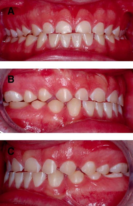
256         Gastroesophageal Reflux Disease, Tooth Erosion, and Prosthodontic Rehabilitation ●   Van Roekel

documenting the type and extent of tissue damage.
Barium esophagography is used to identify any
stricture in patients with severe dysphagia. The
24-hour ambulatory esophageal pH manometry
test is the best study for determining the severity of
the gastric reflux into the esophagus.5 This test is
performed by passing a pH probe sensitive catheter
through the nose into the esophagus. The pH probe
is positioned above the LES. Connected to the other
end of the catheter is a small computer that is worn
around the waist. The pH probe is left in place for
24 hours. During this time the patient may con-
sume a normal diet but is instructed to not swim,
bathe, or shower.
    This report presents the clinical manifestata-
tions, diagnosis, and medical and dental manage-
ment of a patient with GERD with severe tooth
erosion.

                Clinical Report
A 14-year-old Caucasian male first presented to the
author for a dental evaluation in 1998. The patient
was referred by his pediatrician, who had noted the
patient’s significant loss of coronal tooth structure
and made a diagnosis of bruxism. Clinically, the
appearance of the teeth was inconsistent with that
usually seen in people with bruxism. Besides the
                                                              Figure 1. Pretreatment. (A) Front view. (B) Right lat-
occlusal/incisal surfaces, loss of tooth structure also       eral view. (C) left lateral view.
occurred on the buccal and lingual surfaces. This
indicates that erosion, rather than attrition, was
                                                                 This patient needed a multidisciplinary ap-
responsible for the destruction.                              proach to dental rehabilitation. Because of the
   The patient’s medical history revealed that he             large volume of tooth structure that had been lost,
experienced periodic regurgitation of stomach acid            it was necessary to restore the patient’s occlusal
and food from 1993 to 1998. He also felt some                 vertical dimension to permit the fabrication of cast
retrosternal burning several times a month. The               restorations for the anterior and posterior teeth
patient’s previous dentist fabricated a bite guard            (Fig 1 A-C). The diagnostic waxing procedure on
appliance to control tooth wear that the patient had          casts mounted in centric relation on a semiadjust-
used from 1995 to 1998. It was clear that the                 able arcon articulator indicated that orthodontic
patient needed further medical evaluation to ade-             treatment was needed so that a mutually protected
quately address his dental needs.                             occlusion could be developed prosthetically. A semi-
   The patient was referred to a gastroenterologist           adjustable articulator was selected because the oc-
for an upper gastrointestinal (GI) radiograph to              clusal scheme provided anterior disocclusion. The
evaluate possible gastric outlet obstruction and a            patient’s 4 impacted third molars were removed
24-hour ambulatory esophageal pH manometry                    before orthodontic treatment. Crown-lengthening
test. The results of the upper GI radiograph were             procedures were performed surgically on the man-
normal; the results of the 24-hour pH reflux mon-             dibular left second premolar, first molar, and sec-
itoring test indicated episodes of reflux. The patient        ond molar to expose sufficient tooth structure to
was placed on omeprazole, 20 mg/day, and in-                  permit the placement of orthodontic bands. Com-
structed to elevate the head of the bed 6 inches.             posite resin was bonded to the occlusal surfaces of
The patient’s symptoms then resolved.                         the remaining posterior teeth to provide sufficient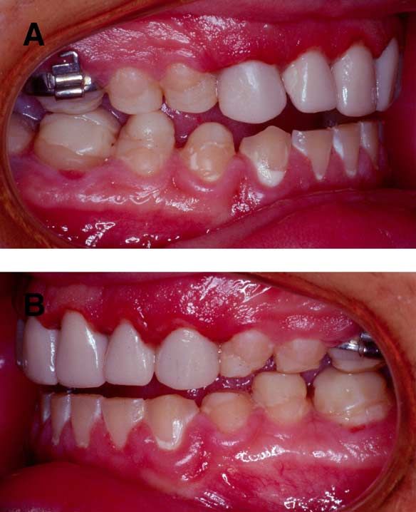
December 2003, Volume 12, Number 4                                    257

                                                                Root canal therapy was performed on both max-
                                                            illary canines and the maxillary left lateral incisor
                                                            to permit fabrication of cast dowel and cores to
                                                            develop adequate retention and resistance form in
                                                            the tooth preparations. All of the teeth in both the
                                                            maxillary and mandibular arches were prepared,
                                                            and provisional restorations were fabricated at the
                                                            new occlusal vertical dimension.
                                                                The patient experienced significant gingival hy-
                                                            perplasia during orthodontic treatment (Fig 3).
                                                            This necessitated performing a gingivoplasty using
                                                            an electrosurgical unit to facilitate tooth prepara-
                                                            tion and impression-making procedures. Noncom-
                                                            pliance with daily oral hygiene procedures made it
                                                            necessary to reduce the hyperplastic tissue both at
                                                            the time of tooth preparation and again when im-
                                                            pressions were made.
                                                                The definitive restorations were fabricated first
                                                            for the maxillary and mandibular anterior teeth,
                                                            then for the opposing posterior teeth on the left
                                                            side, and finally for those on the right side. Metal
                                                            ceramic restorations were placed on all teeth ex-
                                                            cept the second molars. The second molars received
Figure 2. Composite resin bonded to posterior occlusal
surfaces to increase the vertical dimension of occlusion.   full-veneer gold crowns due to restricted interocclu-
(A) Right lateral view. (B) Left lateral view.              sal space and clinical crown length. Prosthetic
                                                            treatment was completed in August 2002 (Fig 4
                                                            A-C).

attachment area for the orthodontic appliances.
The initial increase in occlusal vertical dimension                            Discussion
occurred at this time. All teeth received orthodontic       Dental erosion is defined as the loss of tooth struc-
bands and/or brackets.                                      ture due to a chemical process that does not involve
   As the orthodontic treatment was nearing com-            bacterial action and may be multifactorial in origin.
pletion, another diagnostic waxing procedure was            Erosion is not an uncommon finding during oral
performed to assess the interarch relationship at           examination; a prevalence as high as 42% has been
the desired occlusal vertical dimension. A full con-        reported.6 Erosion may be due to extrinsic sources
toured wax-up on the mounted casts made it pos-             of acid, such as acidic foods, drinks, and acidic
sible to evaluate tooth dimensions to achieve opti-         medications; however, the most common source of
mal esthetics.                                              intrinsic acid in children is regurgitation of gastric
   Additional composite resin was bonded to the             contents into the oral cavity, as occurs in GERD.15
occlusal surfaces of the posterior teeth to achieve
the same increase in the occlusal vertical dimension
that had been developed on the mounted casts (Fig
2 A, B). I prefer to use a processed acrylic bite guard
to assess changes in the occlusal vertical dimension,
but this approach would have interfered with this
patient’s ongoing orthodontic treatment. The pa-
tient had no difficulty adapting to the new occlusal
vertical dimension; he experienced no muscle pain
or discomfort and no difficulty in function. The
orthodontic treatment was completed in December             Figure 3. Front view illustrating postorthodontic gingi-
2001.                                                       val hyperplasia.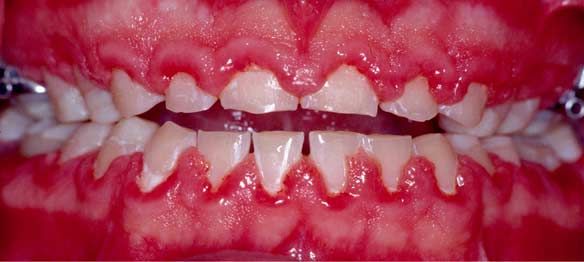
258 Gastroesophageal Reflux Disease, Tooth Erosion, and Prosthodontic Rehabilitation ● Van Roekel
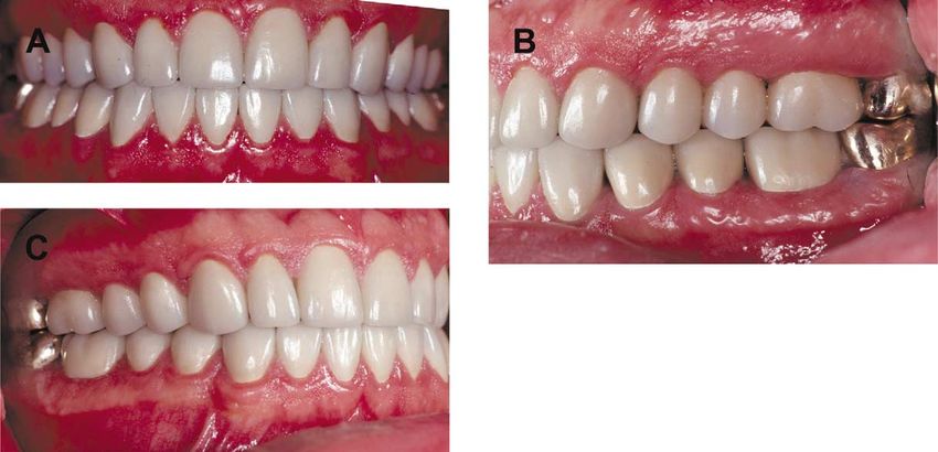
Figure 4. Definitive restorations. (A) Front view. (B)
Right lateral view. (C) Left lateral view.
The pattern of loss of tooth structure is similar patient’s previous dentist was treating him for a
to that seen in bulimia nervosa. The palatal sur- dental condition (bruxism) when in fact a medical
faces of the maxillary teeth are affected first. Ero- condition (GERD) was responsible for the lost tooth
sion of the occlusal surfaces of the posterior teeth in structure. The patient was subsequently referred
both arches and the labial or buccal surfaces results for a medical evaluation. The medical diagnosis was
from an extended period of acid reflux. The lower made, the appropriate pharmacologic agent was
anterior teeth are the last to be affected.16 prescribed, and the GERD was controlled. After
Many patients with GERD do not experience medical control of the GERD was established, the
heartburn, belching, unexplained sour taste, or re- patient’s dentition was restored to correct form,
gurgitation. This condition has been termed “silent function, and esthetics with an expectation of a
GERD.”17 Enamel erosion of the posterior teeth favorable long-term prognosis.
may be the first symptom of GERD. Thorough
history taking and oral examination are essential to
eliminate bulimia nervosa, attrition, and abrasion
References
as possible causes for the lost tooth structure. Re- 1. Gayal RK: Diseases of the esophagus, in Braunwold E, Fauci
AS, Kasper DL, et al (eds): Harrison’s Principles of Internal
ferral to a physician or gastroenterologist for appro-
Medicine, Vol 2 (ed 15). New York, McGraw-Hill, 2001, pp
priate testing is necessary to confirm a diagnosis of 1645-1649
GERD. Dental rehabilitation should not be initi- 2. Jarvinen VK, Rytomaa II, Heinonen OP: Risk factors in
ated until medical treatment has eliminated the dental erosion. J Dent Res 1991;70:942-947
acid reflux. 3. Rodney WM: Gastrointestinal disorders, in Rakel RE (ed):
Textbook of Family Practice (ed 6). Philadelphia, PA, Saun-
ders, 2002, pp 1159-1192
Summary 4. Broussard CN, Richter JE: Treating gastro-oesophageal re-
flux disease during pregnancy and lactation. What are the
Gastroesophageal reflux disease affects all age safest therapy options? Drug Saf 1998;19:325-337
groups. The prosthodontist must take this into con- 5. Eckhardt VF, Dilling B, Bernhard G: The impact of open
sideration when faced with the task of restoring access 24-hour pH manometry on the diagnosis and man-
teeth with significant loss of coronal tooth struc- agement of esophageal reflux disease. Am J Gastroenterol
ture. GERD by itself or in combination with attri- 1999;94:616-621
6. Lussi A, Schaffner M, Hotz P, et al: Dental erosion in a
tion or abrasion may be responsible for the loss. A population of Swiss adults. Commun Dent Oral Epidemiol
thorough diagnostic evaluation is necessary to as- 1991;19:286-290
certain the possible medical and/or dental sources 7. Bartlett D: Regurgitated acid as an explanation for tooth
for the problem. In the case presented here, the wear. Br Dent J 1998;185:210December 2003, Volume 12, Number 4 259
8. Bartlett D, Smith B: The dental relevance of gastro-oesoph- 13. Bartlett DW, Evans DF, Anggiansah A, et al: The role of the
ageal reflux: Part 2. Dent Update 1996;23:250-253 esophagus in dental erosion. Oral Surg Oral Med Oral
9. Schroeder PL, Filler SJ, Ramirez B, et al: Dental erosion and Pathol Oral Radiol Endod 2000;89:312-315
acid reflux disease. Ann Int Med 1995;122:809-815 14. Gregory-Head B, Curtis DA: Erosion caused by gastroesoph-
10. Bartlett DW, Evans DF, Smith BG: The relationship be- ageal reflux: Diagnostic considerations. J Prosthodont 1997;
tween gastro-oesophageal reflux disease and dental erosion. 6:278-285
J Oral Rehabil 1996;23:289-297 15. Linnett V, Seow WK: Dental erosion in children: A litera-
11. Bartlett DW, Evans DF, Smith BG: Oral regurgitation after ture review. Pediatr Dent 2001;23:37-43
reflux-provoking meals: A possible cause of dental erosion? 16. Spigset O: Oral symptoms in bulimia nervosa. A survey of 34
J Oral Rehabil 1997;24:102-108 cases. Acta Odontal Scand 1991;49:335-339
12. Gregory-Head BL, Curtis DA, Kim L, et al: Evaluation of 17. Ali DA, Brown RS, Rodriguez LO, et al: Dental erosion
dental erosion in patients with gastroesophageal reflux dis- caused by silent gastroesophageal reflux disease. J Am Dent
ease. J Prosthet Dent 2000;83:675-680 Assoc 2002;133:734-737You can also read