Double lobectomy in a steatotic liver transplantation rat model
←
→
Page content transcription
If your browser does not render page correctly, please read the page content below
EXPERIMENTAL AND THERAPEUTIC MEDICINE 21: 256, 2021

      Double‑lobectomy in a steatotic liver transplantation rat model
      LIN FAN1*, ZHEN FU1*, YAN XIONG1, SHAOJUN YE1, YANFENG WANG1, GUIZHU PENG1 and QIFA YE1,2

                 1
                  Zhongnan Hospital of Wuhan University, Institute of Hepatobiliary Diseases of Wuhan University,
              Transplant Center of Wuhan University, Hubei Key Laboratory of Medical Technology on Transplantation,
                           Wuhan, Hubei 430071; 2The 3rd Xiangya Hospital of Central South University,
               Research Center of National Health Ministry on Transplantation Medicine Engineering and Technology,
                                              Changsha, Hunan 410013, P.R. China

                                      Received March 28, 2020; Accepted September 16, 2020

                                                      DOI: 10.3892/etm.2021.9687

Abstract. Establishing a steatotic liver transplantation animal        Week 1, 80 vs. 100% and 1 month, 20 vs. 100%. A total of
model can be a challenging process, which requires complex             20 rats were not sacrificed by performing double‑lobectomy
microsurgical technologies. The present study established a            for biopsy. Taken together, the results of the present study
novel rat model of stable steatotic liver transplantation for          suggest that rat liver double‑lobectomy may be safely applied
marginal liver graft research, which notably minimized the             in steatotic liver transplantation without the need to sacrifice a
number of animals used for the experiment. Briefly, male               large number of animals.
Sprague‑Dawley rats (n=90) were fed with a high‑fat diet
(HFD; 60%, kJ) or standard chow diet (SCD) for 8 weeks.                Introduction
The liver enzymes and lipid levels were assessed every week,
and the degree of steatosis was determined via hematoxylin             Liver transplantation (LTx) is the most effective treatment
and eosin and Oil Red O staining. The results demonstrated             for patients with end‑stage liver diseases, including hepatic
that there were no significant differences in alanine amino‑           cirrhosis, hepatocellular carcinoma, hepatic hemangioma and
transaminase and aspartate aminotransferase levels between             hepatic echinococcosis, which has been extensively applied
the SCD and HFD groups (P>0.05), whereas the level of                  in the clinical setting for more than half a century (1‑5).
plasma triglyceride (TG) increased by 1.76‑fold in the HFD             The discrepancy between donor organ supply and demand
group at week 2, and progressively decreased to baseline levels        continues to increase with the growing number of potential
by week 8. Significantly higher levels of TG were observed             candidates on the transplant waiting list (6). Thus, a series
in the HFD group compared with the SCD group at week 2                 of strategies have been advocated to expand the donor pool
(P
2 FAN et al: STEATOTIC LIVER TRANSPLANTATION RAT MODEL
proposed for hepatic steatosis: Liver microcirculation is High‑Tech Co., Ltd.). The lipids included in the HFD consisted
hampered with excessive fat accumulation, which leads to of 90% lard and 10% soybean oil (Trophic Animal Feed
mitochondrial damage, and oxidative stress during reperfusion, High‑Tech Co., Ltd.). The SCD consisted of usual pellet rat
coupled with inflammatory response involving lipid peroxida‑ chow (Trophic Animal Feed High‑Tech Co., Ltd.). In order to
tion and leukocyte adhesion may contribute to the graft failure avoid fatty diarrhea, rats in the HFD group were fed using the
following transplantation (20,22,23). Ploeg et al (18) reported following 6 day schedule: i) 2 days of 30% weight (wt) HFD
that PNF rates increase up to 80% in the severely steatotic liver. and 70% wt SCD; ii) 2 days of 50% wt HFD and 50% wt SCD,
In addition, previous studies have demonstrated that severely and iii) 2 days of 70% wt HFD and 30% wt SCD.
steatotic graft is associated with high PNF rates (0‑66%)
and a 1‑year graft with a survival rate of 25‑90% (18,24‑26). Graft procurement and reduced size procedure. Anesthesia
Currently, macrovesicular steatosis of >30% is considered an during liver procurement and transplantation was maintained
independent predictor for a reduction of PNF and 1‑year graft using isoflurane (cat. no. R510‑22‑4; RWD Life Technology
survival (27). Thus, the evaluation and restoring of organs Co., Ltd.; 4% isoflurane for induction and 2% isoflurane for
from ECD attract increasing attention. maintenance). Briefly, heparin (100 IU) in 2 ml saline solution
Due to difficulties and complexities in microsurgical (cat. no. H8060‑1g; Beijing Solarbio Science & Technology Co.,
technology (28), establishing a rat steatotic liver transplanta‑ Ltd.) was injected into the penile vein, and a 5 mm long stent
tion model can be challenging. In addition, donor livers or prepared from polyethylene tube was inserted into the common
grafts require multiple biopsies at the point of procurement, bile duct (CBD) and secured with 6‑0 sutures. Livers were
during preservation and after transplantation. When using liver flushed in situ with 20 ml of University of Wisconsin (UW)
transplantation models, a single biopsy requires animals to be solution at 4˚C. A total of two hepatic lobes, the papillary
sacrificed, resulting in an increased number of experimental process and the quadrate lobe were procured for assessment,
animals, which is not conducive to animal welfare and ethical using the liver volume reduction method illustrated in Fig. 2.
principles. Thus, the present study established a novel method The papillary process (Fig. 2C) was removed to assess the
to decrease the number of experimental animals. The results of extent of hepatic steatosis before cold storage (CS) at 4˚C. The
the present study suggest that liver tissues from different parts quadrate lobe (Fig. 2D) was cut following preservation with
at different time points may significantly decrease the number UW solution at 2‑4˚C for 4 h. Venous cuffs prepared from two
of experimental animals used. In addition, the proposed method sizes of polyethylene tubes were subsequently placed in the
did not affect post‑operative animal mortality (papillary portal vein (PV) and intrahepatic inferior vena cava (IHVC).
process and quadrate lope excision: Partial liver transplantation The inside/outside diameters of polyethylene tube for CBD,
vs. whole liver transplantation survival; 5/5 vs. 5/5). PV and IHVC were 0.6/1.0, 1.8/2.2 and 2.8/3.2 mm, respec‑
The steatotic liver animal model induced by a high‑fat tively. Grafts were stored in UW solution at 4˚C for 4 h prior to
diet (HFD) is commonly applied to assess hepatic steatosis implantation (Fig. 2E).
in vivo. This model can mimic the etiology of hepatic steatosis
in human beings (29). The present study aimed to establish Serum analyses. Blood was drawn from the rats every week
a stable and reproducible steatotic liver model induced by a during the modeling process to detect hepatocyte injury and
HFD that may be used to investigate the efficacy of reduced serum lipid levels via alanine aminotransaminase (ALT),
size transplantation in MLG research. aspartate aminotransferase (AST), triglyceride (TG), total
cholesterol (TC), free fatty acid, high‑density lipoprotein and
Materials and methods low‑density lipoprotein. These indices were measured at the
Institute for Clinical Biochemistry and Diagnostics, Zhongnan
Animals. A total of 90 male Sprague‑Dawley rats (210±10 g), Hospital of Wuhan University (Wuhan, China). Lipids from
aged 6‑8 weeks, were obtained from Hubei Provincial Center rat livers were prepared using chloroform‑methanol extrac‑
for Disease Control and Prevention in China. All animals tion (30). Plasma glucose concentration was detected using a
were housed in an environment with a temperature of 23±1˚C, glucose analyzer (590; Yuwell), while insulin concentration
relative humidity of 55±10%, air exchange 12‑14 times/h, a was detected using commercially available RIA kits (cat. no.
light/dark cycle of 12/12 h, and were provided with food and E‑EL‑R3034; Elabscience Biotechnology Co., Ltd.).
tap water ad libitum. The present study was approved (IRB
approval no. AF‑177) by Wuhan University Institutional Histological assessment. The papillary process of liver tissues
Animal Care and Use Committee (Wuhan, China). was obtained and divided into two parts immediately after
A total of 10 rats were procured after 7 days of acclimation to collection. One part was fixed in a cold buffered 4% para‑
provide baseline values. A total of five rats in the HFD group or formaldehyde solution or cold buffered 3% glutaraldehyde
standard chow diet (SCD) group were samples each week during solution. Following fixation, the first part of tissues in each
8 weeks of modeling. Rats were randomly divided into 8 groups group was embedded in olefin, cut into 4‑mm thick slices and
(n=10). Body weight and food intake were monitored on a daily stained with hematoxylin (10 min at room temperature) and
basis. The time schedules for blood extraction in relation to the eosin (2 min at room temperature) or toluidine blue (3 min at
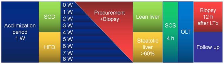
frozen sections used for analysis are presented in Fig. 1. room temperature) (31). The second part of tissues was treated
with Oil Red O (ORO) for 8 min at room temperature to assess
Dietary interventions. The HFD consisted of 60% lipid, the degree of steatosis (32), and for intraoperative assessment
20.6% carbohydrate and 19.4% protein (kJ), and was provided of the steatotic extent to determine whether it can be used
in rods direct from the manufacturer (Trophic Animal Feed as a donor. The degree of steatosis was estimated based on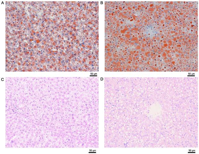
EXPERIMENTAL AND THERAPEUTIC MEDICINE 21: 256, 2021                                                               3

Figure 1. Time schedule of diet‑induced period and orthotopic liver transplantation. SCD, standard chow diet; HFD, high‑fat diet; W, week; SCS, static cold
storage; h, hour; OLT, orthotopic liver transplantation; LTx, liver transplantation.

Figure 2. Reduced size procedure prior to transplantation. (A) Schematic representation of hepatic lobes in rats. (B) Two lobes were cut from the whole liver.
(C) The papillary process was immediately removed following graft procurement. (D) The quadrate lobe was procured after 4 h of cold storage ex vivo. (E) The
remaining five lobes were prepared for transplantation. Liver transplantation process, including (F) pre‑ and (G) post‑ hypothermic perfusion, (H) ex vivo
preparation, (I) pre‑ and (J) post‑ portal vein reperfusion. (K) Bile outflow after reperfusion.

the percentage of hepatocytes containing lipid droplets using                    steatotic change in all groups was determined by calculating
Scoring System Definitions (33) and the following formula:                       the percentage in 10 random areas. A total of two pathologists
Degree of steatotic change (%)=[(number of hepatocytes with                      (Zhongnan Hospital of Wuhan University), blindly assessed
fatty droplets in the all microscopic field)/(number of total                    and confirmed all biopsies. All liver sections were observed
hepatocytes in the all microscopic field)] x100. The degree of                   under a light microscope (magnification, x200; TE2000‑U;

4                                 FAN et al: STEATOTIC LIVER TRANSPLANTATION RAT MODEL

Nikon Corporation) and the number and area of fat droplets in        In addition, the level of plasma TG in the HFD group
hepatocytes were assessed using Image‑Pro Plus (version 6.0;     increased by 1.76‑fold at week 2, and progressively decreased to
Media Cybernetics Inc.).                                         baseline levels by week 8. Significantly higher levels of TG were
                                                                 observed in the HFD group compared with the SCD group by
Orthotopic liver transplantation (OLT). Rats in the HFD          week 2 (P60%, were subjected to intraoperative ORO             (Fig. 4). Notably, the levels of plasma TG in both the HFD and
staining. OLT was performed as previously described by           SCD groups were not time‑dependent (P>0.05; Fig. 4A).
Kamada and Calne (28). Steatotic liver grafts or lean liver          The HFD group exhibited small but significantly higher
grafts were procured and transplanted into healthy adult rat     plasma glucose values (P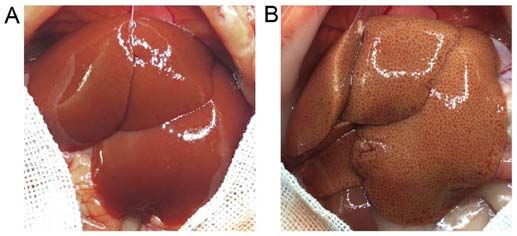
EXPERIMENTAL AND THERAPEUTIC MEDICINE 21: 256, 2021                                                           5

Figure 3. Body weight and serum levels in HFD and SCD groups. (A) The changes in body weight. Plasma (B) glucose, (C) ALT and (D) AST levels.
Plasma insulin concentrations were measured in (E) peripheral and (F) portal blood. n=5 and data are presented as the mean ± standard deviation. *P
6                                         FAN et al: STEATOTIC LIVER TRANSPLANTATION RAT MODEL

Figure 4. Plasma (A) TG, (B) liver TG, (C) plasma TC, (D) HDL, (E) LDL and (F) FFA levels in rats fed a HFD or SCD. Date are presented as the mean ± standard
deviation. SCD compared with HFD, *P

EXPERIMENTAL AND THERAPEUTIC MEDICINE 21: 256, 2021                                                                7

Figure 6. Hematoxylin and eosin stained liver tissues from high‑fat diet observed under a light microscope at week (A) 0, (B) 1, (C) 2, (D) 4, (E) 6 and (F) 8.
Scale bar, 50 µm. Magnification, x200.

                                                                                     Based on this model, strategies such as, ischemic precon‑
                                                                                 ditioning, pharmacological preconditioning and machine
                                                                                 perfusion may be used to enhance graft quality (6,34).
                                                                                 Although the steatotic liver model was stabilized by two
                                                                                 preliminary experiments, the results depended on the
                                                                                 energy intake and individual differences. Regrouping
                                                                                 would be more accurate based on TG content in tissues,
                                                                                 combined with the degree of hepatic steatosis estimated by
                                                                                 ORO staining.
                                                                                     LTx was performed using steatotic grafts or normal liver.
                                                                                 Clinically, hepatic macrovesicular steatosis >60% is consid‑
                                                                                 ered a contraindication for transplantation due to the high
                                                                                 morbidity of PNF (10). With reference to long‑term survival,
                                                                                 rats receiving SCD survived longer than those fed a HFD;
Figure 7. Degree of steatotic change in different groups. Mix refers to hepa‑
tocytes in which both macro and micro vacuoles were observed. n=5 and data       however, no statistically significant differences were observed
are presented as the mean ± standard deviation. *P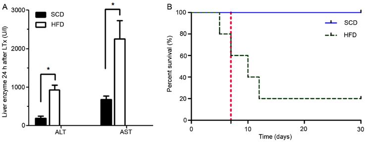
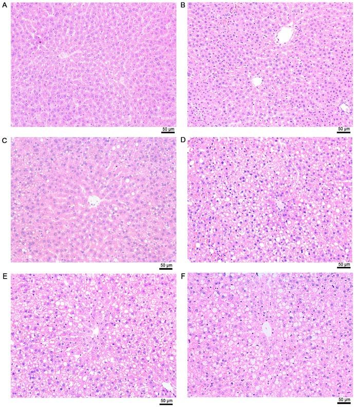
8                                          FAN et al: STEATOTIC LIVER TRANSPLANTATION RAT MODEL

Figure 8. (A and B) Oil Red O, and (C and D) hematoxylin and eosin stained liver tissue sections from the high‑fat diet group at weeks (A and C) 3 and
(B and D) 6. Microvesicular steatosis was observed at week 3, while macrovesicular steatosis was observed at week 6. Scale bar, 50 µm. Magnification, x200.

Figure 9. Post‑transplantation survival of rats and (A) liver enzymes in the HFD and SCD groups. (B) The 1‑ and 4‑week survival rates were 80% (4/5) and
20% (1/5) for recipients of grafts from steatotic liver, whereas the survival rates were 100% (5/5) and 100% (5/5) for recipients of grafts from normal liver,
respectively. n=5 and data are presented as the mean ± standard deviation. *P
EXPERIMENTAL AND THERAPEUTIC MEDICINE 21: 256, 2021                                                  9

PV. The results of the present study demonstrated that fatty         GP contributed to data acquisition and analysis. All authors
LTx displayed poor hepatic morphology following PV reper‑            have read and approved the final manuscript.
fusion compared with the normal graft transplantation. All of
the steatotic grafts appeared to be male‑reperfusion during          Ethics approval and consent to participate
the transplantation. The liver surface appeared to be piebald
due to the uneven blood reperfusion. In addition, combined           The present study was approved (IRB approval no. AF‑177)
with the post‑surgery survival, the intraoperative reperfusion       by Wuhan University Institutional Animal Care and Use
situation of the liver did not have a decisive role in predicting    Committee (Wuhan, China) and all animal experiments
rat prognosis, it was not possible to determine whether the rats     were performed in accordance with the Association for the
would achieve a good outcome just based on the reperfusion           Assessment and Accreditation of Laboratory Animal Care and
morphology. In one case of fatty LTx in the present study,           Institutional Animal Care and Use Committee regulations and
nearly no blood reperfusion was observed on the liver surface        guidelines.
except in the portal vein and its branches. However, despite
the poor performance, this was the only case that survived           Patient consent for publication
more than 6 months, longer than the others that had even
better reperfusion. As a result, the prognosis could not be          Not applicable.
predicted only by intraoperative morphology in steatotic LT.
    The steatotic liver is vulnerable to ischemic injury (21). PNF   Competing interests
was commonly observed in the severely steatotic liver group
that had sustained 4 h CS. The post‑surgery rats only survived       The authors declare that they have no competing interests.
if the liver function was restored, usually within 2 weeks. In
the present study, except for one rat in the HFD group that          References
survived, four rats died 8±2 days after the surgery. In total, 80%
of rats did not recover from PNF until their death. As steatotic      1. Starzl TE, Marchioro TL, Von Kaulla KN, Hermann G,
liver leads to poor prognosis, the authors aim to use machine            Brittain RS and Waddell WR: Homotransplantatin of the liver in
                                                                         humans. Surg Gynecol Obstet 117: 659‑676, 1963.
perfusion in future studies to improve the survival outcomes.         2. Wolfe RA, Merion RM, Roys EC and Port FK: Trends in organ
    In conclusion, the results of the present study suggest that         donation and transplantation in the United States, 1998‑2007.
HFD may be used to induce stable and rapid hepatic steatosis             Am J Transplant 9: 869‑878, 2009.
                                                                      3. Wertheim JA, Petrowsky H, Saab S, Kupiec‑Weglinski JW and
in rats without inducing inflammation. In addition, the lipid            Busuttil RW: Major challenges limiting liver transplantation in
droplets accumulate into fat vacuoles over time, from microve‑           the United States. Am J Transplant 11: 1773‑1784, 2011.
sicular steatosis into macrovesicular steatosis. Furthermore,         4. Durand F, Renz JF, Alkofer B, Burra P, Clavien PA, Porte RJ,
                                                                         Freeman RB and Belghiti J: Report of the Paris consensus
volume reduction, as a self‑control method, may be applied in            meeting on expanded criteria donors in liver transplantation.
steatotic LTx without increasing the number of animals being             Liver Transpl 14: 1694‑1707, 2008.
used, thus promoting animal welfare. However, therapeutic             5. Berg CL, Steffick DE, Edwards EB, Edwards EB, Heimbach JK,
                                                                         Magee JC, Washburn WK and Mazariegos GV: Liver and
approaches or machine perfusion need to be implemented in                intestine transplantation in the United States 1998‑2007.
steatotic grafts to improve the quality of donated organs and            Am J Transplant 9: 907‑931, 2009.
improve the post‑transplantation survival.                            6. Henry SD and Guarrera JV: Protective effects of hypothermic
                                                                         ex vivo perfusion on ischemia/reperfusion injury and transplant
                                                                         outcomes. Transplant Rev (Orlando) 26: 163‑175, 2012.
Acknowledgements                                                      7. Briceño J, Marchal T, Padillo J, Solórzano G and Pera C:
                                                                         Influence of marginal donors on liver preservation injury.
                                                                         Transplantation 74: 522‑526, 2002.
Not applicable.                                                       8. Salizzoni M, Franchello A, Zamboni F, Ricchiuti A, Cocchis D,
                                                                         Fop F, Brunati A and Cerutti E: Marginal grafts: Finding the
Funding                                                                  correct treatment for fatty livers. Transpl Int 16: 486‑493, 2003.
                                                                      9. Tekin K, Imber CJ, Atli M, Gunson BK, Bramhall SR, Mayer D,
                                                                         Buckels JA, McMaster P and Mirza DF: A simple scoring system
The present study was funded by the National Natural Science             to evaluate the effects of cold ischemia on marginal liver donors.
Foundation of China, (grant no. 81970548), the Medical Science           Transplantation 77: 411‑416, 2004.
                                                                     10. Cameron A and Busuttil RW: AASLD/ILTS transplant course:
Advancement Program (Youth Scholars) of Wuhan University                 Is there an extended donor suitable for everyone? Liver Transpl 11
(grant no. TFZZ2018035) and the Zhongnan Hospital of                     (suppl 2): S2‑S5, 2005.
Wuhan University Science, Technology and Innovation Seed             11. Shaker M, Tabbaa A, Albeldawi M and Alkhouri N: Liver trans‑
                                                                         plantation for nonalcoholic fatty liver disease: New challenges
Fund (grant no. ZNPY2018010).                                            and new opportunities. World J Gastroenterol 20: 5320‑5330,
                                                                         2014.
Availability of data and materials                                   12. Rinella ME: Nonalcoholic fatty liver disease: A systematic
                                                                         review. JAMA 313: 2263‑2273, 2015.
                                                                     13. Browning JD, Szczepaniak LS, Dobbins R, Nuremberg P,
The datasets used and/or analyzed during the current study are           Horton JD, Cohen JC, Grundy SM and Hobbs HH: Prevalence
available from the corresponding author on reasonable request.           of hepatic steatosis in an urban population in the United States:
                                                                         Impact of ethnicity. Hepatology 40: 1387‑1395, 2004.
                                                                     14. Neuschwander‑Tetri BA and Caldwell SH: Nonalcoholic steato‑
Authors' contributions                                                   hepatitis: Summary of an AASLD Single topic conference.
                                                                         Hepatology 37: 1202‑1219, 2003.
                                                                     15. Ruhl CE and Everhart JE: Fatty liver indices in the multiethnic
LF, ZF and QY contributed to the conception and design. LF,              united states national health and nutrition examination survey.
ZF, YX and SY performed the experiments. YX, SY, YW and                  Aliment Pharmacol Ther 41: 65‑76, 2015.
10 FAN et al: STEATOTIC LIVER TRANSPLANTATION RAT MODEL
16. Sanyal AJ; American Gastroenterological Association: 32. Mehlem A, Hagberg CE, Muhl L, Eriksson U and Falkevall A:
AGA technical review on nonalcoholic fatty liver disease. Imaging of neutral lipids by oil red O for analyzing the metabolic
Gastroenterology 123: 1705‑1725, 2002. status in health and disease. Nat Protoc 8: 1149‑1154, 2013.
17. McCormack L, Dutkowski P, El‑Badry AM and Clavien PA: Liver 33. Kleiner DE, Brunt EM, Van Natta M, Behling C, Contos MJ,
transplantation using fatty livers: Always feasible? J Hepatol 54: Cummings OW, Fer rell LD, Liu YC, Torbenson MS,
1055‑1062, 2011. Unalp‑Arida A, et al: Design and validation of a histological
18. Ploeg RJ, D'Alessandro AM, Knechtle SJ, Stegall MD, scoring system for nonalcoholic fatty liver disease. Hepatology 41:
Pirsch JD, Hoffmann RM, Sasaki T, Sollinger HW, Belzer FO 1313‑1321, 2005.
and Kalayoglu M: Risk factors for primary dysfunction after 34. de Rougemont O, Breitenstein S, Leskosek B, Weber A, Graf R,
liver transplantation‑a multivariate analysis. Transplantation 55: Clavien PA and Dutkowski P: One hour hypothermic oxygenated
807‑813, 1993. perfusion (HOPE) protects nonviable liver allografts donated
19. Busuttil RW and Tanaka K: The utility of marginal donors in after cardiac death. Ann Surg 250: 674‑683, 2009.
liver transplantation. Liver Transpl 9: 651‑663, 2003. 35. Corominas J, Marchesi JA, Puig‑Oliveras A, Revilla M, Estellé J,
20. Selzner M and Clavien PA: Fatty liver in liver transplantation and Alves E, Folch JM and Ballester M: Epigenetic regulation of the
surgery. Semin Liver Dis 21: 105‑113, 2001. ELOVL6 gene is associated with a major QTL effect on fatty
21. Sun CK, Zhang XY, Zimmermann A, Davis G and Wheatley AM: acid composition in pigs. Genet Sel Evol 47: 20, 2015.
Effect of ischemia‑reperfusion injury on the microcircula‑ 36. Tuvdendorj D, Zhang XJ, Chinkes DL, Wang L, Wu Z,
tion of the steatotic liver of the Zucker rat. Transplantation 72: Rodriguez NA, Herndon DN and Wolfe RR: Triglycerides
1625‑1631, 2001. produced in the livers of fasting rabbits are predominantly stored as
22. Imber CJ, St Peter SD, Lopez I, Guiver L and Friend PJ: Current opposed to secreted into the plasma. Metabolism 64: 580‑587, 2015.
practice regarding the use of fatty livers: A trans‑ Atlantic survey. 37. Katz DL: Ducks, geese, faith, and fatty livers. Child Obes 10:
Liver Transpl 8: 545‑549, 2002. 373‑374, 2014.
23. El‑Badry AM, Moritz W, Contaldo C, Tian Y, Graf R and 38. Awde S, Marty‑Gasset N, Prahkarnkaeo K and Rémignon H:
Clavien PA: Prevention of reperfusion injury and microcircula‑ Relationship between proteolytic activities and cooking loss
tory failure in macrosteatotic mouse liver by omega‑3 fatty acids. variability in liver issued from force‑fed mule ducks. J Agric
Hepatology 45: 855‑863, 2007. Food Chem 62: 3262‑3268, 2014.
24. Todo S, Demetris AJ, Makowka L, Teperman L, Podesta L, 39. Spolding B, Connor T, Wittmer C, Abreu LL, Kaspi A,
Shaver T, Tzakis A and Starzl TE: Primary nonfunction of hepatic Ziemann M, Kaur G, Cooper A, Morrison S, Lee S, et al: Rapid
allografts with preexisting fatty infiltration. Transplantation 47: development of non‑alcoholic steatohepatitis in Psammomys
903‑905, 1989. obesus (Israeli sand rat). PLoS One 9: e92656, 2014.
25. Verran D, Kusyk T, Painter D, Fisher J, Koorey D, Strasser S, 40. Gauthier MS, Favier R and Lavoie JM: Time course of the devel‑
Stewart G and McCaughan G: Clinical experience gained opment of non‑alcoholic hepatic steatosis in response to high‑fat
from the use of 120 steatotic donor livers for orthotopic liver diet‑induced obesity in rats. Br J Nutr 95: 273‑281, 2006.
transplantation. Liver Transpl 9: 500‑505, 2003. 41. Matteoni CA, Younossi ZM, Gramlieh T, Boparai N, Liu YC and
26. Gabrielli M, Moisan F, Vidal M, Duarte I, Jiménez M, Izquierdo G, McCullough AJ: Nonalcoholic fatty liver disease: A spectrum
Domínguez P, Méndez J, Soza A, Benitez C, et al: Steatotic livers. of clinical and pathological severity. Gastroenterology 116:
Can we use them in OLTX? Outcome data from a prospective 1413‑1419, 1999.
baseline liver biopsy study. Ann Hepatol 11: 891‑898, 2012. 42. Flecknell P: Replacement, reduction and refinement. ALTEX 19:
27. Spitzer AL, Lao OB, Dick AA, Bakthavatsalam R, Halldorson JB, 73‑78, 2002.
Yeh MM, Upton MP, Reyes JD and Perkins JD: The biopsied 43. Saltiel AR and Kahn CR: Insulin signalling and the regulation of
donor liver: Incorporating macrosteatosis into high‑risk donor glucose and lipid metabolism. Nature 414: 799‑806, 2001.
assessment. Liver Transpl 16: 874‑884, 2010. 44. Samuel VT, Liu ZX, Qu X, Elder BD, Bilz S, Befroy D,
28. Kamada N and Calne RY: Orthotopic liver transplantation in the Romanelli AJ and Shulman GI: Mechanism of hepatic insulin
rat. Technique using cuff for portal vein anastomosis and biliary resistance in non‑alcoholic fatty liver disease. J Biol Chem 279:
drainage. Transplantation 28: 47‑50, 1979. 32345‑32353, 2004.
29. Hijona E, Hijona L, Arenas JI and Bujanda L: Inflammatory media‑
tors of hepatic steatosis. Mediators Inflamm 2010: 837419, 2010. This work is licensed under a Creative Commons
30. Bligh EG and Dyer WJ: A rapid method of total lipid extraction Attribution-NonCommercial-NoDerivatives 4.0
and purification. Can J Biochem Physiol 37: 911‑917, 1959. International (CC BY-NC-ND 4.0) License.
31. Lattouf R, Younes R, Lutomski D, Naaman N, Godeau G,
Senni K and Changotade S: Picrosirius red staining: A useful
tool to appraise collagen networks in normal and pathological
tissues. J Histochem Cytochem 62: 751‑758, 2014.You can also read