Electrophoresis of Proteins in the Biochemistry Laboratory of the University Hospital of Brazzaville
←
→
Page content transcription
If your browser does not render page correctly, please read the page content below

Open Access Library Journal
                                                                                                        2021, Volume 8, e7902
                                                                                                       ISSN Online: 2333-9721
                                                                                                         ISSN Print: 2333-9705

Electrophoresis of Proteins in the Biochemistry
Laboratory of the University Hospital of
Brazzaville

Fylla Koumou Onanga1,2,3, Jeanne Kibah Gambomi3, Monde Ikia3, Rod Ibara-Okabande3,
Barnes Yoyo3, C. R. Dobhat-Doukakini1, Reine F. Eboka-Loumingou Sakou1,4, Childerick Lekana1,
Aliocha Natuhoyila Nkodila4,5*, Etienne Mokondjimobé1,5, Benjamin Longo Mbenza5,6
1
  Faculty of Health Sciences, Marien Ngouabi University, Brazzaville, Congo
2
  Center National of Reference for Sickle Cell Disease, Brazzaville, Congo
3
  Biochemistry Laboratory, University Hospital Center, Brazzaville, Congo
4
  National Public Health Laboratory, Brazzaville, Congo
5
  Lomo University of Research, Kinshasa, DRC
6
  Department of Medicine, University of Kinshasa, Kinshasa, DRC

How to cite this paper: Onanga, F.K.,         Abstract
Gambomi, J.K., Ikia, M., Ibara-Okabande,
R., Yoyo, B., Dobhat-Doukakini, C.R.,         Background and aim: The electrophoresis of serum proteins is one of the ex-
Eboka-Loumingou Sakou, R.F., Lekana, C.,      aminations requested at the Biochemistry laboratory, with a view to highlight
Nkodila, A.N., Mokondjimobé, E. and           various pathologies. The aim of our study is to analyze the different electro-
Mbenza, B.L. (2021) Electrophoresis of
                                              phoretic profiles encountered in our current practice. Methods: This is a re-
Proteins in the Biochemistry Laboratory of
the University Hospital of Brazzaville.       trospective study of 350 serum samples collected at the biochemistry laboratory
Open Access Library Journal, 8: e7902.        of the University Hospital Center (CHU) of Brazzaville. The electrophoresis of
https://doi.org/10.4236/oalib.1107902         serum proteins was carried out on a Minicap Flex Piercing machine from Sebia.
                                              Results: One hundred and ninety-five (195) sera from women and 155 sera
Received: August 29, 2021
Accepted: September 15, 2021                  from men were collected from patients aged 12 to 85 years. Ninety-one, or 26%
Published: September 18, 2021                 of PSE were normal. Two hundred and fifty nine or 74% were pathological. In-
                                              flammation was noted in 194 (55%) of cases of which 145 (41%) were chronic
Copyright © 2021 by author(s) and Open        and 49 (14%) acute. Forty-two (12%) of our patients had beta-gamma block
Access Library Inc.
                                              and 11 (3%) others presented with nephrotic syndrome. Monoclonal peaks
This work is licensed under the Creative
Commons Attribution International             were observed in 12 patients (3%). Conclusion: This study highlights the plu-
License (CC BY 4.0).                          rality of different electrophoretic profiles, with a predominance of profiles
http://creativecommons.org/licenses/by/4.0/   emanating from the Gastro-Enterology department. It nevertheless reveals the
               Open Access                    question of relevance in the request for these examinations, since no clinical
                                              information is documented for the attention of the clinical practitioner.

                                              Subject Areas
                                              Biochemistry

DOI: 10.4236/oalib.1107902 Sep. 18, 2021                         1                                 Open Access Library Journal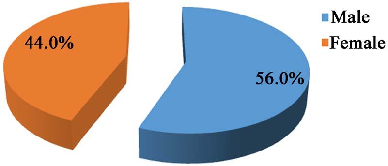
F. K. Onanga et al.

                               Keywords
                               Electrophoresis, Serum Proteins, Minicap, Protein Profiles

                             1. Introduction
                             Serum protein electrophoresis remains a widely used analytical method in clini-
                             cal biology [1] [2]. This examination currently makes it possible to link the elec-
                             trophoretic profile to a certain number of pathologies, including immune, in-
                             flammatory and hepatic diseases, nephrotic syndrome and cancers [3] [4] [5]. It
                             thus helps to refine the diagnosis, treatment and therapeutic monitoring of pa-
                             tients [6] [7] [8] [9]. In Congo Brazzaville, the electrophoretic profile of patients
                             passing through our laboratories is not known. Hence this study proposes an
                             analysis of the different electrophoretic profiles encountered during the analysis
                             of sera. The aim of the study was to show the electrophoretic profile of patients
                             who had been received at the Biochemistry laboratory at the Brazzaville Hospital
                             and University Center.

                             2. Methods
                             This is a retrospective study of 350 patients aged 12 to 85 received at the Bio-
                             chemistry laboratory of the Brazzaville University Hospital from January to De-
                             cember 2015. The electrophoresis of serum proteins was carried out on fasting
                             samples, taken on dry tubes after centrifugation at 3000 revolutions for 5 mi-
                             nutes. Hemolyzed, opalescent and lactescent samples were excluded. These sam-
                             ples were stored at +4˚C and then analyzed within 2 days of receipt in the labor-
                             atory, without exceeding one week. The capillary electrophoresis technique on a
                             Minicap Flex piercing automaton made it possible to define the different profiles
                             and the total protein assay was carried out by the Biuret method on a Cobas
                             C111 automaton from Roche.

                             3. Results
                             Analysis of the 350 identified cases revealed that 275 came from the different
                             departments of the CHU, whose distribution was 155 sera from women and 195
                             sera from men (Figure 1).
                                The ages of our patients ranged from 12 to 85 years (Figure 2). The age group
                             over 55 was more represented with a frequency of 50%.
                                The most demanding departments were the gastroenterology department
                             (33%), rheumatology (31%), hematology (22%) and the pediatric department
                             (14%) (Figure 3).
                                Relevant clinical information was missing in over 80% of requests. When in-
                             dicated, the reasons for prescribing in descending order were liver disease,
                             long-term fever, rheumatoid arthritis and 24 hour proteinuria elevation (Table
                             1).

DOI: 10.4236/oalib.1107902                         2                                   Open Access Library Journal
F. K. Onanga et al.

                                               Figure 1. Distribution of the study population by sex.

                                   Figure 2. Distribution of the study population by age.

                             Figure 3. Distribution of the study population according to the requesting services.

                             Table 1. Distribution of the study population according to the reasons for prescriptions.

                                         Reasons for prescriptions                 Effective                  %

                                        Lack of clinical information                  280                    80.0

                                               Hepatopathy                            41                     11.7

                                             Fever during log                         14                      4.0

                                           Rheumatoid arthritis                       11                      3.1

                                         High 24 hour proteinuria                      4                      1.1

DOI: 10.4236/oalib.1107902                            3                                     Open Access Library Journal
F. K. Onanga et al.
In Table 2, we noted that 26% of the exam performed was normal and 74%
had returned pathological. Inflammation was noted in 194 (55%) of cases of
which 145 (41%) were chronic and 49 (14%) acute. Forty two (12%) of our pa-
tients had beta-gamma block. Eleven (3%) patients presented with a picture of
nephrotic syndrome accompanied by severe hypoalbuminemia. We observed
monoclonal peaks in 12 patients (3%) located in the gamma globulin zone, indi-
cating dysglobulinemia (Table 2).
4. Discussion
Capillary electrophoresis allowed us to observe the morphology of the different
fractions, and to make a consistent interpretation. It is a very sensitive method
which allows a very fine definition of the different peaks but which nevertheless
requires a critical interpretation. Capillary electrophoresis allows a complete au-
tomation of the analysis associated with a fast and resolving free solution separa-
tion into six protein fractions major (albumin, α1-, α2-, β1-, β2-, and
γ-globulins) [1]. This analytical tool makes it possible to detect the major syn-
dromes confronting clinicians throughout their practice in a hospital environ-
ment [2] [3].
Since their application in clinical biology, electrophoresis techniques have
shown all their interest in the detection of monoclonal dysglobulinemia at low
cost [8] and have benefited from significant developments [6].
In our study, 12 monoclonal peaks were found, all of which (100%) migrated
to the gamma globulin zone. A study by Sunita Tripathy et al. carried out on a
series of 150 patients suspected of having multiple myeloma revealed 87.5% of
the monoclonal bands detected in the gammaglobulin area and only 12.5% of the
peaks are detected in the beta area [4] [10] [11] [12] [13]. On the other hand, the
study by Ouardia Bouayadi et al. carried out on 410 patients, 72.5% of monoc-
lonal bands are detected in the gammaglobulin zone and barely 27.5% in the be-
ta zone [5] [14] [15] [16] [17]. Faced with the presence of these monoclon-
al-looking bands, an indication of immunotyping was essential to characterize
the peaks, which was undoubtedly the limitation of this study.
5. Conclusion
Electrophoresis of serum proteins is a frequently prescribed biomedical practice
Table 2. Distribution of the study population according to profile type.
Profile type n = 350 %
Normal 91 26.0
Inflammation 196 56.0
Nephrotic syndrome 10 3.0
Beta-gamma block 43 12.0
monoclonal peak 10 3.0
DOI: 10.4236/oalib.1107902 4 Open Access Library JournalF. K. Onanga et al.
for the demonstration of qualitative and/or quantitative abnormalities of serum
proteins. Its main and indisputable indication is the low-cost screening for mo-
noclonal dysglobulinemia. The interpretation of this examination turns out to be
easy, especially if good medical prescription practices are followed and if the
medical biologist takes into account certain interpretation difficulties.
State of current knowledge on the subject is:
• Routine examination in the medical biochemistry laboratory;
• Diagnosis of monoclonal gammopathies;
Contribution of our study to knowledge is:
• Interest of the clinician-biologist collaboration for a correct interpretation;
• Know how to have EPS interpretation recommendations.
Contributions from the Authors
Principal editors: Fylla Onanga Koumou, Etienne Mokondjimobé.
Critical readers: Benjamin Longo Mbenza, Etienne Mokondjimobé.
Biological analyzes: Aliocha Nkodila, Monde Ikia, Barnes Yoyo, Rod Ibara,
Jeanne Gambomi Kiba.
CR Dobhat-Doukakini, Reine F. Eboka-Loumingou Sakou, Childerick Lekana.
Supervision: Benjamin Longo Mbenza, Etienne Mokondjimobé.
Conflicts of Interest
The authors declare no conflicts of interest.
References
[1] Albert, A., Gaume, M., Ughetto, S., Sapin, V. and Fogli, A. (2010) Évaluation du
couplage protéinémie + électrophorèse des protéines sériques totales par technique
capillaire (Capillarys 2, Sebia): Expérience clermontoise. Annales de Biologie
Clinique, 68, 657-667.
[2] Szymanowicz, A., Cartier, B., Couaillac, J.P., Gibaud, C., Poulin, G., Rivière, H., et
al. (2006) Proposition de commentaires interprétatifs prêts à l’emploi pour
l’électrophorèse des protéines sériques. Annales de Biologie Clinique, 64, 367-803.
[3] Le Carrer, D. (1995) Profils électrophorétiques ou profile protéiques? Intérêts
respectives et limites d’utilisations. Revue Française des Laboratoires, 1995, 79-85.
https://doi.org/10.1016/S0338-9898(95)80258-4
[4] Tripathy, S. (2012) The Role of Serum Protein Electrophoresis in the Detection of
Multiple Myeloma: An Experience of a Corporate Hospital. Journal of Clinical and
Diagnostic Research, 6, 1458-1461.
[5] Ouardia, B., Mohammed, B., Nawal, R., Said, A. and Mohammed, C. (2019)
Electrophorèse des protéines sériques: Etude de 410 profils électrophorétiques. The
Pan African Medical Journal, 32, 161.
https://doi.org/10.11604/pamj.2019.32.161.11455
[6] Karfo, R., Kabré, E., Safir, N., Bouabdellah, M., Benchekroun, L., Sakandé, J., et al.
(2018) Interprétation délicate de l’immunofixation des protéines sériques. The Pan
African Medical Journal, 30, 130. https://doi.org/10.11604/pamj.2018.30.130.13662
[7] Le Carrer, D. and Bach-Ngohou, K. (2004) L’électrophorèse capillaire automatisée
DOI: 10.4236/oalib.1107902 5 Open Access Library JournalF. K. Onanga et al.
en biologie clinique. Colloque du Syndicat national des biologistes des hopitaux
(SNBH) 2004.
[8] Oualla, J. (2018) Profil d’électrophorèse des protéines sériques chez une population
des hémodialysés chroniques. Thèse N˚ 256.
[9] Bissan, T., Diawara, A., Karfo, A., Teguete, A., Tangara, O., Guindo, A., et al. (2020) Une
électrophorèse des protéines sériques insolites dans un contexte de cholangiocarcinome.
The Pan African Medical Journal, 35, 117.
https://doi.org/10.11604/pamj.2020.35.117.20616
[10] Vavricka, S.R., Burri, E., Beglinger, C. and Degen, L. (2009) Électrophorèse des
protéines sériques: Un test sous-utilisé mais très utile. Digestion, 79, 203-210.
https://doi.org/10.1159/000212077
[11] Vijayashree, N. (2009) Le schéma d’électrophorèse des protéines sériques chez les
patients atteints de maladies chroniques dans un hôpital de soins tertiaires. Indian
Journal of Clinical Biochemistry, 24, 204.
[12] Nayak, B.S., Mungrue, K., Gopee, D., Friday, M., Garcia, S., Hirschfeld, E., et al.
(2011) L’épidémiologie du myélome multiple et le rôle de la détection de la bande M
sur l’électrophorèse sérique dans un petit pays en développement. Une étude
rétrospective. Archives of Physiology and Biochemistry, 117, 236-240.
https://doi.org/10.3109/13813455.2011.582875
[13] Kyle, R.A. and Rajkumar, S.V. (2009) Les critères pour le diagnostic, la stadification,
la stratification du risque et l’évaluation de la réponse du myélome multiple.
Leucémie, 23, 3-9.
[14] Katzmann, J., Kyle, R.A. and Lust, J. (2013) Immunoglobulines et reconnaissance en
laboratoire des protéines monoclonales. In: Wiernik, P.H., Goldman, J.M., Dutcher,
J.P., et al., Eds., Maladies néoplasiques du sang, 5, Springer, New York, 565-588.
[15] Tschumper, R.C., Dispenzieri, A. and Abraham, R.S. (2013) L’analyse moléculaire
des gènes d’immunoglobulines révèle une parenté clonale fréquente dans les
gammapathies monoclonales doubles. Journal du cancer du sang, 3, e112.
[16] Guastafierro, S., Ferrara, M.G. and Sica, A. (2012) Doubles composants monoclonaux
sériques et hémopathies malignes: seulement une association fortuite ? Examen de 34
cas. Leukemia Research, 36, 1274-1277.
https://doi.org/10.1016/j.leukres.2012.05.008
[17] Garcia-Garcia, P., Enciso-Alvarez, K. and Diaz-Espada, F. (2015) Gammopathies
biclonales: Etude rétrospective de 47 patients. Revista Clínica Española, 215, 18-24.
https://doi.org/10.1016/j.rce.2014.07.003
DOI: 10.4236/oalib.1107902 6 Open Access Library JournalYou can also read