ENTAMOEBA SP. INFECTION IN A BEARDED DRAGON (POGONA VITTICEPS)
←
→
Page content transcription
If your browser does not render page correctly, please read the page content below
SCHWARTZ Diana et al.: Entamoeba invadens in reptiles

                                                          Veterinarski Glasnik 2020, 74 (1), 77-84
                                                                      UDC: 636.98.09:616.913.12
                                                   https://doi.org/10.2298/VETGL190919007S
Case Report

ENTAMOEBA SP. INFECTION IN A BEARDED DRAGON
(POGONA VITTICEPS)

SCHWARTZ Diana1*, ALI M. Ibne Karim2, ROY Shantanu2, POHLMAN M. Lisa1,
KASTL Brandy1, ESHAR David3

1
 Kansas State University, College of Veterinary Medicine, Department of Diagnostic Medicine/
Pathobiology, Manhattan, Kansas, USA;
2
 Centers for Disease Control and Prevention, 1600 Clifton Road NE, Atlanta, Georgia, 30329, USA;
3
 Kansas State University, College of Veterinary Medicine, Department of Clinical Sciences, Manhattan,
Kansas, USA

Received 19 September 2019; Accepted 18 January 2020
Published online: 17 March 2020

Copyright © 2020 Schwartz et al. This is an open-access article distributed under the Creative Commons
Attribution License, which permits unrestricted use, distribution, and reproduction in any medium,
provided the original work is properly cited

How to cite: Diana Schwartz, Ibne Karim M. Ali, Shantanu Roy, Lisa M. Pohlman, Brandy
Kastl, David Eshar. Entamoeba sp. infection in a bearded dragon (Pogona vitticeps). Veterinarski
Glasnik, 2020. 74 (1), 77-84. https://doi.org/10.2298/VETGL190919007S

       Abstract
       A 3-year-old, male intact, pet inland bearded dragon (Pogona vitticeps) presented with
       a history of diarrhea, progressive inappetence and weight loss. A palpable cranial
       celomic mass was identified on physical examination and confirmed to be hepatic in
       origin by celomic ultrasonography. Hematologic and biochemical abnormalities were
       mild and consistent with inflammation, regenerative anemia, and hepatocellular injury.
       Fine needle aspiration of the liver masses was suggestive of amoebiasis and the patient
       was humanely euthanized. PCR and Sanger DNA sequencing of liver aspirates were
       supportive of Entamoeba infection, although definitive speciation was not possible.
       Pathogenic amoebiasis due to infection by E. invadens has been reported in a wide range
       of reptiles and is an important cause of morbidity and mortality in these species.
       Key words: Entamoeba, enteritis, liver abscess, Pogona vitticeps, reptilia

*Corresponding author – e-mail: diana.schwartz.dvm@gmail.com

                                                                                                   77
Veterinarski Glasnik 2020, 74 (1), 77-84
CASE PRESENTATION
A 3-year-old, male intact, pet inland bearded dragon (Pogona vitticeps) presented to the
Exotics and Zoological Animal Medicine Service at Kansas State University following
several months of diarrhea, decreased appetite progressing to anorexia, and weight
loss. On physical examination, the patient was quiet and alert with a palpable mass
in the cranial celom. Complete blood count revealed a mild heterophilia (7,800/uL)
with mild toxic change, mild monocytosis (2,100/uL) and a mild lymphopenia (1,800/
uL), consistent with inflammation. There was also a mild anemia (PCV 18%) with
mildly increased numbers of immature erythrocytes. Serum chemistry revealed a
mild increase in AST (44 U/L) (Ellman 1997, Tamukai et al. 2011). Two view whole
body radiographs were performed and a large soft tissue opacity mass (41 x 20 mm)
was observed filling the cranial celomic cavity with resultant dorsal displacement and
compression of the trachea. Another large, mid-celomic mass was observed (55 x 30
mm). There was a diffuse interstitial to coalescing nodular pulmonary pattern and
body condition was assessed to be thin given the absence of caudal fat bodies. Celomic
ultrasonography confirmed an irregularly marginated echogenic mass caudal to the
heart and multiple well-circumscribed hypo to mixed echogenic mass lesions within
the liver (Figure 1). A moderate amount of echogenic effusion was also observed
within the celomic cavity and within the pericardial sac. Fine needle aspiration of the
hepatic masses was performed. The sample was of low intact nucleated cellularity
and consisted mostly of necrotic and apoptotic cell debris with frequent vacuolated
Figure 1. Sagital ultrasonographic image of a pet inland bearded dragon (Pogona vitticeps)
demonstrating a mass associated with the liver. The white arrows outline the borders of the
mass. CR indicates the cranial aspect of the coelom, CD indicates the caudal aspect of the
coelom, and V indicates the ventral aspect of the coelom.
78SCHWARTZ Diana et al.: Entamoeba invadens in reptiles
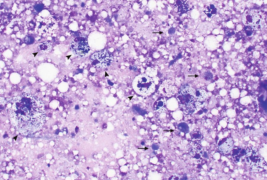
and debris laden macrophages (Figure 2), few intact hepatocytes, and rare structures
that were concerning for, but not diagnostic of, amoeba trophozoites. Based on these
findings Entamoeba invadens infection was suspected and the owners elected humane
euthanasia without necropsy.
Figure 2. Wright-Giemsa stained liver aspirate from a pet inland bearded dragon (Pogona vitticeps).
Highly vacuolated macrophages (black arrowheads) are observed within a purple coarsely
stippled background with acellular lipid droplets and necrotic cellular debris (black arrows). 50x.
The air-dried Wright-Giemsa stained liver aspirates were submitted to the Free-Living
and Intestinal Amebas (FLIA) Laboratory within the Waterborne Disease Prevention
Branch of the U.S. Centers for Disease Control and Prevention (CDC). The
sample was negative for E. histolytica and E. dispar by the diagnostic duplex real-time
polymerase chain reaction (PCR) assay (Qvarnstrom et al., 2005). Generic Entamoeba
sp. conventional PCR assay (Stensvold et al., 2011) was positive, and Sanger sequencing
results (3’-ended 57-bp clean sequence: CCCTTCCGTCAATTTCTTTAAGTTCAG
CCTTGTGACCATACTCCCCCTGAAGTAAG, GenBank accession number MN
879388) were 100% homologous with Entamoeba muris; however, repeated amplification
and sequencing attempts to confirm these findings were unsuccessful.
DISCUSSION
Entamoeba muris is a non-pathogenic species of amoeba that colonizes the intestine of
wild and domestic rodents (Hooshyar, Rostamkhani, and Rezaeian, 2015). E. muris
has been identified in feces of captive tortoises (Wolf et al., 2014), and in this context
is assumed to transit through the intestinal tract of these individuals, presumably
following access in the environment to rodent feces containing cysts. Identification
of E. muris DNA within a liver aspirate in the case of this pet inland bearded dragon
79Veterinarski Glasnik 2020, 74 (1), 77-84 (Pogona vitticeps) would suggest an invasive biological behavior, although these results could not be confirmed, possibly due to poor DNA quality, as the assay was not developed for use with fine needle aspirate samples. It is also possible that the Entamoeba species identified in this sample shares some homology with E. muris but is in fact a distinct species and a pathogen. The clinical presentation and imaging findings in this case were suggestive of E. invadens (Gardhouse et al., 2015), an established pathogen in reptiles, and thus, further discussion will focus on this species. E. invadens is an amoebozoan organism, which can act as either a gastrointestinal commensal or a pathogen, in many species of reptiles. E. invadens has a direct life cycle in which cysts are ingested directly from feces or from a contaminated environment. Excystation occurs within the host intestine releasing the motile trophozoites, which replicate and can either remain within the intestinal lumen feeding on ingesta, or can invade the mucosa resulting in ulcerative enterocolitis and diarrhea. Invasion of adjacent lymphatic and blood vessels can result in embolization of trophozoites to the liver via the portal vein, followed by widespread dissemination to distant sites including lung, spleen, kidney, and brain (Bonner, 2001; Kojimoto et al., 2001; Gardhouse et al., 2015; Park et al., 2019). Secondary localized or disseminated bacterial infections (typically involving enteric organisms) can be present and contribute to morbidity in affected individuals. Factors that influence encystation have not been entirely elucidated, although ambient temperature has been shown to affect cyst formation in experimentally infected Eastern garter snakes (Thamnophis sirtalis) (Meerovitch, 1961). Trophozoites rapidly degrade outside the body (Brewer et al. 2008), but cysts can remain viable and infective in the environment for weeks to months (Bradford et al., 2008). While environmental decontamination protocols have not been established, E. histolytica cysts are rapidly killed when exposed to high temperatures (52°C/126°F), so steam and hot water could be effective against E. invadens cysts (Bonner, 2001). Aquatic reptiles, especially chelonians and crocodilians (Bonner, 2001; Brewer et al., 2008; Garcia et al. 2014), and herbivorous species (Kojimoto et al., 2001) are typically subclinical carriers, although young, old, immunocompromised, or otherwise debilitated individuals (such as following capture and/or transport) of any reptilian species can develop clinical disease. Snakes and lizards appear to be especially prone to clinical illness (Bonner, 2001). E. invadens is highly contagious and has caused outbreaks in juvenile (MacNeill et al., 2002) and wild-caught (Ozaki et al., 2000) chelonians, and in captive pythons (Kojimoto et al., 2001). Pathogenicity is also dependent on differences in strain virulence and ambient host temperature (Meerovitch, 1961; Garcia et al., 2014). The most common presenting complaints in affected reptiles are decreased appetite progressing to anorexia, regurgitation, and diarrhea, although some individuals present dead or acutely moribund (Ozaki et al., 2000; Bonner, 2001; Kojimoto et al., 2001; MacNeill et al., 2002; Brewer et al., 2008; Baseler et al., 2014). Hematologic and biochemical abnormalities associated with E. invadens are poorly characterized. One report in a green iguana documented similar hematologic findings as we observed in our case including decreased PCV with evidence of regeneration 80
SCHWARTZ Diana et al.: Entamoeba invadens in reptiles

and a moderate leukocytosis due to heterophilia and monocytosis, with toxic change
and a left shift (Nikousefat, 2014).
Fresh fecal smears can be used for ante mortem diagnosis of E. invadens (Bonner,
2001; Garcia et al., 2014); however, false negatives are possible as animals can shed
intermittently or have low burden infections. Multiple fecal examinations could be
needed to identify trophozoites and/or cysts; however, in general, fecal examination is
a relatively insensitive test for E. invadens. Iodine wet mounts of fresh feces (Garcia et
al., 2014) or fixation of fecal smears with polyvinyl alcohol followed by trichrome and
iron hematoxylin staining has been reported to highlight the organisms (Bonner, 2001);
however, other non-pathogenic amoeba can have a similar appearance (Bradford et al.,
2008; Garcia et al., 2014; Park et al., 2019). Trophozoites can be cultured from fresh
feces using Robinson’s medium, and this is a more sensitive method than direct fecal
examination, but culture is time consuming and Robinson’s medium can selectively
exclude the growth of concurrent pathogens (Garcia et al., 2014). PCR is a sensitive
technique for identifying E. invadens in feces (Bradford et al., 2008) and tissues (Chia
et al., 2009; Park et al., 2019), although might not be sufficiently specific to exclude
other fecal protozoa, such as Blastocystis species (Garcia et al., 2014). DNA sequencing
is, therefore, recommended to identify species in positive PCR samples. Unfortunately,
there are no commercially available PCR assays for E. invadens. Serologic methods of
detecting E. invadens have yet to be developed and positive fecal samples from reptiles
did not cross-react with an E. histolytica ELISA (Brewer et al., 2008).
For animals that succumb to infection or are euthanized, the most commonly
reported gross findings on necropsy are colonic edema and multifocal to coalescing
gastrointestinal ulcers, sometimes with concurrent mucosal pseudomembrane
formation (Meerovitch, 1961; Ozaki et al., 2000; Bonner, 2001; Kojimoto et al., 2001;
Nikousefat, 2014; Park et al., 2019). Generalized hepatomegaly is also frequently
reported (Ozaki et al., 2000; MacNeill et al., 2002; Chia et al., 2009; Baslet et al.,
2014; Gardhouse et al., 2015; Park et al., 2019). Trophozoites can be readily identified
in histologic sections of affected tissues, and are highlighted by PAS (Park et al.,
2019) and silver stains (Kojimoto et al. 2001; Baseler et al., 2014). PAS and trichrome
staining has also been described as highlighting trophozoites in emulsified and
polyvinyl alcohol-fixed liver samples (MacNeill et al., 2002). Other histologic findings
include necrosis and hemorrhage with mixed inflammation including heterophils,
lymphocytes, macrophages, and eosinophils (Ozaki et al., 2000; Kojimoto et al., 2001;
Baseler et al., 2014). Fluorescent antibodies against E. invadens applied to formalin-
fixed tissues can be helpful in confirming infection, although background staining can
render interpretation challenging (Baseler et al., 2014; Kojimoto et al., 2001).
E. invadens is an important contributor to morbidity and mortality in reptiles. While a
variety of ante mortem diagnostic tests are available, most are insufficiently specific to
distinguish E. invadens from other non-pathogenic amoeba, and thus, PCR followed
by DNA sequencing remains the most reliable ante mortem method for confirming
infection. Unfortunately, there is no commercially available PCR assay for E. invadens.
                                                                                         81
Veterinarski Glasnik 2020, 74 (1), 77-84
Fine needle aspirates of affected tissues can aid in the ante mortem diagnosis of amoebic
infections in reptiles, even if trophozoites cannot be confirmed morphologically, by
providing material for PCR and DNA sequencing. Identification of trophozoites
within histologic sections (collected via biopsy or at the time of necropsy) of intestine,
liver, and other tissues also confirms pathogenic amoeba infection, and trophozoites
can be highlighted by special stains. Unfortunately, once clinical signs have developed,
treatment is often ineffective (Ozaki et al., 2000; MacNeill et al., 2002; Garcia et al.,
2014). Care to avoid potential exposure by limiting mixed-housing of different reptilian
species, especially of chelonians with snakes and lizards, is recommended.
Acknowledgements
The authors thank D. Biller at the Kansas State University Veterinary Teaching Hospital
for providing ultrasound images and interpretation for the publication.
Authors contributions
All authors contributed to manuscript preparation and approval. DS additionally
contributed the photomicrographs of the liver aspirate cytology.
Competing interests
The findings and conclusions in this report are those of the authors and do not
necessarily represent the official position of CDC.
REFERENCES
Baseler L. J., Visvesvara G. S., Ramos-Vara J. A. 2014. Pathology in practice. E invadens infection
in a ball python. Journal of the American Veterinary Medical Association, 245 (5):501-3.
doi: 10.2460/javma.245.5.501.
Bonner B., Denver M., Gamer M., Innis C., Nathan R. 2001. Entamoeba invadens. Journal of
Herpetological Medicine and Surgery, 11(3):6.
Bradford C. M., Denver M. C., and M. R. Cranfield. 2008. Development of a polymerase chain
reaction test for Entamoeba invadens. Journal of Zoo and Wildlife Medicine, 39(2):201-207.
doi: 10.1638/2007-0145.1.
Brewer L. A., Denver M. C., Whitney M., Eichinger D. J. 2008. Analysis of commercial
Entamoeba histolytica ELISA kits for the detection of Entamoeba invadens in reptiles. Journal
of Zoo and Wildlife Medicine, 39(3):493-495. doi: 10.1638/2007-0182.1.
Chia M. Y., Jeng C. R., Hsiao S. H., Lee A. H., Chen C. Y., Pang V. F. 2009. Entamoeba invadens
myositis in a common water monitor lizard (Varanus salvator). Veterinary Pathology,
46(4):673-6. doi: 10.1354/vp.08-VP-0224-P-CR.
Ellman M. M. 1997. Hematology and plasma chemistry of the inland bearded dragon, Pogona
vitticeps. Bulletin of the Association of Reptillian and Amphibian Veterinarians, 7(4):3.
Garcia G., Ramos F., Perez R. G., Yanez J., Estrada M. S., Mendoza L. H., Martinez-Hernandez
F., Gaytan P. 2014. Molecular epidemiology and genetic diversity of Entamoeba species
82SCHWARTZ Diana et al.: Entamoeba invadens in reptiles
in a chelonian collection. Journal of Medical Microbiology, 63:271-283. doi: 10.1099/
jmm.0.061820-0.
Gardhouse S., Pritchard W. T., Eshar D., Biller D. S., Schumacher L., Almes K. 2015. Untitled.
Journal of Exotic Pet Medicine, 24(2):249-253. doi: 10.1053/j.jepm.2015.04.017.
Hooshyar H., Rostamkhani P., Rezaeian M. 2015. An annotated checklist of the human and
animal Entamoeba (Amoebida: Endamoebidae) species – A review Article. Iranian Journal
of Parasitology, 10(2):146-56.
Kojimoto A., Uchida K., Horii Y., Okumura S., Yamaguch R., Tateyama S. 2001. Amebiasis
in four ball pythons, Python reginus. Journal of Veterinary Medical Science, 63(12):1365-8.
MacNeill A. L., Uhl E. W., Kolenda-Roberts H., Jacobson E. 2002. Mortality in a wood turtle
(Clemmys insculpta) collection. Veterinary Clinical Pathology, 31(3):133-6.
Meerovitch E. 1961. Infectivity and pathogenicity of polyxenic and monoxenic Entamoeba
invadens to snakes kept at normal and high temperatures and the natural history of reptile
amoebiasis. Journal of Parasitology, 47(5):791-794. doi: 10.2307/3275473.
Nikousefat Z., Javdani M., Hashemnia M., Rezaei F., A. Chalechale. 2014. Hematology of
a green Iguana infected by Entamoeba invadens. Online Journal of Veterinary Research,
18(11):8.
Ozaki K., Matsuo K., Tanaka O., Narama I. 2000. Amoebosis in the flat-shelled spider tortoise
(Acinixys planicauda). Journal of Comparative Pathology, 123(4):299-301. doi: 10.1053/
jcpa.2000.0417.
Park C. H., Han J. B., Park S. I. 2019. Dual infection with Entameoba invadens and Aeromonas
hydrophila in a captive anaconda (Eunectes murinus) leading to necrotising gastroenteritis and
hepatocyte death. Veterinarni Medicina, 64(3):144-148. doi: 10.17221/140/2018-Vetmed.
Qvarnstrom Y., James C., Xayavong M., Holloway B. P., Visvesvara G. S., Sriram R., da Silva
A. J. 2005. Comparison of real-time PCR protocols for differential laboratory diagnosis of
amebiasis. Journal of Clinical Microbiology, 43(11):5491-7. doi: 10.1128/JCM.43.11.5491-
5497.2005.
Stensvold C. R., Lebbad M., Victory E. L., Verweij J. J., Tannich E., Alfellani M., Legarraga P.,
Clark C. G. 2011. Increased sampling reveals novel lineages of Entamoeba: Consequences of genetic
diversity and host specificity for taxonomy and molecular detection. Protist, 162(3): dio: 10.1016/j.
protis.2010.11.002
Tamukai K., Takami Y., Akabane Y., Kanazawa Y., Une Y. 2011. Plasma biochemical reference
values in clinically healthy captive bearded dragons (Pogona vitticeps) and the effects
of sex and season. Veterinary Clinical Pathology, 40(3):368-73. doi: 10.1111/j.1939-
165X.2011.00329.x.
Wolf D., Vrhovec M. G., Failing K., Rossier C., Hermosilla C., Pantchev N. 2014. Diagnosis
of gastrointestinal parasites in reptiles: comparison of two coprological methods. Acta
Veterinaria Scandinavica, 56:44. doi: 10.1186/s13028-014-0044-4.
83Veterinarski Glasnik 2020, 74 (1), 77-84 ENTAMOEBA SP. INFEKCIJA KOD BRADATE AGAME (POGONA VITTICEPS) SCHWARTZ Diana, ALI M. Ibne Karim, ROY Shantanu, POHLMAN M. Lisa, KASTL Brandy, ESHAR David Kratak sadržaj Trogodišnji nekastrirani mužjak bradate agame (Pogona vitticeps) koji se drži kao kućni ljubimac je doveden na kliniku zbog dugotrajne dijareje, progresivnog gubitka apetita i težine. Kliničkim pregledom je otkrivena palpabilna kranijalna celomska masa i ultrazvukom je utrvrđeno da čini deo jetre. Poremećaji hematoloških i biohemijskih parametara su bili blagi i ukazivali su na inflamaciju, regenerativnu anemiju i hepatocelularna oštećenja. Tankoiglenom biopsijom mase otkrivene na jetri ukazivala je na amebijazu i pacijent je eutaniziran. PCR i DNK sekvencioniranje su takođe ukazivali na infekciju Entamoeba-om, mada definitivno određivanje vrste nije bilo moguće. Patogena amebijaza usled infekcije sa E. invadens je dokazana kod velikog broja vrsta gmizavaca i značajan je uzročnik uginuća kod ovih vrsta životinja. Ključne reči: Entamoeba, enteritis, apsces jetre, Pogona vitticeps, reptilia 84
You can also read