Laparoscopic management of colovesical fistula secondary to sigmoid diverticulitis: case report and the role of intraoperative indocyanine-green ...
←
→
Page content transcription
If your browser does not render page correctly, please read the page content below
Case Report
                                                                                                                                       Page 1 of 7

Laparoscopic management of colovesical fistula secondary to
sigmoid diverticulitis: case report and the role of intraoperative
indocyanine-green fluorescence
Emanuele Asti1, Daniele Bernardi1, Erika Andreatta1, Andrea Conti2, Luca Carmignani2, Luigi Bonavina1^
1
    Division of General Surgery, Department of Biomedical Sciences for Health, University of Milan, Milan, Italy; 2Division of Urology, IRCCS
Policlinico San Donato, San Donato Milanese, Italy
Correspondence to: Prof. Luigi Bonavina. Piazza Malan 1, IRCCS Policlinico San Donato, San Donato Milanese, 20097 Milan, Italy.
Email: luigi.bonavina@unimi.it.

                  Abstract: Colovesical fistula is characterized by multifactorial etiology and the diagnosis is often
                  challenging. Sigmoid diverticulitis accounts for more than two-third of the cases. Pneumaturia and air in the
                  bladder at computed tomography are the most typical clinical-radiologic signs. There is still no evidence-
                  based consensus on the optimal surgical approach for this rare condition. Sigmoid resection with or without
                  bladder resection/enteral stoma are the conventional therapeutic options, but ureter identification may be
                  difficult during the procedure and injury from a direct or indirect trauma can occur. Therefore, double-J
                  stenting is commonplace during open surgical procedures. The laparoscopic approach has been shown to
                  be feasible and effective in case-series, but ureteral protection may be difficult because of the limited tactile
                  feedback. We report the case of a 55-year-old male patient with colovesical fistula complicating repeated
                  episodes of acute left colon diverticulitis and eventually treated with one-stage bladder-sparing laparoscopic
                  sigmoid resection. Indocyanine-green (ICG) fluorescence was used intraoperatively for visualization of
                  the left ureter and for assessment of colonic blood perfusion. The post-operative course was uneventful.
                  Laparoscopy is feasible, safe, and effective for the treatment of colovesical fistula. Magnified vision enhanced
                  by fluorescence imaging allows a precise surgical dissection and has the potential to decrease complication
                  rates and to improve postoperative outcomes.

                  Keywords: Sigmoid diverticulitis; colovesical fistula; laparoscopic sigmoid resection; indocyanine-green
                  fluorescence (ICG fluorescence); case report

                  Received: 25 June 2020; Accepted: 18 December 2020.
                  doi: 10.21037/jovs-20-140
                  View this article at: http://dx.doi.org/10.21037/jovs-20-140

Introduction                                                                     of colovesical fistula is estimated to be three times less
                                                                                 in women because of the protective effect of the uterus
Colonic diverticular disease is widespread in Western                            interposed between the sigmoid colon and the bladder.
countries and is often complicated by recurrent episodes                         Pneumaturia, fecaluria, and urinary tract infections are
of acute diverticulitis and perforation that may require                         the most common features due to the high pressure
emergency surgery and a Hartmann procedure. Colovesical                          gradient between the colonic lumen and the urinary tract
fistula is a rare condition caused by sigmoid diverticulitis                     system. Other symptoms and signs may include lower
in approximately two-thirds of the patients. The incidence                       abdominal pain, fever, and peritonism. Diagnostic methods

^ ORCID: 0000-0002-4880-1670.

© Journal of Visualized Surgery. All rights reserved.                                       J Vis Surg 2021 | http://dx.doi.org/10.21037/jovs-20-140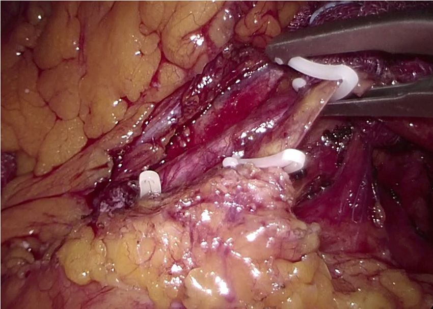
Page 2 of 7                                                                                                    Journal of Visualized Surgery, 2021

                                                                             One-stage laparoscopic sigmoid
                                        Foley catheter, clear liquid diet,   resection with ICG fluorescence
                    E.R.
                                          broad spectrum antibiotics                  May 19, 2020
               admission                                                                                                       Uro-CT
              April 25, 2020                                                                                                July 18, 2020

                                 CT scan                                                                               Cystoscopy
                               April 25, 2020                                                                         June 17, 2020
                                                                                      Discharged home with
                                Cystoscopy
                                                                                          Foley catheter
                               April 27, 2020
                                                                                          May 25, 2020
                               Colonoscopy
                               April 27, 2020

Figure 1 Timeline including work-up, patient preparation, laparoscopic management, and follow-up.

include abdomino-pelvic CT scan, MR, cystoscopy and                             room with acute lower abdominal pain and a 1-week history
colonoscopy, all with a rather low sensitivity (1). Although                    of refractory constipation. He did not report stranguria,
conservative treatment is feasible in selected high-risk                        pneumaturia, or fecaluria. Past medical history was
patients, surgical therapy offers the best chance of cure                       unremarkable except for multiple recurrent episodes of acute
given the risk of bladder outlet obstruction and urosepsis                      sigmoid diverticulitis. Blood pressure was 130/78 mmHg,
that require repeated courses of antibiotics and urinary                        heart rate 100 bpm, and oxygen saturation 97% on room air.
catheterization.                                                                Body temperature was normal. Physical exam showed mild
   The laparoscopic approach has been reported to be                            abdominal distension and tenderness in the lower abdominal
feasible and effective, but conversion rate to an open                          quadrants. Abdominal ultrasound showed a small fluid
procedure has been reported in up to 50% of patients                            collection in the left lower quadrant. Blood tests showed
depending on the degree of bladder infiltration (2).                            increased C-reactive protein (13.7 mg/dL) and white blood
Traditionally, especially in open surgery, use of ureteral                      count (12,500/uL with 86% neutrophils). Plain abdominal
stents was common practice for intraoperative identification                    X-ray did not reveal free air nor air-fluid images. A CT scan
of the ureters. During laparoscopic pelvic surgery, ureteral                    showed a complicated distal left colon diverticulitis with a
injuries occur in up to 10% of patients and are often                           long segment (7 cm) wall thickening, luminal narrowing
diagnosed post-operatively (3). Near-infrared fluorescence                      (Figure 2A), and multiple perivisceral abscesses (Figure 2B).
with indocyanine-green (ICG) injected intravenously is                          Moreover, air bubbles inside the bladder were visible
a technology most commonly used for the intraoperative                          (Figure 3), raising the suspicion of a colovesical fistula
assessment of biliary anatomy and bowel perfusion. Local                        (Figure 4A,B). The patient underwent a cystoscopy, which
ICG injection through a ureteral probe rapidly stains the                       confirmed the presence of a fistula on the posterior-lateral
urothelium, allows prompt visualization of the ureter and                       wall of the bladder 2 cm above the vesical trigone (Figure 5).
may reduce morbidity associated with ureteral stents. We                        A colonoscopy ruled out the presence of malignancy. The
present the following case in accordance with the Case                          patient was treated with a Foley catheter, clear liquid diet,
Report (CARE) reporting checklist (available at http://                         and broad spectrum antibiotics. Blood tests normalized in a
dx.doi.org/10.21037/jovs-20-140).                                               few days, and the patient was discharged home. Readmission
                                                                                for surgical treatment was planned in 3 weeks.

Case presentation
                                                                                Pre-operative preparation
The clinical course and timeline of diagnosis and treatment
is summarized in Figure 1.                                                      The patient maintained the urinary catheter for 4 weeks
                                                                                to reduce intra-bladder pressure and to allow spontaneous
                                                                                healing of the fistula. Low-residue diet was allowed.
Patient selection and workup
                                                                                Oral bowel preparation with polyethylene glycol was
A 55-year-old Caucasian male presented to the emergency                         administered 24 hours before surgery. Cefazoline 2 g and

© Journal of Visualized Surgery. All rights reserved.                                         J Vis Surg 2021 | http://dx.doi.org/10.21037/jovs-20-140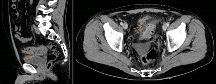
Journal of Visualized Surgery, 2021 Page 3 of 7
A B
Figure 2 CT scan showing distal left colon wall thickening and luminal narrowing (A), and pelvic collection (red mark) (B).
Wayne, Pennsylvania, USA);
Contour® Stapler (Ethicon, Somerville, New Jersey,
USA);
Echelon® circular powered stapler 29 mm (Ethicon,
Somerville, New Jersey, USA);
Alexis® wound retractor (Applied Medical, Rancho
Santa Margarita, CA, USA);
ICG Verdye® 5 mg/mL (Diagnostic Green GmbH,
Aschheim, Germany).
Figure 3 Air bubbles are visible inside the bladder (b), strictly Procedure
adherent to the colonic wall (c).
All procedures performed in this study involving human
participants were in accordance with the ethical standards of
metronidazole 500 mg were given as antibiotic prophylaxis. the institutional research committee and with the Helsinki
Declaration (as revised in 2013). Written informed consent
was obtained from the patient.
Equipment preference cart The patient was positioned supine on the operative
Visera Elite II OTV-S300® video system (Olympus, table with the legs abducted. Under general anesthesia,
Shinjuku, Tokyo, Japan); a cystoscopy was performed, and a 6 Fr Pollack catheter
Visera Elite II CLV-S200-IR® light source (Olympus, was introduced in the left ureter to allow intraoperative
Shinjuku, Tokyo, Japan); injection of ICG. Pneumoperitoneum was induced through
12-mm and 5-mm laparoscopic trocar; a Veress needle placed in the umbilicus. Four ports
10-mm 30° laparoscope; (two 12-mm and two 5-mm) were placed as for laparoscopic
Laparoscopic Croce-Olmi grasper (Karl Storz, left colectomy (Figure 6). Upon exploration of the
Tuttlingen, Germany); abdominal cavity, the sigmoid was tenaciously adherent to
Laparoscopic single-use Endo Clinch (Covidien, the posterior-lateral aspect of the bladder (Figure 7). The
Dublin, Ireland); small bowel was retracted away from the operative field.
Laparoscopic peanuts; Partial mobilization of the descending and sigmoid colon
Laparoscopic Endopath Electrosurgery Probe Plus II® was performed using the monopolar hook and harmonic
(Ethicon, Somerville, New Jersey, USA); scalpel. The inferior mesenteric artery and vein were
Laparoscopic Ultracision Harmonic Scalpel® (Ethicon, clipped with Hem-o-lok and sectioned (Figure 8). The left
Somerville, New Jersey, USA); ureter was visually identified and followed along its route
Hem-o-lok clip (Weck Closure Systems, Teleflex, toward the pelvis, then it could not be clearly distinguished
© Journal of Visualized Surgery. All rights reserved. J Vis Surg 2021 | http://dx.doi.org/10.21037/jovs-20-140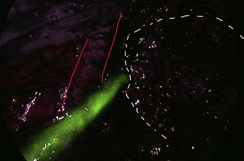
Page 4 of 7                                                                                                Journal of Visualized Surgery, 2021

                       A                                B

Figure 4 CT scan showing colovesical fistula (arrow) on sagittal (A) and axial plane (B).

                                                                                                           Monitor

                                                                                                     3rd

                                                                                                                          Scrub
                                                                                                                          nurse
                                                                                                   1st
                                                                                                                2nd

Figure 5 Cystoscopy showing fecal leakage from the left posterior         Figure 6 Laparoscopic setting and trocar positioning.
wall of the bladder.

from the pseudotumor involving the sigmoid colon and                      Griffen colorectal anastomosis was performed using a
the bladder. Infusion of 5 mL of ICG through the ureteral                 29-mm circular stapler. Two abdominal drains were left in
catheter allowed to visualize the left ureter (Figure 9) and              the pelvis, and the abdominal incisions were closed. Total
guided the dissection of the sigmoid from the bladder                     duration of the procedure was 220 minutes, and blood loss
                                                                          was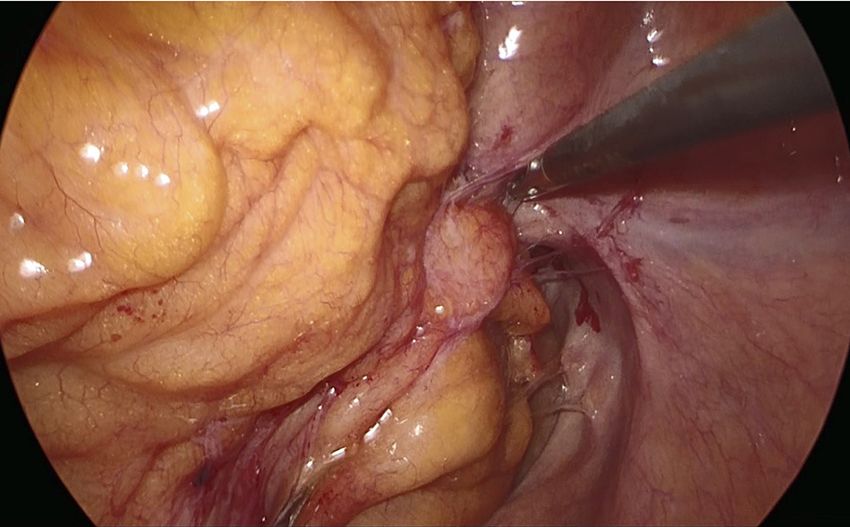
Journal of Visualized Surgery, 2021 Page 5 of 7
Bladder
Pseudotumor
IIiac Artery
Figure 7 Laparoscopic view of the inflammatory pseudotumor
Figure 9 Use of ICG-fluorescence to identify the left ureter. ICG,
surrounding the colovesical fistula.
indocyanine-green.
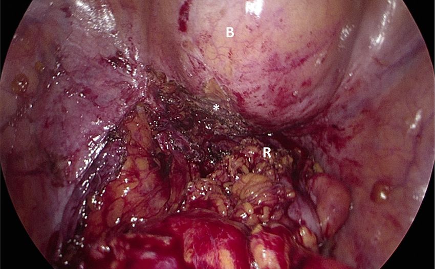
Figure 10 Laparoscopic view of the pelvis after dissection of the
Figure 8 Dissection of the inferior mesenteric artery using Hem-
colovesical fistula (*). The bladder (B) is filled with methylene blue
o-lok clips.
solution, and there is no evidence of leaks. R, rectal stump.
ureter cannulation and remained on call to provide assistance Tips, tricks, and pitfalls
in case a partial cystectomy could have become necessary.
An integrated surgical and urological team is a pre-
requisite for a successful outcome of the procedure.
Post-operative management In case of acute diverticulitis (Hinchey 1–2), leave
an indwelling urinary catheter and proceed with
The ureteral catheter was removed at the end of the
broad spectrum antibiotic therapy for at least
procedure. The postoperative course was uneventful. 3 weeks before surgery to facilitate recovery from
Liquid diet was initiated on the second postoperative day. inflammation/infection and spontaneous fistula
The patient was discharged on the 6th postoperative day healing.
with a Foley catheter in place. An outpatient cystoscopy Laparoscopy may allow a bladder-sparing, one-stage
was performed three weeks later by the consultant urologist minimally invasive sigmoid resection.
to confirm the complete healing of the bladder, and the Bladder leak testing with methylene blue should be
urinary catheter was removed. Histopathology examination routinely performed intraoperatively.
of the surgical specimen confirmed the inflammatory nature ICG fluorescence through a ureteral catheter helps
of the pseudotumor. At the 3-month follow-up the patient to visualize the ureter and may reduce the risk of
reported normal urinary and bowel function, was very intraoperative injuries.
pleased with the outcome of the procedure and returned to Intravenous ICG is useful to visualize colonic
his usual business activity. A URO-CT scan showed normal perfusion after rectal stapling and to choose the ideal
findings (Figure 11). site for the colorectal anastomosis.
© Journal of Visualized Surgery. All rights reserved. J Vis Surg 2021 | http://dx.doi.org/10.21037/jovs-20-140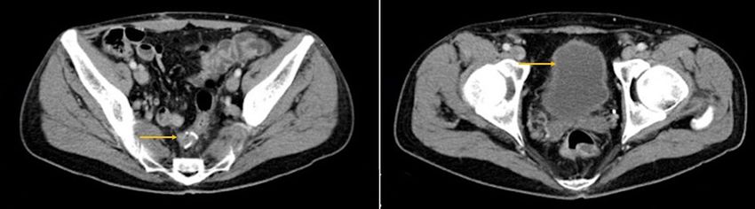
Page 6 of 7                                                                                        Journal of Visualized Surgery, 2021

            A                                                     B

Figure 11 Postoperative CT scan showing normal findings. Arrows indicate the mechanical colorectal anastomosis (A) and the bladder
without air-fluid levels (B).

Discussion                                                            it has already been shown that intra-ureteral ICG allows
                                                                      to visualize the ureters during a variety of laparoscopic
This case study is exemplary of the ideal, bladder-sparing
                                                                      procedures for up to 6 hours (9-11). Moreover, this
management of a benign colovesical fistula, and adheres
                                                                      technique avoids the potential complications associated
to the CARE guidelines (4). Our patient had a prior
                                                                      to double-J stenting by simply inserting the tip of an
history of recurrent episodes of sigmoid diverticulitis, and
                                                                      intra-ureteral catheter into the vesico-ureteric junction.
presented with acute abdominal pain and no urological
                                                                      On the other hand, the disadvantages of using ICG may
symptoms/signs. The diagnosis of colovesical fistula was
                                                                      be related to the need of direct ureteral injection which
suspected on CT scan and subsequently confirmed by
                                                                      requires expertise and leads to additional surgical time (12).
cystoscopy.                                                           In our experience, close cooperation with the urologists
   Laparoscopic colorectal resection for sigmoid diverticulitis       allowed to safely manage our patient without significant
has been shown to reduce postoperative morbidity and                  prolongation of the operative time. We are convinced that
to increase quality of life compared to the open approach             the magnified laparoscopic vision enhanced by fluorescence
in a randomized trial (5). Although formal comparative                allows a safe dissection of colovesical fistula and a safe
studies of surgical therapy in colovesical fistula are lacking,       colorectal anastomosis, and this may translate into a
the laparoscopic approach has been shown to be safe and               decrease frequency of bladder resection and defunctioning
effective in case series and associated to less pain, shorter         ileostomy.
hospital stay, faster return to normal activity, and more
cosmetic incisions (6). A retrospective cohort study from the
American College of Surgeons NSQUIP database found                    Acknowledgments
that, out of 512 patients with colovesical fistula (85.5%             This work was supported by AIRES (Associazione Italiana
diverticulitis), 152 (29.7%) had a laparoscopic procedure.            Ricerca ESofago).
No bladder repair was needed in 36% of the patients.                  Funding: None.
Interestingly, an open surgical approach was an independent
predictor of postoperative morbidity (OR 2.56, 95% CI,
                                                                      Footnote
1.35–4.84) in this study (7). In a large single-center series,
36 (40.5%) patients were treated laparoscopically, with               Reporting Checklist: The authors have completed the CARE
an overall 33.3% morbidity rate and no bladder leaks (8).             reporting checklist. Available at http://dx.doi.org/10.21037/
Ureteral identification and protection is critical during             jovs-20-140
minimally invasive pelvic surgery, and early recognition of
injury can reduce postoperative morbidity associated with             Conflicts of Interest: All authors have completed the ICMJE
secondary procedures. Although there is no conclusive                 uniform disclosure form (available at http://dx.doi.
evidence supporting the effectiveness of near-infrared                org/10.21037/jovs-20-140). The authors have no conflicts
fluorescence using ICG based on this single case-report,              of interest to declare.

© Journal of Visualized Surgery. All rights reserved.                             J Vis Surg 2021 | http://dx.doi.org/10.21037/jovs-20-140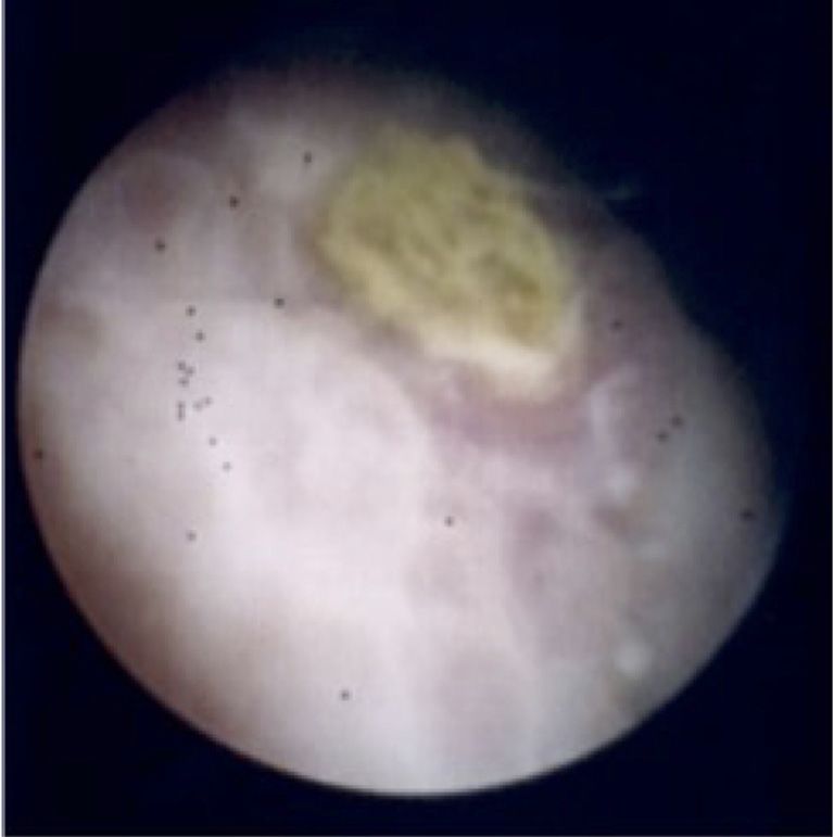
Journal of Visualized Surgery, 2021                                                                                            Page 7 of 7

Ethical Statement: The authors are accountable for all                      guideline development. J Med Case Rep 2013;7:223.
aspects of the work in ensuring that questions related                  5.  Klarenbeek BR, Bergamaschi R, Veenhof AA, et
to the accuracy or integrity of any part in the work are                    al. Laparoscopic versus open sigmoid resection
appropriately investigated and resolved. All procedures                     for diverticular disease: follow-up assessment of
performed in this study involving human participants were                   the randomized control Sigma trial. Surg Endosc
in accordance with the ethical standards of the institutional               2011;25:1121-6.
research committee and with the Helsinki Declaration (as                6. Cirocchi R, Arezzo A, Renzi C, et al. Is laparoscopic
revised in 2013). Written informed consent was obtained                     surgery the best treatment in fistulas complicating
from the patient.                                                           diverticular disease of the sigmoid colon? A systematic
                                                                            review. Int J Surg 2015;24:95-100.
Open Access Statement: This is an Open Access article                   7. Aydinli HH, Benlice C, Ozuner G, et al. Risk factors
distributed in accordance with the Creative Commons                         associated with postoperative morbidity in over 500
Attribution-NonCommercial-NoDerivs 4.0 International                        colovesical fistula patients undergoing colorectal surgery: a
License (CC BY-NC-ND 4.0), which permits the non-                           retrospective cohort study from ACS-NSQIP database. Int
commercial replication and distribution of the article with                 J Colorectal Dis 2017;32:469-74.
the strict proviso that no changes or edits are made and the            8. Dolejs SC, Penning AJ, Guzman MJ, et al. Perioperative
original work is properly cited (including links to both the                Management of Patients with Colovesical Fistula. J
formal publication through the relevant DOI and the license).               Gastrointest Surg 2019;23:1867-73.
See: https://creativecommons.org/licenses/by-nc-nd/4.0/.                9. Siddighi S, Yune JJ, Hardesty J. Indocyanine green for
                                                                            intraoperative localization of ureter. Am J Obstet Gynecol
                                                                            2014;211:436.e1-2.
References
                                                                        10. Mandovra P, Kalikar V, Patankar RV. Real-Time
1.   Tomizawa K, Toda S, Tate T, et al. Laparoscopic surgery                Visualization of Ureters Using Indocyanine Green During
     for colovesical fistula associated with sigmoid colon                  Laparoscopic Surgeries: Can We Make Surgery Safer?
     diverticulitis: a review of 39 cases. J Anus Rectum Colon              Surg Innov 2019;26:464-8.
     2019;3:36-42.                                                      11. Kanabur P, Chai C, Taylor J. Use of Indocyanine Green
2.   Kitaguchi D, Enomoto T, Ohara Y, et al. Laparoscopic                   for Intraoperative Ureteral Identification in Nonurologic
     surgery for diverticular colovesical fistula: single-center            Surgery. JAMA Surg 2020;155:520-1.
     experience of 11 cases. BMC Res Notes 2020;13:177.                 12. Slooter MD, Janssen A, Bemelman WA, et al. Currently
3.   Burks FN, Santucci RA. Management of iatrogenic                        available and experimental dyes for intraoperative near-
     ureteral injury. Ther Adv Urol 2014;6:115-24.                          infrared fluorescence imaging of the ureters: a systematic
4.   Gagnier JJ, Kienle G, Altman DG, et al. The CARE                       review. Tech Coloproctol 2019;23:305-13.
     guidelines: consensus-based clinical case reporting

 doi: 10.21037/jovs-20-140
 Cite this article as: Asti E, Bernardi D, Andreatta E, Conti A,
 Carmignani L, Bonavina L. Laparoscopic management of
 colovesical fistula secondary to sigmoid diverticulitis: case report
 and the role of intraoperative indocyanine-green fluorescence. J
 Vis Surg 2020.

© Journal of Visualized Surgery. All rights reserved.                               J Vis Surg 2021 | http://dx.doi.org/10.21037/jovs-20-140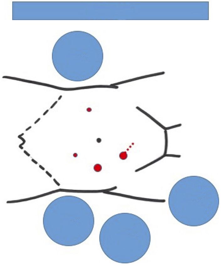
You can also read