Biocompatibility and osteoconductivity of scaffold porous composite collagen-hydroxyapatite based coral for bone regeneration
←
→
Page content transcription
If your browser does not render page correctly, please read the page content below
Open Chemistry 2020; 18: 584–590
Research Article
Siswanto Siswanto*, Dyah Hikmawati, Umi Kulsum, Djony Izak Rudyardjo, Retna Apsari,
Aminatun Aminatun
Biocompatibility and osteoconductivity of scaffold porous
composite collagen–hydroxyapatite based coral for bone
regeneration
https://doi.org/10.1515/chem-2020-0080 patients. This can be caused by trauma, tumors, or other
received July 15, 2019; accepted March 17, 2020 bone diseases [1]. The Ministry of Health of the Republic of
Abstract: The synthesis of collagen–hydroxyapatite compo- Indonesia in 2013 stated that traffic accidents in Indonesia
sites has been carried out, and the biocompatibility and have increased every year by 21.8% in 5 years. There are
osteoconductivity properties have been tested. This research many victims of injuries and bone fractures from the
was conducted to determine the ability of hydroxyapatite– incidents that occurred. According to the data from the
collagen composites to support the bone growth through the Hospital Information System in Indonesia in 2018, cases of
graft surface. Hydroxyapatite used in this study was synthe- fractures have increased by 105% compared to that in 2013
sized from coral with a purity of 96.6%, while collagen was [2]. Under these conditions, a biomaterial is needed as a
extracted from the chicken claw. The process of forming a substitute for the bone (bone graft). The need for
scaffold of collagen–hydroxyapatite composites was carried biomaterials as a substitute for the bone will increase with
out using the freeze-drying method at −80°C for 4 h. The the increasing number of cases of bone damage due to
biocompatibility characteristics of the sample through the trauma, tumors, congenital abnormalities, infections, and
cytotoxicity tests showed that the percentage of viable cells in bone resorption due to complications in the installation of
collagen–hydroxyapatite biocomposite was 108.2%, which is joint prostheses [3].
higher than the percentage of viable cells of hydroxyapatite Several types of bone grafts are used in the
or collagen material. When the viable cell is above 100%, treatment of bone defects – autograft, allograft, and
collagen–hydroxyapatite composites have excellent osteo- xenograft. Autograft is the “gold standard” for bone
conductivity as a material for bone regeneration. growth in the spine because autograft has osteoconduction,
osteoinduction, and osteogenesis properties [4]. Although
Keywords: biocompatibility, osteoconductivity, composite, autograft is an ideal bone graph, autograft cannot be
coral used in large bone defects. A large size bone will cause
large morbidity at the place where the bone was taken
[5]. In addition, several autograft limitations that need to
1 Introduction be considered include blood loss, wound infections and
complications, prolonged pain, and local sensory loss.
The bone defect is a disease associated with the functional Allograft and xenograft are alternatives to the use of
disability that has a serious impact on the quality of life of autograft. Both these procedures do not require surgery
to get a bone graft from another part of the patient’s
body. So they do not require two surgeries similar to
autograft and can reduce pain. However, an allograft
* Corresponding author: Siswanto Siswanto, Department of can be rejected by the immune system and the
Physics, Faculty of Science and Technology, Universitas Airlangga,
transmission of diseases such as HIV and hepatitis C,
Kampus C, Jalan Mulyorejo, Surabaya, East Java, 60116, Indonesia,
e-mail: siswanto@fst.unair.ac.id while xenograft has an immunogenic risk and risk of the
Dyah Hikmawati, Djony Izak Rudyardjo, Retna Apsari, Aminatun transmission of zoonotic diseases [6]. In addition, the
Aminatun: Department of Physics, Faculty of Science and sterilization process carried out in allograft and xeno-
Technology, Universitas Airlangga, Kampus C, Jalan Mulyorejo, graft also results in the nature of osteogenesis being
Surabaya, East Java, 60116, Indonesia
completely lost, and hence, all bone cells would have
Umi Kulsum: Biomedical Engineering Program Study, Department
of Physics, Faculty of Science and Technology, Universitas
died [7].
Airlangga, Kampus C, Jalan Mulyorejo Surabaya, East Java, 60116, The requirements for synthetic bone grafts that must
Indonesia be met are being acceptable to the body (biocompatible)
Open Access. © 2020 Siswanto Siswanto et al., published by De Gruyter. This work is licensed under the Creative Commons Attribution 4.0
Public License.
Biocompatibility and osteoconductivity of composite collagen–hydroxyapatite 585
and advantageous to the osteoconduction process (guiding The most appropriate synthesis method for fabricating
the reparative growth of natural bone), osteoinduction porous biomaterials is the freeze-drying method. In this
(encouraging differentiated cells to active osteoblasts), and method, controlling the growth of ice crystals is very
osteogenesis (living bone cells in the material bone graft important to obtain the appropriate pore diameter and
that contributes to bone remodeling) [4]. Osteoconductive shape because the pore structure is a replication of the ice
and osteoinductive are the most important properties of crystal dendrite entrapment.
resorbable biomaterials to direct and encourage the forma-
tion of the tissue growth [8]. Osteoconductive and
osteointegration of a bone graft are related to the porosity
and pore size [9]. 2 Materials and methods
Based on the previous research, the minimum
requirements for the pore size are around 100 µm [10].
2.1 Synthesis of hydroxyapatite from
This corresponds to the cell size, requirements for
migration, and cell transport. However, a pore size of marine coral
300 µm is recommended for increasing new bone
formation and capillary formation [11]. High macro- In this study, hydroxyapatite was synthesized from
porosity can increase bone formation, but porosity that marine coral. Fossilized coral is used because this coral
is higher than 50% can result in the decreasing of the has a higher calcium content than that of a marine coral.
mechanical properties of a biomaterial [6]. The coral is taken from the southern coast of Java Island
A good synthetic bone graft is a bone graft that has a (Popoh Tulungagung beach) as shown in Figure 1. Then,
chemical structure and composition similar to the natural the coral is cleaned and dried in an open space. Corals
bone [12]. The collagen–hydroxyapatite (HA) composite is are manually crushed to become smaller particles. The
a synthetic bone graft that has similar properties to the crushed corals are calcined at 900°C to remove some
natural bone. As is already known, bone’s main compo- unnecessary elements or compounds and form CaO
nents are hydroxyapatite (67%) and collagen (28%) and compounds. Then, the calcined coral was mashed using
the other component is noncollagen protein [13]. Hydroxy- a mortar so that the size of the coral became powdered
apatite has a modulus elasticity of 73–117 GPa, a com- and filtered using a 200 mesh or 74 m sieves. After that,
pressive strength of 600 MPa, and a density of 3.1 g/cm3, it is smoothed using high-energy milling (HEM type E3D)
while the modulus elasticity of collagen is 46.5 MPa [14]. for 2 h with dry milling, so that the particles are reduced
Based on its mechanical properties, hydroxyapatite pro- to a smaller size and formed as Ca(OH)2. The synthesis of
vides stiffness and hardness, while collagen provides hydroxyapatite is carried out by reacting calcium
tensile strength and flexibility. Collagen–HA composites hydroxide and phosphoric acid (H3PO4, 85% Aldrich-
have good biocompatibility and are osteoconductive, so Sigma) as stated in equation (3).
collagen–HA composites are suitable for using as a
scaffold for the bone tissue engineering [15].
The method to obtain composites that have the same
structure and the composition as a natural bone is by 2.2 Collagen extraction from chicken claws
collaborating several methods of synthesis. One of
the keys to the synthesis of macroporous composites In this study, collagen was extracted from broiler
can be done through variations in the freezing rates [2]. chicken paws obtained at the Wonokromo Surabaya
Figure 1: The process of scouring the coral becomes powder.
586 Siswanto Siswanto et al.
market, East Java, Indonesia. Chicken claws were 2.4 Biocompatibility test
separated from the bones by being cut using a knife to
facilitate the destruction process. The claw pieces were The biocompatibility of biomaterials was tested by the
crushed with a blender. Then, the crushed chicken claws MTT assay method. The MTT method tests (3-[4,5-
were soaked in HCl solution for 24 h with 5 wt/vol% dimethylthiazol-2-yl]-2,5 diphenyl tetrazolium bromide) to
ratio. Soaking process was carried out in the cold environ- measure the number of cells that still survive without the
ment of the refrigerator and then filtered. Liquid filtrate need for calculations. Cells that live with active meta-
was mixed with 0.1 N NaOH solution until it reached a bolism can convert MTT into purple formazan products
neutral pH and was then allowed to stand until the with maximum absorption at a wavelength of 570 nm,
collagen agglomerated. When approaching neutral pH, while dead cells lose the ability to convert MTT to
collagen clots can be observed and the fibers began to formazan. Thus, the formation of color serves as a marker
form and coalesce, and hence, clots appeared more clearly. for viability cells [16]. The results of the MTT assay are shown
Collagen clots formed perfectly at neutral pH (pH 7). The in Figure 2. Biomaterials are considered toxic if the cell death
chemicals used were Sigma–Aldrich HCl 37% and NaOH is more than 50% and the number of viable cells is less than
with 97% purity. 50%. The increase or decrease in the number of viable cells
indicates the level of toxicity of a biomaterial. Biomaterials
have better biocompatibility if the number of proliferating
cells is higher or the inhibitory value is lower [17,18].
2.3 Synthesis of collagen–hydroxyapatite
composite Ethical approval: The conducted research is not related
to either human or animal use.
The synthesis of collagen–hydroxyapatite composite
begins by forming hydroxyapatite solution and collagen
solution. Collagen solution is a dissolved collagen in
acetic acid, while the hydroxyapatite solution is hydro- 3 Results and discussion
xyapatite, which was dissolved in the phosphoric acid.
The hydroxyapatite solution and the neutral collagen 3.1 Results of hydroxyapatite made from
solution are mixed while manually slowly stirring them.
coral
The mixture was put into a cylindrical tube and then
frozen at −80°C in freeze-drying with a variation of
freezing time of 2, 4, and 6 h. Dried collagen–hydrox- The preparation of coral-based hydroxyapatite is carried
yapatite composites were removed from the mold for out using the precipitation method. Coral is used
characterization. because this material contains CaCO3 of 95.5%, MgSiO3
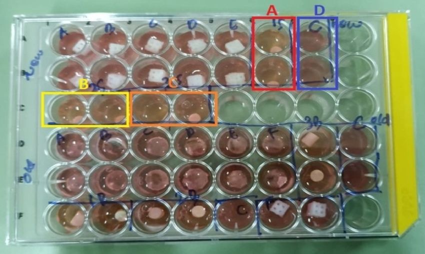
Figure 2: Incubation of collagen-composite biomaterials.
Biocompatibility and osteoconductivity of composite collagen–hydroxyapatite 587
Figure 3: XRD of (a) coral and (b) heated coral at 900°C for 3 h.
of 3.8%, and FeSi of 0.7%, as indicated by the X-ray 10Ca (OH)2 (s) + 6H3 PO4 (l) → Ca10 (PO4)6 (OH)2 (s)
(3)
diffraction (XRD) spectrum in Figure 3a. + 18H2 O (l)
Coral that is heated at 900°C for 3 h produces new
compounds, namely, calcium oxide (Lime, syn) CaO and Sintering of precipitation results is carried out at
calcium hydroxide Ca(OH)2. The presence of Ca(OH)2 in this 900°C. The identification of the XRD spectrum in Figure 4b
process is thought to be due to the furnace not in a vacuum produces hydroxyapatite compounds of 96.6% and tri-
state, and hence, when the temperature is reduced to the calcium phosphate compounds of 3.4%.
ambient temperature, a reaction between water vapor and
CaO occurs. The remaining CaCO3 (aragonite) compound in
this heating is 8.9%. Silicate compounds are not detected in
XRD peaks as in the results of the pure coral spectrum; 3.2 Results of collagen extraction
however, they have good thermostability. The invisible
height of the silicate compound is assumed to be of an Collagen in this study was obtained by extracting
insignificant quantity and the amount of calcium phosphate chicken claws. Fourier-transform infrared spectroscopy
which is very dominant. To improve the efficiency of the (FTIR) collagen extraction from chicken paws is shown
reaction of hydroxyapatite formation, milling for 20 h is in Figure 5.
carried out on coral powder, so the powder size becomes The main markers used for the collagen identifica-
smaller. During the milling process, CaO reacts with H2O to tion are amide A, amide I, amide II, and amide III. The
form Ca(OH)2 as shown in Figure 4a. peaks of 3369.64, 3439.08, 3126.61, and 2445.74 cm−1 are
The formation of the coral hydroxyapatite by the the amide A absorption area at the wavenumber of
precipitation method can be described as follows. 3,600–2,300 cm−1, which indicates the presence of N–H
bond. The peak at the wave number 1649.14 cm−1
CaCO3 (s) → CaO(s) + CO2 (g) (1) indicates the presence of C]O bonds, which is the
CaO(s) + H2 O(l) → Ca (OH)2 (s) (2) amide I absorption area. The amide I absorption area is
in
Figure 4: XRD of (a) coral milling for 20 h and (b) synthesized hydroxyapatite.
588 Siswanto Siswanto et al.
3.3 Results of the collagen–hydroxyapatite
composite
Collagen–hydroxyapatite composites were synthesized
using the freeze-drying method with a freezing time of
4 h. The FTIR results of this composite are stated in
Figure 5. The characteristic peak of hydroxyapatite is at
a wavelength of 500–600 cm−1. The FTIR of the
collagen–hydroxyapatite composite was found at a
wavelength of 553 cm−1. The characteristic peak of
collagen was found at 2,873 cm−1 for CH stretching,
1,716 cm−1 for group C]O, and above 3,000 cm−1 for
N–H. Amide I bending occurs between a wavelength of
1,600–1,700 cm−1, and (PO4)3− bending occurs between
900 cm−1–1,200 cm−1.
Figure 5: FTIR of HA, collagen, and collagen–HA composite. The possible bond between collagen and hydroxya-
patite is a hydrogen bond because collagen including
protein and hydroxyapatite is ceramic. Hydrogen bonds
the wavenumber 1,636–1,661 cm−1. Meanwhile, the peak
occur between the hydrogen atom in collagen with the
at the numbers 1544.98, 1454.33, and 1402.25 cm−1 is the
oxygen atom in hydroxyapatite as shown in Figure 7.
amide II absorption area. The wave number of
Mixing hydroxyapatite and collagen results in miner-
1544.98 cm−1 indicates the presence of N–H bonds, while
alized collagen by hydroxyapatite. Mineralized collagen
wavenumbers of 1454.33 and 1402.25 cm−1 also indicate
occurs because of interactions that arise between the
the amide II uptake regions that have CH2 and CH3
structure of collagen and hydroxyapatite crystals. This
bonds. The four markers are also found at the peak of
interaction occurs between the carboxylic group and the
absorption of pure collagen, especially collagen type I,
Ca2+ cation, which is illustrated in Figure 8. This hypothesis
as shown in Figure 6.
is supported by the FTIR data, which is explained based on
the C–O and C]O band spectra of pure collagen and
collagen–hydroxyapatite composites [19].
The synthesis of the collagen–hydroxyapatite scaf-
fold was carried out using the freeze-drying method. The
freezing time during freeze-drying affects the porosity of
the scaffold formed. In this research, the freezing time is
carried out in 2, 4, and 6 h. The measurement data and
the calculation of porosity with freezing time are
presented in Table 1. Composite porosity formed by
freezing for 2 h has the highest porosity compared to
porosity formed by freezing for 4 and 6 h. The greater the
percentage of porosity, the easier the cell proliferation,
Figure 6: FTIR of pure collagen [17]. but it is recommended not to exceed 60% because it will
Figure 7: Bonding of hydrogen atoms in collagen.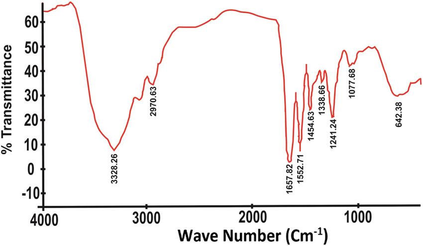
Biocompatibility and osteoconductivity of composite collagen–hydroxyapatite 589
Figure 8: The mezomeric form of the stable carboxylic group during mineralization.
Table 1: Porosity scaffold with various freezing time the cell growth above 100% can be interpreted that
the collagen–hydroxyapatite composite is a very good
Quantity Freezing Freezing Freezing medium for the growth and the development (osteocon-
time, 2 h time, 4 h time, 6 h ductive) of bone cells. To strengthen this suspicion,
Porosity 52% 35% 27% currently, researchers are still conducting in vitro collagen–
hydroxyapatite composites using the alkaline phosphatase
test.
affect the physical and mechanical properties. The presence
of pores in the scaffold is useful for facilitating the transport
of oxygen and cell nutrition [20].
The results of cytotoxicity tests on samples of collagen,
hydroxyapatite, and collagen–hydroxyapatite composites are
4 Conclusion
shown in Figure 9. The graph of the MTT test results shows
From the results of the preparation of hydroxyapatite-
that collagen and hydroxyapatite are not toxic because the
based coral and its composites with collagen, the
percentage of living cells is above 100%. The percentage of
following conclusions can be made. First, coral can be
living cells above 100% also indicate cell proliferation in the
used as a bone replacement biomaterial because it can
MTT process. The occurrence of cell proliferation in collagen
produce calcium phosphate in the form of 96.6%
and hydroxyapatite is thought to be caused by two
hydroxyapatite and 3.4% tricalcium phosphate. These
components that have osteoinductive factors, namely,
two compounds are essential parts of the compact bone
bone morphogenetic proteins (BMPs) [21]. Type 1 collagen
and the cancellous bone. Second, the MTT assay test
has BMP7 and hydroxyapatite has BMP2 [22].
results showed that cell viability in hydroxyapatite,
Collagen–hydroxyapatite composites increase the
collagen, and collagen–hydroxyapatite composites were
percentage of living cells [22]. This proves that using
103.6%, 105.6%, and 108.9%, respectively. This shows
collagen and hydroxyapatite together has benefits in
that collagen and hydroxyapatite and its composites are
terms of the cell growth. These MTT data also show that
nontoxic and biocompatible. Cell viability percentage
hydroxyapatite, collagen, or combined collagen–hydroxya-
values above 100% also indicate the cell proliferation in
patite has an excellent biocompatible property. In addition,
the MTT process. Cell proliferation in collagen and
hydroxyapatite is thought to be caused by the two
ingredients having osteoconductive and osteoinductive
factors, namely, BMPs.
Conflict of interest: The authors declare no conflict of
interest.
Acknowledgments: The authors would like to thank Uni-
versitas Airlangga for the support through its Penelitian
Unggulan Fakultas (PUF) funds, and the Ministries of
Research, Technology, and Higher Education for its support
through the Exceptional Applied Research in Higher Educa-
tion program (Penelitian Terapan Unggulan Perguruan Tinggi
Figure 9: MTT assay of samples. or PTUPT), 2020 fiscal year.590 Siswanto Siswanto et al.
References for biomedical application. J Miner Mater Charact Eng.
2011;10(8):727–34, doi: 10.4236/jmmce.2011.108057.
[1] Chen L, Wu Z, Zhou Y, Li L, Wang Y, Wang Z, et al. Biomimetic [13] Ficai A, Andronescu E, Voicu G, Ficai D. Advances in collagen/
porous collagen/hydroxyapatite scaffold for bone tissue hydroxyapatite composite materials. In: Attaf B, editors.
engineering. J Appl Polym Sci. 2017;134(37):45271, Advances in composite materials for medicine and nano-
doi: 10.1002/app.45271. technology. London, UK: IntechOpen; 2011, doi: 10.5772/13707.
[2] Al Bakki AH. Public Expose, BPJS Health Performance [14] Manssor NAS, Radzi Z, Yahya NA, Mohamad Yusof L, Hariri F,
Achievements in 2018. Jakarta: BPJS (Social Security Khairuddin NH, et al. Characteristics and young’s modulus of
Administering Agency); 2019. collagen fibrils from expanded skin using anisotropic
[3] Laurencin C, Jiang T. Bone Graft Substitutes and Bone controlled rate self-inflating tissue expander. Skin Pharmacol
Regenerative Engineering. Pennsylvania: ASTM International Physiol. 2016;29(2):55–62, doi: 10.1159/000431328.
Publisher; 2014. [15] Ficai A, Albu MG, Birsan M, Sonmez M, Ficai D, Trandafir V,
[4] Cuomo AV, Virk M, Petrigliano F, Morgan EF, Lieberman JR. et al. Collagen hydrolysate based collagen/hydroxyapatite
Mesenchymal stem cell concentration and bone repair: composite materials. J Mol Struct. 2013;1037:154–9,
potential pitfalls from bench to bedside. J Bone Joint Surg Am. doi: 10.1016/j.molstruc.2012.12.052.
2009;91(5):1073–83, doi: 10.2106/JBJS.H.00303. [16] Riss TL, Moravec RA, Niles AL, Duellman S, Benink HA,
[5] Ferdiansyah RD, Rantam FA. Aulani’am, regeneration of Worzella TJ, et al. Cell viability assays. Assay Guidance Manual
massive bone defect with bovine hydroxyapatite as scaffold of [Internet]. Bethesda (MD): Eli Lilly & Company and the National
mesenchymal stem cells. JBP. 2011;13(3):179–95. Center for Advancing Translational Sciences; 2013. Available
[6] Oryan A, Alidadi S, Moshiri A, Maffulli N. Bone regenerative from: https://www.ncbi.nlm.nih.gov/books/NBK144065/
medicine: classic options, novel strategies, and future [17] Kamal AF, Iskandriati D, Dilogo IH, Siregar NC, Hutagalung EU,
directions. J Orthop Surg Res. 2014;9(1):18, doi: 10.1186/ Susworo R, et al. Biocompatibility of various hydroxyapatite
1749-799X-9-18. scaffolds evaluated by proliferation of rat’s bone marrow
[7] Wang J, Wang L, Zhou Z, Lai H, Xu P, Liao L, et al. mesenchymal stem cells: an in vitro study. Med J Indonesia.
Biodegradable polymer membranes applied in guided bone/ 2013;22(4):202–8, doi: 10.13181/mji.v22i4.600.
tissue regeneration: a review. Polymers. 2016;8(4):1–20, [18] Kirubanandan S, Sehgal PK. Regeneration of soft tissue using
doi: 10.3390/polym8040115. porous bovine collagen scaffold. J Optoelectron Biomed
[8] Wang C, Huang W, Zhou Y, He L, He Z, Chen Z, et al. 3D Mater. 2010;2(3):141–9.
printing of bone tissue engineering scaffolds. Bioact [19] Murphy CM, Haugh MG, O’Brien FJ. The effect of mean pore
Mater. 2020;5(1):82–91, doi: 10.1016/j.bioactmat.2020. size on cell attachment, proliferation and migration in
01.004. collagen–glycosaminoglycan scaffolds for bone tissue
[9] Radulescu M, Ficai D, Oprea O, Ficai A, Andronescu E, engineering. Biomaterials. 2010;31(3):461–6, doi: 10.1016/
Holban AM. Antimicrobial chitosan based formulations with j.biomaterials.2009.09.063.
impact on different biomedical applications. Cur Pharm [20] Azhar FF, Olad A, Salehi R. Fabrication and characterization of
Biotechnol. 2015;16(2):128–36, doi: 10.2174/ chitosan–gelatin/nanohydroxyapatite–polyaniline composite
138920101602150112151157. with potential application in tissue engineering scaffolds. Des
[10] Pellin G, Pelin C-E, Ştefan A, Dincă I, Ficai A, Andronescu E, Monomers Polym. 2014;17(7):654–67, doi: 10.1080/
et al. Influence of nanometric silicon carbide on phenolic resin 15685551.2014.907621.
composites properties. Bull Mater Sci. 2016;39:769–75, [21] Narbat MK, Orang F, Hashtjin MS, Goudarzi A. Fabrication of
doi: 10.1007/s12034-016-1185-z. porous hydroxyapatite–gelatin composite scaffolds for bone
[11] Karageorgiou V, Kaplan D. Porosity of 3D biomaterial tissue engineering. Iran Biomed J. 2006;10(4):215–23.
scaffolds and osteogenesis. Biomaterials. 2005;26(27): [22] Yetmez M, Erkmen ZE, Kalkandelen C, Ficai A, Oktar FN.
5474–91, doi: 10.1016/j.biomaterials.2005.02.002. Sintering effects of mullite-doping on mechanical properties
[12] Agrawal K, Singh G, Puri D, Prakash S. Synthesis and of bovine hydroxyapatite. Mater Sci Eng C. 2017;77:470–5,
characterization of hydroxyapatite powder by sol–gel method doi: 10.1016/j.msec.2017.03.290.You can also read