European Journal of Pharmaceutical Sciences - Department of Chemical and ...
←
→
Page content transcription
If your browser does not render page correctly, please read the page content below
European Journal of Pharmaceutical Sciences 162 (2021) 105812
Contents lists available at ScienceDirect
European Journal of Pharmaceutical Sciences
journal homepage: www.elsevier.com/locate/ejps
Impact of gastrointestinal tract variability on oral drug absorption and
pharmacokinetics: An UNGAP review
Zahari Vinarov a, b, Mohammad Abdallah c, José A.G. Agundez d, Karel Allegaert e, f,
Abdul W. Basit g, Marlies Braeckmans a, Jens Ceulemans h, Maura Corsetti i, j, Brendan T. Griffin k,
Michael Grimm l, Daniel Keszthelyi m, Mirko Koziolek n, Christine M. Madla g,
Christophe Matthys o, p, Laura E. McCoubrey g, Amitava Mitra q, Christos Reppas r,
Jef Stappaerts h, Nele Steenackers o, Natalie L. Trevaskis c, Tim Vanuytsel s, Maria Vertzoni r,
Werner Weitschies l, Clive Wilson t, Patrick Augustijns a, u, 1, *
a
Drug Delivery and Disposition, Department of Pharmaceutical and Pharmacological Sciences, KU Leuven, Leuven, Belgium
b
Department of Chemical and Pharmaceutical Engineering, Sofia University, Sofia, Bulgaria
c
Drug Delivery, Disposition and Dynamics, Monash Institute of Pharmaceutical Sciences, Monash University, Parkville, Australia
d
University Institute of Molecular Pathology Biomarkers, UEx. ARADyAL, Instituto de Salud Carlos III, Cáceres, Spain
e
Department of Hospital Pharmacy, Erasmus MC, University Medical Center Rotterdam, Rotterdam, the Netherlands
f
Department of Development and Regeneration and Department of Pharmaceutical and Pharmacological Sciences, KU Leuven, Leuven, Belgium
g
UCL School of Pharmacy, University College London, London, United Kingdom
h
Pharmaceutical Sciences, Janssen Research & Development, Beerse, Belgium
i
NIHR Nottingham Biomedical Research Centre (BRC), Nottingham University Hospitals NHS Trust and the University of Nottingham, Nottingham, United Kingdom
j
Nottingham Digestive Diseases Centre, School of Medicine, University of Nottingham, Nottingham, United Kingdom
k
School of Pharmacy, University College Cork, Cavanagh Pharmacy Building, Cork, Ireland
l
Department of Biopharmaceutics and Pharmaceutical Technology, Center of Drug Absorption and Transport, University of Greifswald, Greifswald, Germany
m
Division of Gastroenterology-Hepatology, Department of Internal Medicine, NUTRIM, Maastricht University Medical Center, Maastricht, the Netherlands
n
NCE Formulation Sciences, AbbVie Deutschland GmbH & Co. KG, Ludwigshafen, Germany
o
Clinical and Experimental Endocrinology, Department of Chronic Diseases and Metabolism, KU Leuven, Leuven, Belgium
p
Department of Endocrinology, University Hospitals Leuven, Leuven, Belgium
q
Clinical Pharmacology and Pharmacometrics, Janssen Research & Development, Spring House, Pennsylvania, United States
r
Department of Pharmacy, School of Health Sciences, National and Kapodistrian University of Athens, Athens, Greece
s
Translational Research Centre for Gastrointestinal Disorders (TARGID), Department of Chronic Diseases, Metabolism and Ageing (ChroMeTa), KU Leuven, Leuven, Belgium
t
Strathclyde Institute of Pharmacy and Biomedical Sciences (SIPBS), University of Strathclyde, United Kingdom
u
Chairperson of the UNGAP network, COST CA 16205
Abbreviations: 5-FU, 5-fluorouracil; ADME, Absorption, distribution, metabolism and excretion; AIDS, Acquired immunodeficiency syndrome; AUC, Area under
the curve; BCS, Biopharmaceutics classification system; BMI, Body mass index; CD, Cyclodextrin; CETP, Cholesterylester transfer protein; CI, Confidence interval;
CIPO, Chronic intestinal pseudoobstruction; Cmax, Maximum plasma concentration; CRA, Cannabinoid receptor agonist; CTT, Colon transit times; CV, Coefficient of
variance; CYP, Cytochrome P450; DAG, Diacylglycerides; DDI, Drug-drug interactions; Dv50, Median particle size by volume; EHC, Enterohepatic circulation; FaHIF,
Fasted state human intestinal fluids; FaSSIF, Fasted state simulated intestinal fluids; FDA, U.S. Food and drug administration agency; FFA, Free fatty acids; GC,
Glycocholate; GCDC, Glycochenodeoxycholate; GDC, Glycodeoxycholate; GET, Gastric emptying time; GIT, Gastrointestinal tract; HIV, Human immunodeficiency
virus; HPMC, Hydroxypropyl methylcellulose; HPMC-AS, Hydroxypropyl methylcellulose acetate succinate; HPMCP, Hydroxypropyl methylcellulose phtalate; IBD,
Inflammatory bowel disease; IMMC, Inter-digestive migrating motor complex; IR, Immediate release; L-DOPA, Levodopa; Lyso-PC, Lyso-phosphatidylcholine; MAG,
Monoacylglycerides; MRI, Magnetic resonance imaging; NAPQI, N-acetyl-p-benzoquinone imine; NSAID, Nonsteroidal anti-inflammatory drug; OHM, 1-hydroxy-
midazolam; PBPK, Physiologically based pharmacokinetic modelling; PC, Phosphatidylcholine; PD, Pharmacodynamic(s); PEG, Polyethylene glycol; PK, Pharma
cokinetic(s); PPI, Proton pump inhibitors; RYGB, Roux-en-Y gastric bypass; SBWC, Small bowel water content; SIBO, Small intestinal bacterial overgrowth; SITT,
Small intestinal transit time; TAG, Triacylglycerides; TC, Taurocholate; TCDC, Taurochenodeoxycholate; TDC, Taurodeoxycholate; TKI, Tyrosine kinase inhibitors;
tmax, Time to reach maximum plasma concentration; UDC, Ursodeoxycholate; UNGAP, European network on understanding gastrointestinal absorption-related
processes; VEGF-C, Vascular endothelial growth factor C.
* Corresponding author at: Drug Delivery and Disposition, Department of Pharmaceutical and Pharmacological Sciences, KU Leuven, Gasthuisberg O&N II,
Herestraat 49, Box 921, 3000 Leuven, Belgium.
E-mail address: patrick.augustijns@kuleuven.be (P. Augustijns).
1
www.ungap.eu.
https://doi.org/10.1016/j.ejps.2021.105812
Received 5 December 2020; Received in revised form 19 February 2021; Accepted 16 March 2021
Available online 20 March 2021
0928-0987/© 2021 The Authors. Published by Elsevier B.V. This is an open access article under the CC BY license (http://creativecommons.org/licenses/by/4.0/).
Z. Vinarov et al. European Journal of Pharmaceutical Sciences 162 (2021) 105812
A R T I C L E I N F O A B S T R A C T
Keywords: The absorption of oral drugs is frequently plagued by significant variability with potentially serious therapeutic
Variation consequences. The source of variability can be traced back to interindividual variability in physiology, differ
Fasted and fed state ences in special populations (age- and disease-dependent), drug and formulation properties, or food-drug in
Physiology
teractions. Clinical evidence for the impact of some of these factors on drug pharmacokinetic variability is
Pediatrics and geriatrics
Diseases
mounting: e.g. gastric pH and emptying time, small intestinal fluid properties, differences in pediatrics and the
Drug formulation elderly, and surgical changes in gastrointestinal anatomy. However, the link of colonic factors variability (transit
time, fluid composition, microbiome), sex differences (male vs. female) and gut-related diseases (chronic con
stipation, anorexia and cachexia) to drug absorption variability has not been firmly established yet. At the same
time, a way to decrease oral drug pharmacokinetic variability is provided by the pharmaceutical industry:
clinical evidence suggests that formulation approaches employed during drug development can decrease the
variability in oral exposure. This review outlines the main drivers of oral drug exposure variability and potential
approaches to overcome them, while highlighting existing knowledge gaps and guiding future studies in this
area.
1. Introduction perform a meta-analysis of the available data. However, as most results
available in the literature are based on small scale explorative clinical
The oral intake of drugs remains the preferred administration route studies, a statistical comparison is usually not justified. In addition,
because of its non-invasive character and convenience for the patient, differences in the experimental protocols and instrumentation used to
which increases drug regimen compliance. However, ensuring sufficient gather the data would undermine any attempts at such analysis.
and predictable systemic drug exposure when developing oral drug Therefore, results are reported in a descriptive way.
products is not straightforward (Basavaraj and Betageri, 2014; Kor Furthermore, the impact of methodology, clinical study design and
stanje, 2003; Li et al., 2016): issues with bioavailability and pharma statistics on the measurement and quantification of variability is an issue
cokinetics (PK) are among the top 3 reasons for attrition of oral on its own (Augustijns et al., 2020; Evans, 2010; Pocock et al., 2015) and
small-molecule new drug candidates. In particular, the effectiveness of will not be addressed in the current paper. The effect of drug absorption
oral drug products in clinical practice can be plagued by significant variability on clinical performance and therapeutic outcome is also not
variability in drug exposure with serious therapeutic consequences in the scope of the current review.
(Pasipanodya et al., 2012).
It is logical to expect that the factors, which control drug absorption 2. Physiological inter- and intraindividual variability in the
and PK, are also responsible for the variability of drug exposure fasted and fed state
observed in the clinic. Hence, the impact of physiological differences (in
special populations and gastrointestinal tract (GIT) regions), drug and 2.1. Gastric conditions
formulation properties, and food-drug interactions on drug absorption
was recognized and described by the European Network on Under This section focuses on the variability of data in healthy fasted and
standing Gastrointestinal Absorption-related Processes (UNGAP)(Boyd fed state adults. Fasted state is defined as an overnight fast, followed by a
et al., 2019; Koziolek et al., 2019; Stillhart et al., 2020; Vertzoni et al., glass of water in the morning. The fed state data reported was generated
2019). Considering the complexity and the lack of awareness about by using the standard U.S. Food and drug administration agency (FDA)
variability, the current review expands beyond the state-of-the-art to meal: a high-calorie (900–1000 kcal) breakfast with approximately 150,
provide a focused description and analysis of the subject. Particular 250, and 500–600 calories originating from protein, carbohydrate and
attention was paid to examples, which demonstrate the link between fat, respectively (EMA 2010; FDA 2002).
GIT variability and drug absorption/PK, in both fasted and post-prandial
conditions. 2.1.1. pH and buffer capacity
All anatomical, physiological and pharmaceutical factors that were In the fasted state, median gastric pH values 10–20 min and
considered as a source of variability in the current paper are listed in 30–40 min after a glass of water have been reported to be 1.7–3.3 and
Table 1. 1.6–2.7, respectively (Kalantzi et al., 2006; Koziolek et al., 2014; Petrakis
The drug transit times (gastric emptying time (GET), intestinal et al., 2015). The resistance of gastric fluids to increase with one pH unit
transit time) and fluid volumes (including intestinal fluid pockets) in the when titrating with NaOH (buffer capacity) varies with gastric pH and
GIT have been identified as considerable sources of variability and are becomes close to zero in adults treated with famotidine (Figure 1).
the first factors discussed in the current review. The body of literature, Oncology drugs constitute an important example of the impact of
which describes the compositional and physicochemical (e.g. pH and gastric pH in PK variability, as about half of oral cancer therapies are
buffer capacity (buffer capacity)) variability of human gastric and in weak bases and display solubility-limited dissolution properties (Sme
testinal fluids, will also be discussed from the angle of their contribution lick et al., 2013). The implications of chronic use of PPI inhibitors on oral
to oral absorption variability. Further down the GIT, the impact of the drug absorption are considerable and are examined in detail in section
gut microbiota and bacterial drug degradation on variability will be 4.3.3.
addressed. The specific effect of formulation-related parameters are In the fed state, the median pH values reported in literature at 30 min
described in a separate section. Next, a number of additional factors, and 60 min after administration of the standard meal are around 3.5
which are usually disregarded, but can play a huge role in the context of (Dressman et al., 1990; Koziolek et al., 2015b) and 3 (Dressman et al.,
variability will be examined. These include the influence of age (pedi 1990; Koziolek et al., 2014; Pentafragka et al., 2020b), respectively. pH
atrics, geriatrics), sex and disease-specific differences. Finally, an in values return to baseline levels (median pH value lower than 2) at about
dustry perspective on how drug product development takes into account 3 hours after the meal (Dressman et al., 1990; Koziolek et al., 2014).
the aforementioned challenges and succeeds to limit variability and Interestingly, intraindividual variability in pH values is low. The
provide drug exposure at therapeutic levels, will be presented. average difference in pH values at specific time points during the first
In various sections, results obtained from different studies are re three hours after meal administration in a given individual ranges from
ported. In order to compare the study results, the best option would be to -0.3 to 0.9 pH units (Pentafragka et al., 2020b). During the first four
2
Z. Vinarov et al. European Journal of Pharmaceutical Sciences 162 (2021) 105812
hours after initiation of meal administration, the average buffer capacity reason may be the non-homogenous distribution of lipid components in
of antral contents is about 20 mmol/L/ΔpH (Pentafragka et al., 2020b). the gastric contents (Koziolek et al., 2014). On average, triacylglycerides
In line with pH data, the average difference in buffer capacity values (TAG), diacylglycerides (DAG), free fatty acids (FFA) and phosphati
during the first three hours after meal administration in a given indi dylcholine (PC) are comparatively the most abundant lipids (Penta
vidual is low and ranges from -3.4% to 15% (Pentafragka et al., 2020b). fragka et al., 2020a). It has been shown that modelling of intragastric
lipolysis is necessary for simulating felodipine release from extended
2.1.2. Volumes release tablets in the fed stomach (Diakidou et al., 2009a), whereas
Based on various magnetic resonance imaging (MRI) studies per simulation of colloidal species seems to be key for the prediction of
formed after at least 8 h of fasting, the volume of gastric contents is intragastric apparent solubility of lipophilic molecules in the fed stom
typically below 50 mL (Koziolek et al., 2016). Interestingly, interindi ach (Diakidou et al., 2009b).
vidual and intraindividual variability is comparable, suggesting that the
variability within the studies is mainly resulting from intraindividual 2.1.5. Gastric emptying of drugs
day-to-day variations (Grimm et al., 2018a). On the other hand, direct
aspiration of gastric contents in the fasted state indicates very limited Fasted state. Gastric emptying of aqueous drug solutions administered in
resting gastric volumes (mean and median values less than 10 mL, a total volume of about a glass of water (200–250 mL) is an apparent
n = 15) (Vertzoni et al., 2020a). first-order process (Grimm et al., 2018b; Mudie et al., 2014). Interindi
In the fed state, intragastric volumes are similar or slightly higher vidual and intraindividual variability of the process is comparable,
than the volume of the standard meal (slightly more than 500 mL) for suggesting that the variability within the studies was mainly resulting
more than two hours after administration of the standard meal (Koziolek from intraindividual day-to-day variations (Grimm et al., 2018a). Based
et al., 2014; Pentafragka et al., 2020b), implying that gastric emptying of on published data, gastric emptying half-life can be estimated to be
meal contents is balanced by intragastric secretions. Inter-individual 11 min (85% of initial gastric volume is emptied after 30 min (Grimm
variability in volumes 15 minutes after intake of the standard meal is et al., 2018a)) or slightly longer (15 min (Kourentas et al., 2016b)). A
low (579.6 ± 38.1 mL, n=12)(Koziolek et al., 2014). slightly increased half-life of gastric emptying may be expected in case
the administered aqueous solution is acidic or with increased caloric
2.1.3. Viscosity and osmolality value (Cooke and Hunt, 1970).
Both under fasting and under fed state conditions the gastric contents Drug suspensions in aqueous media seem to empty with water unless
show pseudoplastic behavior, i.e. viscosity decreases with increasing intragastric dispersion of the particles is problematic. In the latter case,
shear rate. Especially after the standard meal, intragastric viscosity is gastric emptying can be delayed substantially (Kourentas et al., 2016a).
highly variable (Pentafragka et al., 2020a). On average, the viscosity of Disintegrating solid dosage forms typically empty from stomach after
gastric contents in the fasted state at 37 ◦ C (1.4–6.4 mPa•s at a shear rate disintegration. Rupture times of less than 10 min have been reported for
of 100 s− 1 (Pedersen et al., 2013)) is 80–800 times lower than the vis hard gelatine capsules (Digenis et al., 2000). On average, disintegration
cosity at a shear rate of 100 s− 1, after the standard meal (Pentafragka times in the stomach after administration of the dosage form with a glass
et al., 2020a). Similar observations have been made at shear rates of 50 of water have been reported to be 10–20 min for tablets (Kelly et al.
and 200s− 1. A non-caloric hydroxy-propyl-methyl-cellulose aqueous 2003) and little less than 30 min for the immediate release (IR) layer of a
meal, with a viscosity similar to intraluminal fed state values (Penta modified release product (Weitschies et al., 2008). It should be noted,
fragka et al. 2020b), has been shown to significantly decrease indinavir however, that disintegration times depend to a significant extent not
plasma concentrations after administration of Crixivan® capsules, as only on physiological conditions (liquid volumes, mixing), but also on
compared to the fasted state administration with an equal volume of the type of formulation and excipients used (Quodbach and Kleine
water (Carver et al., 1999). budde, 2016). As most of the water will be emptied until complete
In the fasted state, contents are highly hypoosmotic (Pentafragka et al., disintegration of tablets, emptying of disintegrating particles will be
2019). After the standard meal, osmolality increases to reach iso-osmotic affected by the inter-digestive motility pattern and the density of the
levels (Pentafragka et al., 2020a). The potential impact of such difference disintegrated particles. One of the first relevant studies in adults was
on gastric emptying rates in humans has not been investigated, however, performed by Aoyagi et al (Aoyagi et al., 1992). After an overnight fast,
hyperosmolarity delays the gastric emptying of liquid caloric meals three adults received together with 200 mL water at two different oc
(Paraskevopoulos et al., 1988; Vermeulen et al., 2011). casions on a crossover basis: (A) 5 spherical enteric coated tablets of
barium sulfate and (B) 50 cylindrical granules of barium sulfate coated
2.1.4. Composition with ethylcellulose. No food was consumed until 4 h after dosing. The
In the fasted state, bile salt levels are highly variable and concen number of tablets and granules remaining in the stomach were deter
trations are up to 0.15 mM on average (Pentafragka et al., 2019). In the mined by periodic roentgenography. Data after administration of the
fed state, no data after the standard meal have been reported. tablets in one of the adults indicated 80 % gastric retention at 2 h. The
There are no data on lipid levels in the fasted state (Pentafragka et al., same volunteer retained about 90 % of the granules in the stomach 1.5 h
2019). After the standard meal, lipid concentrations are highly variable after dosing, with the granules appearing to lie along the gastric wall, as
both between and within subjects (Pentafragka et al., 2020a). One if trapped in the mucus layer. Authors concluded that the strength of the
Table 1
Anatomical, physiological and pharmaceutical factors discussed in the frame of oral absorption variability in the current paper.
Stomach Small intestine Colon Special populations Pharmaceutical factors
Gastric pH Small intestinal transit time Colonic transit times Pediatrics Drug properties
Volume of gastric fluids Intestinal fluid volume Colonic luminal composition Geriatrics Formulation effects
Viscosity and osmolality Viscosity and osmolality Microbiome Sex differences Fasted vs. fed state
Composition of gastric fluids Intestinal fluid composition Post-bariatric surgery changes
Gastric emptying time Intestinal pH Surgical resection
Epithelial permeability Chronic constipation
Absorption into blood and lymph Small intestinal motility disorder
Enterohepatic circulation Patients on proton pump inhibitors
Anorexia and cachexia patients
3
Z. Vinarov et al. European Journal of Pharmaceutical Sciences 162 (2021) 105812
phasic contractions in addition to the time of occurrence of the phasic 2.2. Small intestinal phase
gastric contraction seemed to increase the variability of gastric emptying
of non-digestible solids, at least of those with somewhat increased 2.2.1. Intestinal transit times
density. Although the small intestinal transit time (SITT) of oral drug prod
Non-disintegrating dosage form (e.g. enteric coated or certain ucts is often reasonably predictable, the presentation of the drug for
extended release tablets) are emptied from the stomach mainly during absorption depends on various factors. These include the properties of
Phase III of the inter-digestive migrating motor complex (IMMC), i.e. the drug product (e.g. disintegration behavior), matrix characteristics as
their bolus emptying is difficult to manage, and can remain in the well as the individual gastrointestinal physiology (e.g. motility, dietary
stomach up to 2 h or even longer (Koziolek et al., 2016). A significant habits). In particular, for drugs with low permeability (BCS class 3 and 4
consequence of this variability relates to the fact that safe and effica drugs) and those with limited absorption in the colon, the SITT may be of
cious gastroretentive dosage forms remain an unmet goal, especially critical importance for drug absorption (Sugihara et al., 2015). Specif
under fasting conditions, although they are considered as promising ically, a drug product may have a SITT too short for dissolution or for
drug delivery systems (Lopes et al., 2016). absorption. Furthermore, there may also be a preferred region for drug
absorption in the small intestine (absorption window). This is usually
Fed state. Aqueous drug solutions administered after the standard meal encountered in the upper small intestine, where the drug dissolved in the
are typically emptied as if they were administered in the fasted state stomach remains supersaturated as it enters the intestine in chyme for a
(Grimm et al., 2017). For IR tablets or IR layers of modified release variable time before nucleation and precipitation. Moreover, the
tablets, time for complete disintegration in the human stomach after a expression of uptake and efflux transporter as well as of
high-caloric, high-fat meal has been reported to range on average from drug-metabolizing enzymes also varies along the GIT, which can lead to
slightly more than 10 min to about one hour (Kelly et al., 2003; Weits regional differences of drug absorption (Drozdzik et al., 2014; Fritz et al.,
chies et al., 2008; Rubbens et al., 2019). For hard gelatine capsules, 2018).
rupture time in the fed state is typically slightly longer than 10 min Various methods are available to assess the variability of intestinal
(Digenis et al., 2000). After disintegration of the dosage form, gastric transit times for liquids as well as solid objects. These include methods
emptying half-lifes of Biopharmaceutics classification system (BCS) class based on PK markers [e.g. combined use of paracetamol (gastric
1 and class 2 drugs administered after the standard meal have been emptying marker) and sulfasalazine (colon arrival marker)], scintig
estimated to be around 40 min (Pentafragka et al., 2020b). Data on raphy, magnetic marker monitoring (magnetic marker monitoring),
disintegration times and subsequent gastric emptying half-lifes are in magnetic resonance imaging as well as telemetric capsules (Hens et al.,
line with earlier data: the half GET of paracetamol administered as 2017). It should be noted that the in vivo technique itself as well as the
Panadol IR tablets after the standard meal varies from 77 to 106 min study protocol can also contribute to the variability (see Table 2).
(Kelly et al., 2003) with obvious consequences on the variability in the Typically, a range of 3 – 4 h is presented in the literature for SITT,
onset of paracetamol absorption. The large variation could be explained being less variable as compared to gastric and colonic transit time. This
by the length of the lag period prior to emptying, which is related to the value is in line with a meta-analysis on SITTs of single- and multiple-unit
principal motor reaction of the stomach after food ingestion, i.e. the dosage forms that was recently published (Abuhelwa et al., 2016). In this
accommodation reflex and the retropulsive antral contractions (Hasler, work, the meta-mean SITT was 3.49 h and the meta-SD was 1.02 h.
2008). Non-disintegrating dosage forms will be retained in the stomach Thereby, the mean SITT was neither affected by the prandial state nor
until the recurrence of the IMMC activity in the upper GIT, i.e. for more the type of dosage forms. Overall, similar values were observed for
than 4 h after administration of the standard meal (Koziolek et al., multiple and single-unit dosage forms. Further studies have also shown
2016). that SITT is not affected by the size of the object investigated, age and
sex (Davis et al., 1986; Khosla et al., 1989).
However, by having a closer look into different studies it is obvious
that the SITT can also vary considerably and that certain aspects must be
taken into deeper consideration. Published data from telemetric
Fig. 1. Buffer capacity (BC) of antral contents
of healthy adults in the fasted state vs. the
corresponding pH values estimated after titra
tion with NaOH (data from (Kalantzi et al.,
2006; Litou et al., 2016), squares and circles,
respectively, n=60 individual samples mea
surements). The insert shows individual data
estimated after titration of aspirates collected
from healthy adults who had been treated with
famotidine (data from (Litou et al., 2016), n=16
individual measurements) (modified from
(Litou et al., 2020)).
4
Z. Vinarov et al. European Journal of Pharmaceutical Sciences 162 (2021) 105812
Table 2
In vivo methods used to characterize SITTs.
Technique Short description Limitations
PK markers Determination of SITT of Less suitable for solid objects
solutions by calculating the as drug release occurs with a
difference between the onset of certain delay
plasma concentrations for
gastric emptying marker (e.g.
paracetamol) and colon arrival
marker (e.g. sulfasalazine)
Scintigraphy Assessment of SITT based on Lack of anatomical
image analysis. Useful for information in scintigraphic
monitoring dispersion of drug images
formulation in the GIT
Magnetic Assessment of SITT based on Limited availability of the
Marker analyzing the tracking data of a equipment, lack of
Monitoring magnetically labelled object anatomical information,
limited to single object
Magnetic Assessment of SITT based on Often limited by the
resonance image analysis frequency of imaging
imaging timepoints, contrasting agent Fig. 2. Variability of transit times of Bravo® pH capsule (left hand side) and a
typically needed tablet (diameter: 8 mm; right hand side) coated with Eudragit® S in 8 fasted,
Telemetric Assessment of SITT based on Colon arrival not always healthy subjects. GET – gastric emptying time, Upper SI – upper small intestinal
capsules characteristic pH changes in clearly detectable, no transit time, ICV - residence time at the ileocaecal valve, CA – caecal arrival
the GIT lumen information about transit time. The figure is based on data published by Ibekwe and colleagues (Ibekwe
behavior within the small et al., 2008).
intestine as there are no pH
‘landmarks’.
regional differences in absorption (e.g. levodopa (L-DOPA), furose
mide). Unfortunately, little is known about regional transit time as the
capsules as well as from scintigraphy have demonstrated that the SITT visualization of dosage forms along with exact localization within the
can vary between less than 1 h and more than 10 h (Aburub et al., 2018; small intestine is hampered by the complex anatomy and therefore,
Davis et al., 1986; Fallingborg et al., 1990; Koziolek et al., 2015b; Peh often not possible. Nonetheless, imaging techniques such as scintigraphy
and Yuen, 1996). Solid objects larger than 5 mm are typically emptied and magnetic marker monitoring have been used in the past for the
from the stomach only by strong peristaltic waves occurring during purpose of determining regional transit times. For instance, magnetic
phase III of the Migrating Motor Complex in the fasted state. Since these marker monitoring data have shown that the transfer through the duo
peristaltic waves can move down to the ileum, their presence may denum is typically fast (Weitschies et al., 1999). In addition, the appli
explain exceptionally short SITT as they have been observed in some cation of these techniques has also revealed that the transit of dosage
studies. Additionally, the aforementioned meta-analysis revealed that forms through the small intestine is not a continuous process but char
an increased caloric content of the test meal results in a reduced vari acterized by alternating phases of low and high transit velocities.
ability in SITT of solid dosage forms. Moreover, Adkin et al. have shown Thereby, dosage forms are in rest most of the time (Weitschies et al.,
that some excipients (e.g. mannitol) used in oral drug delivery can also 2005). Multiple-unit formulations as well as formulations disintegrating
affect the SITT of oral dosage forms (Adkin et al., 1995a). An excellent in the stomach were further shown to spread within the small intestine
summary of the effects of various excipients on SITT was provided in a before re-grouping again in the ileum (Khosla et al., 1989). The disper
review by Yuen (Yuen, 2010). Moreover, certain drugs (e.g. opioids, sion within the small intestine was mainly affected by the kinetics of
metoclopramide erythromycin, laxatives) are known to affect GI gastric emptying (Yuen, 2010).
motility and by this, they may also affect the SITT (Deane et al., 2009; Apart from SITTs of solid objects, pharmaceutical scientists are also
Litou et al., 2019). Interestingly, some drugs such as erythromycin can interested in assessing the transit of solutions and suspensions. A meta-
accelerate gastric emptying, but seem to have little effect on small in analysis of scintigraphic data in 1986 suggested that SITT of solutions,
testinal transit time (Deane et al., 2019). It can be expected that absolute multiple-unit and single-unit formulations are in the same range (Davis
variability will be reduced in case of prokinetic drugs, but conclusive et al., 1986). In contrast, Kellow and colleagues administered a sus
literature on this question could not be identified. pension containing 2 g of sulfasalazine directly into the duodenum of
An interesting study has been published by Ibekwe et al., in which the three healthy subjects. Interestingly, the time until sulfapyridine, a
transit behavior of the Bravo pH capsule and a tablet coated with degradation product indicating colon arrival, could be detected varied
Eudragit S was studied by scintigraphy (Figure 2) (Ibekwe et al., 2008). between 90 and 140 min (Kellow et al., 1986). This data set suggests that
As can be seen from Figure 2, the variability that was observed for the liquids may be transferred quicker through the small intestine as
SITT in the aforementioned studies represents the sum of the variability compared to solid objects. However, further information is needed to
of transit through the upper small intestine as well as the residence time confirm this hypothesis.
at the ileocecal junction, an area serving as a reservoir. Both times can be Although large variability in SITTs has been shown for different oral
variable and depend on further factors. The residence time at the ileo dosage forms, only a few studies have linked changes in SITT with
cecal valve is mainly controlled by the gastroileal reflex, which repre changes in PK parameters. It seems obvious that for drugs with poor
sents a physiological mechanism that enables the transfer of small permeability longer exposure times would lead to higher drug absorp
intestinal contents into the caecum upon intake of food and drinks tion, but despite its relevance, this effect has not been well studied yet.
(Deiteren et al., 2010). Therefore, the dietary regime, in particular Riley and co-workers have shown that for two poorly permeable drugs,
subsequent meals or drinks, can have dramatic consequences for the atenolol (BCS class 3) and hydrochlorothiazide (BCS class 4), an
SITTs and orocecal transit times in clinical studies as it affects the increased osmotic load results in limited drug absorption (Riley et al.,
transfer of material from the ileum to the cecum (Priebe et al., 2006; 1992). As compared to administration with water, the AUC of these
Priebe et al., 2004). drugs is clearly lower if they are co-administered with solutions con
The variability of transit times through certain parts of the small taining high osmotic loads. However, it should be noted that high os
intestine is also of interest, in particular for drugs with pronounced motic loads can also change the fluid volumes present in the small
5
Z. Vinarov et al. European Journal of Pharmaceutical Sciences 162 (2021) 105812
intestine and thus, may also result in changes of intraluminal drug
concentration (Grimm et al., 2018b). The effects seen in this study may
not only result from changed transit times and therefore, further studies
are needed to investigate the link between transit time and drug ab
sorption in humans. In addition, in silico investigations can also be
applied to study the effect of variability in SITT on drug absorption
(Willmann et al., 2009).
2.2.2. Intestinal fluid volumes and pockets
The described transit of dosage forms or drug substances always
needs to be evaluated considering the available amount of dissolution
medium and its physicochemical properties. It is very likely that not only
different transit times, but also different media volumes and composi
tions contribute to PK variability.
The available fluid volume plays a key role in dissolution and ab
sorption processes as it determines the concentration arising from a
specific dose of a drug (Koziolek et al., 2016; Sutton, 2009; Yu et al., Fig. 3. Median of SBWC (black) with interquartile range (the difference be
2017). This role of fluid volume represents a general consent mainly tween 75th and 25th percentiles) (grey) before (t = -1 min) and after intake of
based on physical laws like the Noyes-Whitney equation for dissolution 240 mL of water under fasted conditions (n=6)(Grimm et al., 2018b).
rate and Fick’s laws of diffusion. Depending on dosage form and phys
icochemical properties of the drug, a change in the available intestinal 2005). MRI studies revealed a highly variable and dynamic distribution
volume can lead to increased or decreased bioavailability (Grimm et al., of fluid pocket quantity and pocket volume, as illustrated in Figure 4. A
2018b; Karsdal et al., 2008; Koziolek et al., 2016; Sunesen et al., 2005). study by Schiller et al. reported a median number of 4 fluid pockets with
Thus, the pronounced variability of freely available fluid in the small a median volume of 12 mL (Schiller et al., 2005). In contrast, Mudie et al.
intestine is of high relevance and needs to be considered accordingly. reported a mean of 8 ± 1 pockets with a mean volume of 4 ± 1 mL
After overnight fasting, very different average values of small bowel (Mudie et al., 2014). These highly variable but small coherent volumes
water content (SBWC) within representative young and healthy study will also have implications on dissolution rate, local drug concentrations
populations were observed with values of 51 ± 34 mL (n=24), 91 ± and eventually on absorption.
68 mL (n=16), 43 ± 14 mL (n=12) and 105 ± 72 mL (n=12), respec Irrespective of the real number of pockets and their volume, these
tively (Grimm et al., 2018b; Marciani et al., 2010; Mudie et al., 2014; observations show that a dosage form in the small bowel might not
Schiller et al., 2005). Although all measurements were performed by T2 necessarily have contact with a relevant amount of dissolution medium.
weighted MRI, differences might not only be attributed to interindi It has been reported that, about 30 % of monolithic dosage forms had no
vidual variability or differences between subject groups, but also contact and additional 20 % only had partial contact to intestinal fluids
methodical differences in volume quantification. Thus, statistical com during transit after fasted administration (Schiller et al., 2005). Espe
parison of these studies or meta-analysis are not meaningful. Moreover, cially for enteric coated dosage forms, this variable contact with fluid
individual data would be lacking. Repeated measurements of the same pockets might explain the high variability of disintegration time after
subject group of six individuals revealed no significant differences in the gastric emptying of the dosage forms (Al-Gousous et al., 2017;
resting SBWC in fasted state between four consecutive study days by use Grimm et al., 2019; Wilding et al., 1992).
of Friedman test (Grimm et al., 2018b). Nonetheless, the ranges from Regarding small intestinal volumes, the postprandial intake of
minimum to maximum volume in the aforementioned studies were dosage forms under conditions representing typical clinical studies is
impressive. The ranges amounted to 45–319 mL (Schiller et al., 2005), insufficiently studied until now. Nonetheless, particular food compo
12–253 mL (Marciani et al., 2010) and 5–159 mL (Mudie et al., 2014) at nents were shown to have a specific effect not only on small intestinal
starting conditions of common clinical studies. The range might be a volumes but also on its variability. For example, glucose is able to
better predictor for variability than standard deviations, as it depends decrease variability of SBWC, whereas fructose can drastically increase
less on the sample size. The rapid but variable gastric emptying not only it (Grimm et al., 2018b).
leads to an increase in SBWC after intake of water with the dosage form, Moreover, a decrease in the volume of free fluid and high interin
but also in its variability. In a study by Grimm et al., the mean SBWC dividual variability was observed after a meal, together with an increase
amounted to 54 ± 42 mL (n=6) with a range of 18–121 mL after over in the number of fluid pockets (Schiller et al., 2005). But those evalua
night fasting. Nine minutes after intake of 240 mL of water, the vari tions of free intestinal fluids in fed state conditions need to be inter
ability was peaking with a range of 41–204 mL. This increase is only preted and used with care. Since the small intestine is filled with an
short lived, since, after 24 min, a mean SBWC of 45 mL with a range of inhomogeneous slurry of partially digested food components, the
18-134 mL was observed (Grimm et al., 2018b). Due to the rapid dis determination of freely available water in fluid pockets in this chyme
tribution of fluid from the stomach through the jejunum and subsequent and water bound to chyme is highly dependent on imaging and analysis
absorption, the available volume, its variability and its distribution are procedures. It is to be expected that the variability of free small intes
comparable to conditions before intake after approximately 45 min tinal media is increased not only in terms of amount but also in distri
(Grimm et al., 2018b; Mudie et al., 2014). The SBWC and its variability in bution. On the other hand, the small bowel might be filled more
clinical trials under fasted conditions can be seen in Figure 3. quantitatively after a large meal, so that variations due to erratic fluid
Data on intraindividual variability under clinically relevant condi contact could be reduced.
tions is lacking, but referring to data obtained for gastric volumes and The intake of oral dosage forms with or without food not only
gastric water emptying, it is very likely that also for SBWC, intra changes the volume available and its distribution, it can also cause
individual variability is comparable to interindividual variability dramatic changes in the physicochemical properties of the media.
(Grimm et al., 2018a).
Besides the variability of cumulative small intestinal fluid volume, 2.2.3. Chemical composition and colloidal aggregates in the small intestinal
the small intestinal fluid distribution is characterized by variability. It is fluids
known that there is not one coherent volume in the small bowel, but that The composition of human intestinal fluids changes constantly dur
the volume is distributed in several discrete fluid pockets (Schiller et al., ing the day, leading to great inter- and intraindividual variability. In
6
Z. Vinarov et al. European Journal of Pharmaceutical Sciences 162 (2021) 105812
Fig. 4. Clear differences in amount and distribution of fluids in 3D reconstructions of gastrointestinal water from four different subjects 9 min after intake of oral
dosage form with 240 mL water in fasted state conditions.
addition, the intake of food leads to different biliary/pancreatic secre concentrations found in the fasted duodenum compared to the fasted
tions, which further complicates the intestinal environment. Reported jejunum (Table S1C in the Supporting information)(Bergstrom et al.,
concentrations of the chemical composition of the GIT also depend on 2014; Fuchs and Dressman, 2014; Riethorst et al., 2016b). In post
the study design (e.g. amount and timing of water intake, composition of prandial conditions, higher levels of phospholipids can be detected as
the administered meal) and how the samples are handled and analyzed they can be present in food. The distribution of total phospholipids was
(Fuchs and Dressman, 2014). An overview of the chemical composition reported to vary less after intake of a liquid meal but still, a large range
of small intestinal fluids in the fasted and fed state (without drug from 0.16 to 14.39 mM was observed (Table S2C in the Supporting in
co-administration) and the associated variability is summarized in formation)(Riethorst et al., 2016b). Phospholipids are known to influ
Tables S1 and S2 (available in the Supporting information). ence the solubility of many compounds in a positive way. For example,
the solubility of danazol, itraconazole, probucol, felodipine and fenofi
Bile salts. Bile salts are secreted into the upper GIT to aid in the solu brate increased with increasing phospholipid levels in simulated intes
bilization of lipophilic compounds. In the fasted duodenum, the total tinal fluids (Madsen et al., 2021; Zhou et al., 2017). The ratio of bile salts
bile salt concentration reported in the literature ranges from 0.03 to to phospholipids is also highly variable, ranging from 4.5 to 39 in the
36.18 mM (Table S1A in the Supporting information) (Riethorst et al., fasted state and from 1.3 to 16 in the fed state (Kleberg et al., 2010),
2016b). However, bile salt levels are generally considered to be low (< influencing the formation and structures of mixed micelles. The solu
5 mM) in the fasted state, resulting in an overall mean of 3.3 mM in the bility of aprepritant was influenced positively by the bile salt to phos
duodenum and 3 mM in the proximal jejunum (Fuchs and Dressman, pholipid ratio (Madsen et al., 2018), while that of fenofibrate, felodipine,
2014). Reported high values are thought to be caused by sampling zafirlukast and danazol was influenced negatively (Madsen et al., 2018;
immediately after gall bladder emptying. In the fed state, a larger range Madsen et al., 2021). The effect on permeability, however, was not
in total bile salt levels was reported, namely between 0.74 and investigated. In an Ussing chambers experiment with rat tissue, phos
86.14 mM (median 9.59 mM) (Riethorst et al., 2016b). This range is pholipids decreased the permeation of propranolol and indomethacin
broader compared to Bergström et al., who reported a range of 3.6 to (Riethorst et al., 2018a). However, to the best of our knowledge, a direct
24 mM (median 11.8 mM)(Bergstrom et al., 2014). In addition, the influence of phospholipids on drug pharmacokinetics is not known.
qualitative and quantitative composition of individual bile salts in the
small intestine is highly variable (Table S1B in the Supporting infor Dietary lipids. In fasted state, cholesterol is only present in a low amount
mation). Taurocholate (TC), glycocholate (GC) and glycochenodeox in the duodenum, ranging between 0 and 1.8 mM (Table S1C in the
ycholate (GCDC) together represent about 70 – 75 % of the total bile salt Supporting information) (Heikkila et al., 2011; Psachoulias et al., 2011;
concentration in the fasted state small intestine (Fuchs and Dressman, Riethorst et al., 2016b). In postprandial conditions, cholesterol con
2014; Persson et al., 2006). In both duodenum and jejunum, TC is most centrations increase due to bile secretions but also due to the presence of
prevalent, followed by almost equivalent amounts of GC and GCDC cholesterol in the meal. The range of cholesterol measured in the duo
(Fuchs and Dressman, 2014; Perez de la Cruz Moreno et al., 2006). In denum after intake of 400 mL of Ensure Plus (0.45 mM cholesterol
general, glyco-conjugated bile salts are more abundant than content) was between 0 and 3.29 mM (Table S2C in the Supporting in
tauro-conjugated bile salts (70% vs. 30%, respectively) (Riethorst et al., formation) (Riethorst et al., 2016b).
2016b). The relative abundance of individual bile salts was reported to In fasted state human intestinal fluids (FaHIF), only FFA (0–3.86 mg/
remain nearly constant over time for all volunteers in both fasted and fed mL) and monoacylglycerides (MAG; 0–1.09 mg/mL) were detected
states (Riethorst et al., 2016b). (Table S1C in the Supporting information). The FFA were probably
derived from hydrolyzed phospholipids with C16/C18 chain lengths
Phospholipids. Besides bile salts, phospholipids are also secreted with (Riethorst et al., 2016b). In the fed state, TAG and DAG originating from
bile into the duodenum to aid in the solubilization of lipophilic com food degrade very rapidly and effectively to FFA and MAG in the small
pounds. The most prevalent phospholipids found in the GIT are PC and intestine. Hence, the majority of lipids present in duodenal fluids are
its hydrolysis product lyso-phosphatidylcholine (lyso-PC). PC is hydro degradation products. After intake of a liquid meal, the range of FFA,
lyzed in the small intestine to lyso-PC and a free fatty acid by MAG, DAG and TAG were 0.53–15 mg/mL, 0–11.36 mg/mL,
phospholipase-A2 and nonspecific lipases secreted by the pancreas. 0–3.64 mg/mL, and 0–6.76 mg/mL, respectively (Table S2C in the
Persson et al. showed that 98.4 % of the total phospholipid content Supporting information). Intake of a meal with a higher lipid content
consists of lyso-PC (Persson et al., 2005). In the fasted state, a range of does not result in higher duodenal lipid concentrations (Armand et al.,
total phospholipids between 0.01 and 6.33 mM is reported with higher 1996).
7
Z. Vinarov et al. European Journal of Pharmaceutical Sciences 162 (2021) 105812
The combination of increased lipid, phospholipid and bile salt con tacrolimus (Böttiger et al., 2002). In vitro solubility and permeability
tent in the fed state generally enhances the solubility of lipophilic drugs studies using blank FaHIF and FeHIF are more abundant in literature,
due to their poor aqueous solubility. In particular, the intake of a high fat which provides an idea on how drugs will behave in vivo. However, the
meal (e.g. the FDA breakfast) can lead to drastically improved oral high variability in solubility of different compounds not only across
bioavailability in case of oral anticancer agents lapatinib (4-fold), volunteers and sampling sites but also in function of time in different
vemurafenib (5-fold)(Koziolek et al., 2019) and abiraterone acetate nutritional states is reported (Clarysse et al., 2009a; de la Cruz-Moreno
(10-fold)(Stappaerts et al., 2015). et al., 2017). This reported variability is the highest for highly lipo
philic compounds and for compounds with a pKa value within the
Enzymes. To aid in the digestion of various nutrients, the pancreas se physiological pH range. Augustijns et al. summarized solubility data of
cretes enzymes, such as lipase, amylase, trypsin and chymotrypsin. 59 different compounds in FaHIF and 28 different compounds in FeHIF
Enzyme secretions peak 20–60 min postprandially, especially after a (Augustijns et al., 2014). Postprandial conditions increased the solubi
high fat meal as lipids are the strongest stimulants of pancreatic enzyme lizing capacity of 24 of the 28 compounds tested but no correlation could
secretion (Keller and Layer, 2005). Fasted state pancreatic lipase con be found between the solubility and presence of bile acids and/or
centrations ranged between 23 and 86 µg/mL (corresponding to an ac phospholipids. A more pronounced interindividual variability in solu
tivity between 184 and 690 IU/mL) in the duodenum, whereas in the fed bility in the more complex fed state was also observed for different
state, they increased 5- to 10-fold (Riethorst et al., 2016b). Pancreatic protease inhibitors, which suffer from a low bioavailability due to
lipase is responsible for 40 to 70 % of TAG hydrolysis (Armand, 2007). In intraluminal solubility and dissolution issues and first-pass elimination
addition, amylase and trypsin concentrations increase 3–6 fold after a (Wuyts et al., 2013). This interindividual variability in both fasted and
meal (Keller and Layer, 2005). Duodenal phospholipase-A2 concentra fed state is visualized in Figure 5. The average coefficient of variance
tions ranged between 3 and 6 ng/mL in the fasted state and increased (CV) amounted to 40.4% and 60.6% in the fasted and fed state,
5-fold in the fed state (Riethorst et al., 2016b). In addition, an increase in respectively (Wuyts et al., 2013). The extent of drug solubilization is not
esterases can be observed, influencing the conversion of ester prodrugs only dependent on the chemical composition of intestinal fluids, but also
(Riethorst et al., 2016b). Although a considerable variation in the on the ultrastructure (Riethorst et al., 2018b). The solubility of different
enzyme levels both in the fasted and the fed state is described in liter lipophilic compounds increased when including the lipid layer of HIF,
ature, studies that determine the enzyme activity in parallel with drug meaning that the structures in the lipid layer (e.g. vesicles, lipid drop
pharmacokinetics are required to reveal the clinical impact of enzyme lets) play a significant role in solubilizing these compounds. It is, how
variability. ever, unclear what the fate of compounds solubilized in these larger
colloidal structures is. Entrapment of lipophilic compounds in colloidal
Colloidal structures. Besides the variable nature of the intestinal structures has been shown in vitro (Stappaerts et al., 2014; Wuyts et al.,
composition, the assembly of different colloidal structures in the GIT has 2015a, 2015b). The negative food effect observed in vitro for metoprolol
a highly dynamic nature, influenced by variables such as motility, se and darunavir could be attributed to entrapment by lipid structures
cretions, intestinal transfer and digestion. Bile salts are able to associate
in aqueous media, forming micelles. In the presence of phospholipids,
cholesterol and lipolytic hydrolysis products, mixed micelles are formed
with higher solubilization capacity. PC generally forms vesicles, lipo
somes, bilayer sheets or lamellar structures in the GIT due to the very
low aqueous solubility. Lyso-PC, however, is able to form micelles,
giving rise to more stable colloidal structures compared to PC.
In FaHIF, only simple (bile salt/phospholipid) micelles ranging be
tween 10 and 50 nm in size could be detected but no vesicles (Riethorst
et al., 2016a). In another study, few multi- and oligolamellar vesicles
were observed 30 and 60 min after administration of an aqueous
dipyridamole solution in the fasted state (Mullertz et al., 2015). The
intraluminal environment gets more complex in the fed state, containing
(mixed-)micelles (10 and 100 nm), large clusters of mixed-micelles and
vesicles (100 and 500 nm), and lipid droplets (≥ 2 µm) (Riethorst et al.,
2016a). Riethorst et al. found far less multilaminar vesicles than re
ported by Müllertz et al (Mullertz et al., 2012; Riethorst et al., 2016a).
Multicompartmental vesicles, however, were common (Riethorst et al.,
2016a). However, the intestinal composition and ultrastructure of in
testinal aspirates vary over time and amongst individuals. In general, fed
state human intestinal fluids (FeHIF) containing high concentrations of
bile salts were found to display a more complex ultrastructure, including
small mixed-micelles, whereas FeHIF containing high lipid concentra
tions was characterized by vesicles and lipid droplets (Elvang et al.,
2019; Riethorst et al., 2018b). Nevertheless, composition does not
directly translate into ultrastructure (Riethorst et al., 2016a).
Effect of chemical composition on solubility and ultrastructure. Data link
ing a specific factor of the chemical composition of intestinal fluids to
drug release and dissolution in the human GIT are scarce. One way to
investigate this is to aspirate intestinal fluids over time after drug intake
or to look at specific disease states, which will be discussed later on (see Fig. 5. Interindividual variability of the solubility of human immunodeficiency
section 4). For instance, the absence of intestinal bile flow in patients virus (HIV) protease inhibitors in (A) fasted and (B) fed human intestinal fluids.
undergoing liver transplantation did not influence the absorption of Data from fluids aspirated from four individuals and pooled intestinal fluids
(Wuyts et al., 2013).
8
Z. Vinarov et al. European Journal of Pharmaceutical Sciences 162 (2021) 105812
rather than influence by the bile salts and phospholipids present in SIF Regardless of the dosing conditions, the pH in the distal ileum
(Riethorst et al., 2018a). Using β-blockers with different physicochem (20–30 cm from the ileocecal valve) is about pH = 8.0 and the buffer
ical properties, Stappaerts et al. observed that micellar entrapment capacity is within the range of values reported for the upper small in
increased with increasing lipophilicity, causing a decrease in absorptive testine in the fasted state (Reppas et al., 2015). However, data from more
flux in the fed state compared to the fasted state in the in situ rat individuals are needed to confirm these findings.
perfusion model (Stappaerts et al., 2014). It should be noted that the in
vivo intestinal environment is highly dynamic and colloids change as 2.2.5. Viscosity and osmolality of small intestinal contents
digestion progresses, possibly resulting in higher absorptive flux. For the upper small intestine, viscosity data in the fasted state are
Nevertheless, predicting the in vivo behavior of drugs remains chal limited, however, values seem to be similar or slightly higher than that
lenging due to the highly variable nature of both chemical composition of water (Pentafragka et al., 2020a). After the standard meal, rheological
and ultrastructure of intestinal fluids. characteristics are pseudoplastic and viscosity is highly variable.
Compared at a shear rate of 100 s− 1, the average viscosity is at least 100
2.2.4. Intestinal pH and buffer capacity times higher than in the fasted state (Pentafragka et al., 2020a). In vitro
As in the corresponding section of the gastric phase, this section fo and in silico data suggest that concomitant food intake can diminish oral
cuses on the variability of data in fasted adults, after a glass of water absorption of drugs with limited permeability and an absorption win
(fasted state) and the variability of data in fasted adults after the stan dow in the proximal intestine, due to viscosity-mediated decrease in
dard meal (fed state). dosage form disintegration time and drug dissolution rates (Cvijic et al.,
In the fasted state, the average pH in the upper small intestine is near 2014). In the distal ileum, the liquid fraction and the size of non-liquid
neutral with reported median values ranging from 6.1 to 7.0. Reported particles has been measured in the fasted (glass of water) and fed
pH values seem to not follow normal or log-normal distributions (Pyper (standard FDA meal) state, 5 h after liquid or food ingestion (Reppas
et al., 2020). However, during the first hour after water administration, et al., 2015). The liquid fraction was significantly lower in the fed state
variability is high and pH values as low as 3 could be occasionally (69 %) compared with the fasted state (90 %). The volume mean
observed (Vertzoni et al., 2019). The resistance of contents of the upper diameter of non-liquid particles was slightly higher than 200 µm,
small intestine to decrease in one pH unit when titrating with HCl does regardless of the dosing conditions.
not seem to be related to the pH and it is highly variable (Figure 6). In Contents of the upper small intestine in the fasted state are almost
vitro data indicate that both the buffer capacity and the pH of bicar iso-osmotic (Pentafragka et al., 2019). After the standard meal, contents
bonate solutions up to 30 mM are affected by subjecting the samples to a become hyperosmotic (values are on average less than 400 mOsm/kg)
freeze-thaw cycle (Litou et al., 2020). Since subjecting aspirates to a between t = 90 and 180 min after food ingestion (Pentafragka et al.,
freeze-thaw cycle does not significantly affect the pH of aspirates from 2020a). Low osmolarity of a nutrient solution mediates an increase in
the upper small intestine, it appears that species other than bicarbonates water absorption from the small intestine and it lowers water flow along
e.g. enzymes and/or mucin glycoproteins, may play an important role in the upper small intestine (Pfeiffer et al., 1998).
regulating the intraluminal pH (Litou et al., 2020). This possibility is also In the distal ileum, contents are generally hypoosmotic with the
supported by data concerning the importance of bicarbonates in bio mean value in the fasted state (60 mOsmol/kg) being significantly lower
relevant media simulating the conditions in the stomach under elevated than the mean value in the fed state (252 mOsmol/kg) (Reppas et al.,
gastric pH conditions and in the upper small intestine in the fasted state 2015).
(Litou et al., 2016; Litou et al., 2017). When changes in osmolality are mediated primarily via changes in
In the fed state, the overall median pH value for the period between ionic strength, drug release characteristics from certain modified release
60 and 240 min, after initiation of administration of the standard meal, products may (Mikac et al., 2010; Verhoeven et al., 2006) or may not (Li
has been reported to be 6.3 (Dressman et al. 1990) and 5.3 (Pentafragka et al., 2013; Rahmouni et al., 2001) be significantly affected.
et al., 2020b). Intraindividual variability in pH values has been reported
to be low and similar to that in the stomach (Pentafragka et al., 2020b). 2.2.6. 2.2.6. Epithelial permeability
During the first four hours after initiation of meal administration, buffer The absorption of a drug following oral administration is a complex
capacity values in the upper small intestine in the fed state are similar to process that depends on the physicochemical properties of the drug,
those measured in the stomach in the fed state. Also, during the first four pharmaceutical formulation, and physiological and anatomical vari
hours after meal administration, the average difference in buffer ca ables in the GIT (Koziolek et al., 2019; Williams et al., 2013). Once a drug
pacity values between two administrations in a given individual ranges is in solution in the gastrointestinal lumen, the absorption of the drug
from -50 % to 30 % in the upper small intestine, i.e. intraindividual depends to a large extent on permeability of the epithelium of the small
variability of buffer capacity can be quite high. intestine since the small intestine is the major site of absorption of most
drugs (Williams et al., 2013). Drugs can pass across the intestinal
epithelial layer via either paracellular, transcellular, or carrier-mediated
facilitated transport. Transport across the intestinal epithelial layer can
be enhanced by the manipulation of drug physicochemical properties
and structure (Laksitorini et al., 2014). For example, increasing lip
ophilicity and reducing ionization can enhance passive permeability
(Williams et al., 2013) whereas conjugation of drugs with endogenous
substrates for intestinal transporters can lead to facilitated transport
(Zhang and Wu, 2014).
Any factor (physiological, pathological, or pharmacological) that
alters intestinal epithelial permeability might affect the oral absorption
of drugs. This is particularly true for drugs with permeability (rather
than solubility) limited absorption (Williams et al., 2013). Many disease
states are associated with altered intestinal epithelial permeability as
Fig. 6. Buffer capacity (BC) of contents in the upper small intestine in the further discussed in section 4, including intestinal inflammatory diseases
fasted state vs. the corresponding pH values estimated after titration with HCl (Buchman et al., 2005), infections (Allam et al., 2018), short bowel
(data from two studies, squares and circles, n=45 individual samples mea syndrome (Tappenden, 2014), critical illness (Fink, 2003) and Alz
surements) (modified from Litou et al. 2020). heimer’s disease (Jin et al., 2020). Whilst pathological changes in
9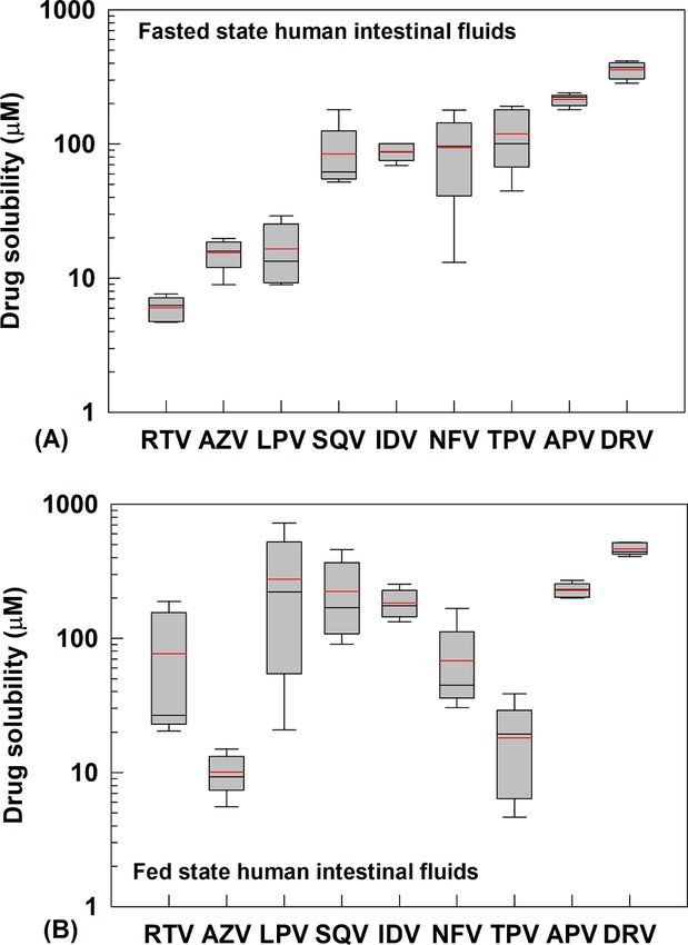
You can also read