EUthyroid recommendation on conducting thyroid ultrasound in population studies
←
→
Page content transcription
If your browser does not render page correctly, please read the page content below
EUthyroid recommendation on conducting
    thyroid ultrasound in population studies

This project has received funding from the European Union’s Horizon 2020 research and innovation programme under grant agreement No 634453   1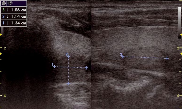
EUthyroid Consortium
1.    University Medicine Greifswald, DE                       12. The Barcelona Institute for Global Health –                          24. Centre for Regional Policy Research and
      Prof. Henry Völzke, MD                                       Campus Mar, ES                                                           Cooperation Studiorum Zdruzenje, MK
                                                                   Ass. Res. Prof. Mònica Guxens, MD                                        Prof. Borislav Karanfilski, MD
2.    National Institute for Health and
      Welfare, FI                                              13. University Hospital Center                                           25. The Queen’s University of Belfast, GB
      Dr. Iris Erlund                                              “Sestre Milosrdnice”, HR                                                 Prof. Jayne Woodside
                                                                   Prof. Zvonko Kusič
3.    Iodine Global Network, CA                                                                                                         27. Uppsala University, SE
      Prof. John H. Lazarus, MD                                14. Free University of Brussels, BE                                          Prof. Mehari Gebre-Medhin, MD

4.    University of Patras, GR                                     Prof. Rodrigo Moreno-Reyes, MD                                       28. Landspitali University Hospital, IS
      Prof. Kostas B. Markou, MD                               15. Sofia University, Faculty of Medicine,                                   Prof. Ingibjorg Gunnarsdottir

5.    Erasmus Medical Centre Rotterdam, NL                         “Lozenetz”, BG                                                       29. Toulouse University Hospital, FR
      Assoc. Prof. Robin Peeters, MD                               Assoc. Prof. Ludmila Ivanova, MD                                         Prof. Philippe Caron, MD

6.    University of Latvia, LV                                 16. University of Pisa, IT                                               30. Endocrinology Centre, EE
      Prof. Valdis Pirags, MD                                      Prof. Paolo Vitti, MD                                                    Toomas Podar, MD

7.    University of Surrey, UK                                 17. The Hebrew University of Jerusalem, IL                               31. Institute of Endocrinology, CZ
      Prof. Margaret Rayman                                        Dr. Aron Troen                                                           Prof. Vaclav Zamrazil, MD

8.    UMIT – University for Health Sciences, Medical           18. National Institute of Nutrition and Seafood                          32. Ministry of National Defense, Armed Forces
      Informatics and Technology, AT                               Research, NO                                                             University Hospital, PT
      Prof. Uwe Siebert, MD, MPH, MSc, ScD                         Dr. Lisbeth Dahl                                                         João J de Castro, MD

9.    The Capital Region of Denmark, Research Centre           19. University of Debrecen, HU                                           33. University of Gothenburg, SahlgrenskaAcademy,
      for Prevention and Health, DK                                Prof. Endre V. Nagy, MD                                                  Institute of medicine, SE
      Betina H. Thuesen, PhD                                                                                                                Ass. Prof. Helena Filipsson Nyström, MD
                                                               20. University Medical Centre Ljubljana, SI
10. Swiss Federal Institute of Technology Zurich, CH               Prof. Simona GaberšĆĆek, MD
    Prof. Michael Zimmermann, MD                               21. Jagiellonian University, PL
11.   biolution GmbH, AT                                           Prof. Alicja Hubalewska-Dydejczyk, MD
      Dr. Iris Grüner                                          22. The Health Sciences Research Institute of the
                                                                   “Germans Trias i Pujol” Foundation, ES
                                                                   Prof. Manel Puig Domingo, MD

                                                       This project has received funding from the European Union’s Horizon 2020 research and innovation programme under grant agreement No 634453   2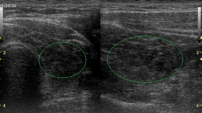
1 Introduction                                                                4     4 Diagnostic Findings                                                                               18
1.1 Aims                                                                      4     Endemic goitre (iodine-deficiency induced goitre)                                                   18
1.2 General Information                                                       4     Autoimmune disorders of the thyroid                                                                 18
                                                                                     Autoimmune thyroiditis (Hashimotos’ thyroiditis)                                                    19
2 Anatomy of the thyroid gland                                                5
                                                                                     Focal lesions                                                                                       20
Oesophagus                                                                    6
                                                                                     Post-treatment thyroid	                                                                             21
Vessels                                                                       7
Lymph nodes                                                                   7     5 Literature                                                                                        22

Muscles                                                                       7

3 Examination                                                                 8
3.1. Examination preparation                                                  8
3.2 Thyroid gland echo pattern                                                9
3.3 Performing the thyroid ultrasound examination                            9
  3.3.1 Participant set-up                                                   9
  3.3.2 Examination                                                         10
Locating the thyroid gland                                                  11

Evaluation of the thyroid echo pattern                                        11
Volume measurement                                                            13
Nodules                                                                     15
  3.3.3 Examination Wrap-Up                                                 16
3.4 Flow-Chart                                                              17

                                         This project has received funding from the European Union’s Horizon 2020 research and innovation programme under grant agreement No 634453            3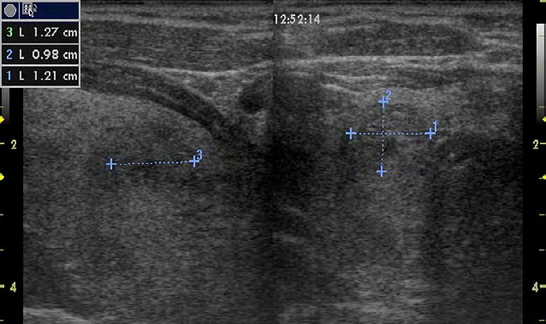
1 Introduction
1.1 Aims

This guidance is indented as a practical tool for study nurses and ultrasound readers conducting population based studies to monitor
iodine deficiency disorders (IDD). The guidance may be useful when writing a study manual and may be used for educational as well
as training purposes. In addition to this guidance, training efforts can be supplemented with the EUthyroid ultrasound training video
(https://vimeo.com/168037156) and the EUthyroid training application ARCUS (https://arcus.euthyroid.medizin.uni-greifswald.de), a
web-based ultrasound reader certification tool (registration required).

The guidance was written within the EUthyroid project, which aims to harmonize iodine and thyroid-related population based studies
in Europe. This can be realized by ensuring comparability of results therefore, improving the overall quality of studies. The objective of
thyroid ultrasound in epidemiological studies is to track the prevalence and incidence of thyroid disorder (e.g. goitre, thyroid nodules)
in a study population. In particular, changes of the thyroid gland due to improved iodine supply can be of interest in prevalence trend
analyses. In addition, knowledge of risk factors and correlations with other organ systems can be obtained.

1.2 General Information

The ultrasound enables three-dimensional morphological structure analysis of the thyroid gland free from superposition. Sonographic
volumetric is the gold standard for determining the organ size. The sonographic method currently used in clinical practice is based on
the principle that ultrasound is radiated into the tissue and, once reflected from tissue structures; its sound components are captured
and processed electronically (pulse-echo method, B-mode sonography).
The transmission frequencies used for this purpose are usually between 7.5-10MHz, the choice of the respective frequency represents a
compromise between resolution and penetration depth. Although a higher frequency improves the resolution of the image displayed, it
allows for a smaller penetration depth into the tissue.

                                          This project has received funding from the European Union’s Horizon 2020 research and innovation programme under grant agreement No 634453   4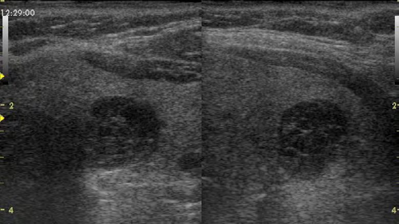
2 Anatomy of the thyroid gland
The thyroid gland is shaped like the letter ‘H’, with an oval lobe on each side of the trachea connected by the isthmus (Figure 1). The
pyramidal process, originating from the isthmus and developing in the midline upwards, is a rare inborn variant. Abnormalities, such as
the sublingual thyroid or a unilateral gland, are also very rare. The two lower arteries (inferior thyroid artery) enter the capsule of the
thyroid on the dorsal side of the lower poles.

                                                                                      The two upper arteries (superior thyroid artery) originating from
                                                                                      the external carotid artery enter the upper poles. The thyroid
                                                                                      gland consists of small lobules, each containing around 25 follicles.
                                                                                      The content of the follicles depends on their function. It also
                                                                                      determines the echo pattern (Table 1).

                                                                                      The echo pattern of the thyroid gland is homogeneous and
                                                                                      hyperechoic, at normofollicular stage, giving a strong contrast
                                                                                      to the hypoechoic pattern of the surrounding muscles, which
                                                                                      can serve as a reference. The outline of the gland is regular and
                                                                                      smooth. The bright echoes behind the isthmus are caused by air
                                                                                      within the trachea. The section of the lobes is round or triangular
                                                                                      in transverse planes and oval in longitudinal planes. The size of
                                                                                      each lobe is 5–6 cm (length (a)) * 2–4 cm (width (b)) * 1–2.5 cm
                                                                                      (thickness (c)). The volume of the whole gland is up to 18 ml in
                                                                                      females and 25 ml in males according to Gutekunst et. al., 1993.
                                                                                      Please refer to the Section 4 – Endemic goitre and Zimmermann
                                                                                      et al., 2004 on specific reference values for children. The gland
Figure 1: Thyroid Gland                                                               volume is determined by adding the volumes (V) of both lobes,
                                                                                      each calculated from the formula:

                                                                                       V = 0.479 x length x width x depth (Brunn et al., 1981)

Lutz, H., & Buscarini, E. (Eds.), 2013    This project has received funding from the European Union’s Horizon 2020 research and innovation programme under grant agreement No 634453   5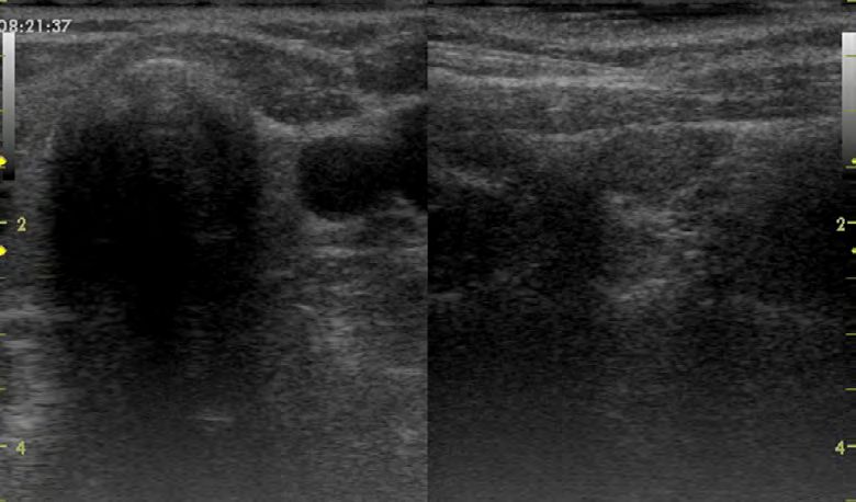
Please note that the cut-off to determine goitre is only useful for epidemiological studies. Reference values for thyroid volumes
        vary depending on the iodine status under investigation. Therefore, cut-off shall not be used in a clinical setting. By experience,
        slightly enlarged thyroid glands are rarely accompanied with symptoms. By using the definition of goitre established by
        Gutekunst results of current studies can be compared with findings of population surveys and of monitoring studies performed
        in the last decades.

                                                                                        Oesophagus

                                                                                        The cervical oesophagus (Figure 2) can be seen in the transversal
                                                                                        plane behind the thyroid as a tubular or target-like structure if this
                                                                                        area is scanned with the probe moved to the left-hand side to avoid
                                                                                        the shadow of air within the trachea.

Figure 2: Oesophagus

Lutz, H., & Buscarini, E. (Eds.), 2013      This project has received funding from the European Union’s Horizon 2020 research and innovation programme under grant agreement No 634453   6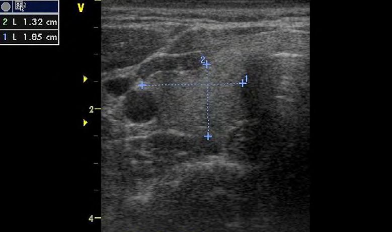
Vessels

                                         The large vessels (the carotid artery and the jugular vein) are behind the sternocleidomastoid
                                         muscles and lateral to the thyroid gland. The jugular vein shows as an oval shape and may
                                         collapse in certain phases of respiration or from the pressure of the probe. The carotid artery
                                         is round and shows stronger wall echoes than the vein.

                                         Lymph nodes

                                         The number of lymph nodes is especially high in the neck. High-frequency ultrasound
                                         frequently demonstrates normal lymph nodes, which are usually oval, with a maximum short
                                         axis diameter of 8 mm. The pattern is hypoechoic with a hyperechoic central hilum. In older
                                         people, the pattern becomes increasingly hyperechoic due to fatty degeneration, mostly
                                         starting from the hilum.

                                         Muscles

                                         The muscles are hypoechoic with a striated structure. They are important as anatomical
                                         landmarks and serve as a reference point for evaluating the echo pattern of the thyroid.

Lutz, H., & Buscarini, E. (Eds.), 2013   This project has received funding from the European Union’s Horizon 2020 research and innovation programme under grant agreement No 634453   7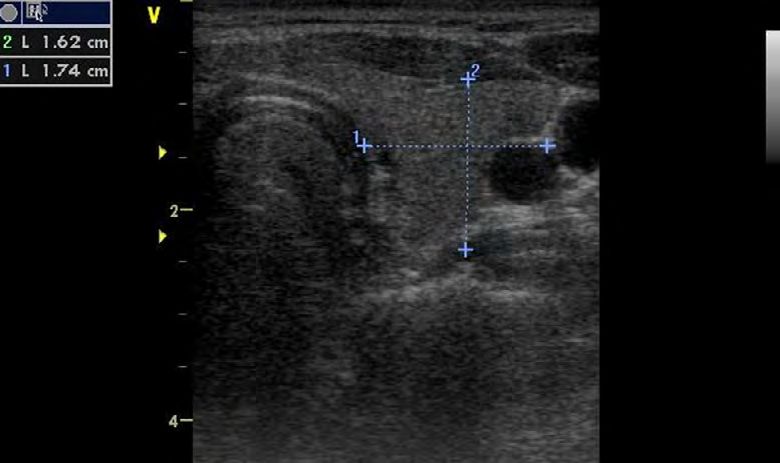
3 Examination
                                        3.1. Examination preparation

                                            SPECIFICATION FOR EXAMINATION ROOM

                                          • Temperature: approx. 26°C ± 2°C
                                          • Sufficient space for equipment (10m2 recommended)
                                          • Room has to be shaded by blinds

                                            EQUIPMENT

                                          • Treatment table
Figure 3: Example of examination room
                                          • Height adjustable sonographer chair
                                          • Ultrasound system, 5-12 MHz linear array (most frequently 7.5 MHz)
                                          • Knee pillow for support
                                          • Neck roll to hyper-extend the neck

                                            SUPPLIES

                                          • Probe gel
                                          • Sheet for treatment table
                                          • Disinfectant

                                        This project has received funding from the European Union’s Horizon 2020 research and innovation programme under grant agreement No 634453   8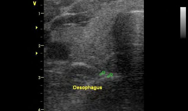
3.2       Thyroid gland echo pattern

Number and size of the thyroid follicles and its colloidal content influence the echo pattern.
The assessment of nodule echo patterns follows the echo pattern guidelines of the thyroid gland.
Table 1: Differentiation of thyroid echo patterns ro

     Echo-normal                       normofollicular, normally functioning thyroid gland

     Hypoechoic                        micro-follicular, the image appears dark

     Hyperechoic                       macro-follicular, the image appears bright

                                       concurrent echo-normal/hypoechoic/hyperechoic patterns in one
     Echo-complex
                                       structure with blurred margins

     Echo-free                         dark areas, possible fluid (cyst)

     Echo-dense                        Possible calcifications

3.3       Performing the thyroid ultrasound examination

3.3.1 Participant set-up

After the participant has entered in the ultrasound room, the sonographer reviews the
examination with the participant to make sure he/she understands the procedure and to
gain his/her cooperation. The participant will also be asked about any thyroid surgery and
radioiodine therapy. The year of the last operation and the operated side and the year of the
last radio-iodine therapy should be documented.

This project has received funding from the European Union’s Horizon 2020 research and innovation programme under grant agreement No 634453   9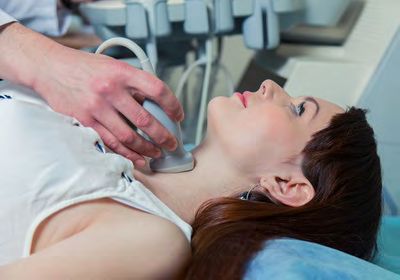
The participant will be asked to undress the neck and remove necklaces and large earrings.
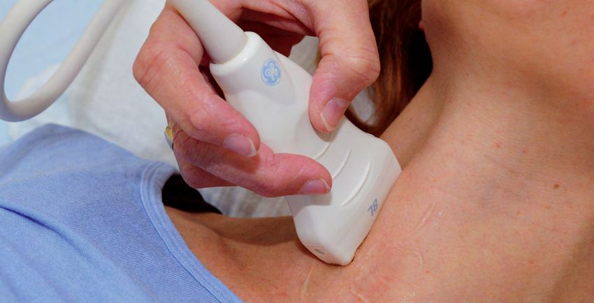
The thyroid ultrasound is performed in the supine position with slightly overstretched cervical
spine. In support of the cervical spine a neck roll placed underneath (Figure 4). The head
portion of the examination table is not to be raised. In exceptional cases, breaks during
the ultrasound examination can be offered. The neck area will be covered with a cloth and
ultrasound gel is applied to the throat. Scars should be documented.
3.3.2 Examination
Please use the following image sequence when examining the thyroid gland.
Figure 4: Position of neck roll room
Examination Documentation//labelling
Evaluation of the echo pattern right
Split image // Right lobe
Transverse and longitudinal scan right
Volume measurement right Transverse and longitudinal image with
Transverse scan right, longitudinal scan right measurements // Volume right
Evaluation of the echo pattern left
Split image // Left lobe
Transverse and longitudinal scan left
Volume measurement left Transverse and longitudinal image with
Transverse scan left, longitudinal scan left measurements // Volume left
Nodules Split Image // Right lobe, nodule 1...5
Transverse and longitudinal scan right Split Image // Left lobe, nodule 1…5
Transverse and longitudinal scan left
This project has received funding from the European Union’s Horizon 2020 research and innovation programme under grant agreement No 634453 10Please note, when thyroid ultrasound is performed in a clinical setting, cervical lymph
nodes should be assessed. Locating the thyroid gland Locating the thyroid gland
Locating the thyroid gland
Locating the thyroid gland
After a brief orientation the probe is applied in a coronal plane above the cricoid without any
additional pressure. It is then moved slowly and continuously towards the sternum. The trachea
is clearly visible in the image centre. Thus, the transverse axis of both thyroid lobes can be
followed.
The echo pattern of the thyroid gland is homogeneous and hyperechoic giving a strong
contrast to the hypoechoic pattern of the surrounding muscles, which can serve as a reference.
The outline of the gland is regular and smooth.
Evaluation of the thyroid echo pattern
The trachea, the accompanying vessels (carotid artery, jugular vein) and the echo pattern of
the surrounding muscle groups (Smm (sternocleidomastoid muscle)) serve as a reference point
for assessing the thyroid tissue echo pattern.
The examination starts in the coronal plane and the transverse scan image is captured first.
Then the probe is rotated at the point of the largest expansion in width of the thyroid gland by
90 ° and the lobe is examined in a longitudinal scan. The longitudinal scan image is added to
the already recorded transverse image (split).
In a hypoechoic pattern, the coloration of the thyroid tissue is identical to the muscle tissue.
Muscle and thyroid tissue can only be visibly separated by the connective tissue of the thyroid
capsule.
This project has received funding from the European Union’s Horizon 2020 research and innovation programme under grant agreement No 634453 11Documentation/labelling: A split image of the transverse and longitudinal scan should be saved for each lobe. In case of enlarged
thyroid glands, a non-complete longitudinal scan image is sufficient. The image is labelled either “right lobe” or “left lobe” in the bottom
right corner.
Figure 5: Documenting echo pattern, split image of left lobe; transverse scan left, Figure 6: Documenting echo pattern, split image of left lobe after subtotal resection
longitudinal scan right
After subtotal resection, small thyroid tissue remnants may be hard to identify, especially if they are not hyperechoic.
Lack of the isthmus is typical example of a post-operative situation.
This project has received funding from the European Union’s Horizon 2020 research and innovation programme under grant agreement No 634453 12Volume measurement
To determine the volume of each lobe, the width and depth are measured first in the transverse image. The width is measured at the
lobe largest expansion. The depth is measured axial from the top edge to the bottom edge of the lobe.
Documentation/labelling: The transverse image with measurements is saved labelled either “volume right” or “volume left” in the
bottom right corner of the image. The transverse image should be saved even if the participant has an operated thyroid gland.
Figure 7: Diameter measurements of left lobe; 1) width, 2) depth Figure 8: Diameter measurement of right lobe; 1) width, 2) depth
To capture the largest expansion in length, the lobe needs to be measured in a slight angle extending towards the cranium.
For accuracy, a complete image of the longitudinal scan is preferred. The volume for each lobe is calculated according to the formula:
V = 0.479 x length x width x depth (Brunn et al., 1981)
This project has received funding from the European Union’s Horizon 2020 research and innovation programme under grant agreement No 634453 13Between the volume measurements of both thyroid lobes the depth of the isthmus is measured
and recorded as well. The isthmus thickness should always be examined. If the isthmus
measures > 1cm, it should be examined for nodules.
Documentation/labelling: The longitudinal image with measurements is saved labelled either
“volume right” or “volume left” in the bottom right corner of the image. The longitudinal image
should be saved even if the participant has an operated thyroid gland.
Table 2: Plausible measurements for each dimension of a thyroid lobe and the isthmus
Dimension Minimum (in cm) Maximum (in cm)
Width 0.2 4.8
Depth 0.5 4.4
Length 1.2 10.3
Isthmus 0.1 1.5
This project has received funding from the European Union’s Horizon 2020 research and innovation programme under grant agreement No 634453 14In rare occasions the gland is enlarged so that it cannot be shown in one longitudinal scan or the probe used is insufficient in size to
depict the full length of lobe. The following procedure is a recommendation on how to estimate the length if not despicable in one
ultrasound image:
• a. The caudal tip of the lobe is adjusted in the longitudinal scan, so that it is exactly on the right hand side of the screen.
• b. The sonographer remembers the structures on the left hand side of the screen.
• c. The probe is moved slowly along the longitudinal axis of the thyroid gland in cranial direction until the memorized structure
from the left hand side is just visible on the right hand side of the screen.
• d. Freeze the image.
• e. In case the thyroid extension exceeds the length of the probe, the sonographer measures the length from the right hand side
of the screen to the cranial tip of the lobe, and adds 4 cm to this measurement to estimate the complete length of the lobe.
The length measurement must be performed at least twice. If the two measurements differ substantially, a third assessment is required.
The length measurements are averaged and used to calculate the volume for each lobe according to formula listed previously.
Documentation/labelling: Since the longitudinal scan cannot be depicted within the screen, no image will be saved.
Nodules
Thyroid nodule is a structure, which can be differentiated from thyroid parenchyma by an imaging method. Each nodular structure (up
to five) with a diameter larger than 1 cm in at least one dimension should be documented in a split image. Width, depth and length are
measured. Based on the nodule’s contour, the distinction between well definable margin or blurred margin and the echo pattern should
be documented with the measurements. In comparison, cysts and cystic lesions have a hypoechoic pattern. All three dimensions should
always be measured and documented as well.
Documentation/labelling: A split image of the transverse and longitudinal scan with measurements should be saved for each nodule.
The image is labelled in the bottom right corner with the following information: either “right lobe” or “left lobe” and nodule number
(1 through 5).
This project has received funding from the European Union’s Horizon 2020 research and innovation programme under grant agreement No 634453 15Figure 9: Left lobe; Nodule; 1) width; 2) depth; 3) length Figure 10: Right lobe; Nodule; 1) width; 2) depth; 3) length
3.3.3 Examination Wrap-Up
After removing the ultrasound gel and the cloth from the participant’s upper body, the participant gets up and dressed.
The sonographer can briefly review the examination results pointing out goitre, noticeable echo patterns and nodules.
This project has received funding from the European Union’s Horizon 2020 research and innovation programme under grant agreement No 634453 163.4 Flow-Chart
• Checking on all supplies
• Verbally preparing the participant
• Inquire about any thyroid surgery and radioiodine therapy
Participant set-up
• Ask participant to undress neck area and remove necklaces and earrings
(Duration: 5min)
• Dimming lights or closing blinds
• Preferred position for examining a patient is supine
• Look for scars in the thyroid area
Examination • Assessment of each lobe’s echo pattern and homogeneity
(Duration: 6-12 min; depending on • Volume measurement of each lobe
indications and quality of images) • Monitoring and recording of pathological findings
Examination wrap-up • Ask participant to get dressed
(Duration: 5 min) • Review examination results and findings
This project has received funding from the European Union’s Horizon 2020 research and innovation programme under grant agreement No 634453 174 Diagnostic Findings Endemic goitre (iodine-deficiency induced goitre) Endemic goitre is very common in areas of iodine deficiency At this stage, goitre may regress if iodine deficiency is corrected. and is defined by an enlarged thyroid volume (>18 ml in women, The pattern becomes inhomogeneous (nodular goitre), mainly >25 ml in men). In children aged 6 to 12 years, a thyroid volume in middle-aged patients, because of inhomogeneous growth of above 97th percentile from international reference distributions the thyroid follicles and the development of macro-follicular (age-, sex-, and body surface-area- specific) is defined as goitre hyperechoic nodules. Degenerative alterations, such as cystic (Zimmermann et al., 2004). A moderate thyroid enlargement is degeneration of the nodules and calcification, may also occur. rarely accompanied by symptoms. Liquid parts of the nodules are echo-free and may imitate cysts. Endemic goitre shows a homogeneous, hyperechoic pattern in the Calcifications cause strong echoes, sometimes with acoustic early stage of the disorder. shadows. Autoimmune disorders of the thyroid A common sonographic finding in autoimmune disorders of the thyroid is a hypoechoic pattern. Graves’ disease In this disease, autoantibodies against the thyroid-stimulating corresponding histologically to hyperplastic but empty follicles. hormone receptor stimulate the thyroid in similar way as TSH. Colour and power Doppler examinations demonstrate striking The disease occurs more often in young female adults, but can hypervascularity. Focal lesions (nodules) within the hypoechoic be seen at any age and also in men. Ultrasound can be used to thyroid gland in Graves-Basedow’s disease are independent of the visualize a moderately enlarged thyroid with a characteristic basic disease and should be considered separately. hypoechoic homogeneous or slightly inhomogeneous pattern Lutz, H., & Buscarini, E. (Eds.), 2013 This project has received funding from the European Union’s Horizon 2020 research and innovation programme under grant agreement No 634453 18
Autoimmune thyroiditis (Hashimotos’ thyroiditis)

In this disease, lymphocytic infiltration slowly destroys the gland. This infiltration causes a hypoechoic or heterogeneous pattern of the
thyroid, which develops over a long time.

                                                                                                    In the initial stage, the gland might be slightly enlarged; in the
                                                                                                    later (atrophic) stage, however, the thyroid gland becomes small,
                                                                                                    with a volume of less than 10 ml, and blood flow can remain normal
                                                                                                    or decrease.
                                                                                                    In some patients, though, the thyroid gland remains enlarged,
                                                                                                    with heterogeneous structures. Typical focal lesions should be
                                                                                                    considered for biopsy. Ultrasound imaging alone cannot confirm
                                                                                                    Hashimoto’s thyroiditis. Additional blood analysis is required to
                                                                                                    confirm the diagnosis.

Figure 11: Hypoechoic pattern typical for Hashimotos’ thyroiditis

Lutz, H., & Buscarini, E. (Eds.), 2013                  This project has received funding from the European Union’s Horizon 2020 research and innovation programme under grant agreement No 634453   19
Focal lesions
Cysts and cystic lesions
True cysts of the thyroid are very rare, and most cystic echo-free lesions are cystic-degenerated nodules (Figure 12). In these cases,
ultrasound demonstrates a cloud of echoes floating in the fluid. The puncture shows brownish fluid (‘chocolate cyst’).
Solid nodules
Nodules in the thyroid are common findings and are often
asymptomatic. Malignant tumours of the thyroid are relatively rare.
The differentiation of benign and malignant nodules is a challenge
in the ultrasound diagnosis of the thyroid gland.
Benign lesions
Adenomas are encapsulated, grow slowly and are mainly micro-
follicular. Adenomas vary in their function. Toxic adenomas are
highly differentiated and can accumulate iodine independently
of thyroid-stimulating hormone regulation. Depending on the
follicular structure, adenomas are mostly hypoechoic (micro-
follicular) or more hyperechoic and rather homogeneous. Their
contour is smooth. A hypoechoic halo (B-scan) with vessels
around the nodule is characteristic of a benign adenoma and is,
Figure 12: Cyst in the right lobe (transverse scan left, longitudinal scan right) therefore, best demonstrated with colour Doppler. Toxic adenomas
often show hypervascularity. Hyperplastic nodules develop over
several years in cases of endemic goitre, and are caused by the
differential growth ability of the thyroid cells. These lesions are not
true neoplasms. Owing to their normofollicular or macro-follicular
structure, they are often hyperechoic with a hypoechoic halo.
Degenerative alterations, such as cystic parts or calcifications, are
common, causing a complex sonographic pattern with echo-free
areas and strong echoes.
Lutz, H., & Buscarini, E. (Eds.), 2013 This project has received funding from the European Union’s Horizon 2020 research and innovation programme under grant agreement No 634453 20Malignant tumours Post-treatment thyroid
Papillary and follicular carcinomas are the commonest thyroid
carcinomas, with prevalence of 75% and 16%, respectively. They Providing the patient with information about the treatment
are highly differentiated and grow slowly. Anaplastic carcinoma (disease and method) is a prerequisite for post-treatment
is rare, especially in iodine-sufficient areas (ca. 1–2%), and is a check-ups. This is especially important after surgery. During
carcinoma of older patients (> 65 years). Medullary carcinoma the preparation of the patient’s neck for thyroid ultrasound,
arises from the C cells. It occurs either sporadically, in a hereditary the examiner should look for scars. After subtotal/non-radical
form (RET mutation; isolated familial MTC), or as part of multiple resection of an endemic goitre, relatively large nodular relapses
endocrine neoplasia type 2 (2A or 2B). Metastases, mainly from with inhomogeneous echo patterns on both sides of the trachea
lung cancer and malignant lymphomas, may involve the thyroid may be seen. Lack of the isthmus is typical in the post-operative
gland. The malignant tumours are hypoechoic and slightly situation. After subtotal resection, it is sometimes difficult
inhomogeneous, although an inhomogeneous pattern is difficult to identify small thyroid remnants, especially if they are not
to recognize in smaller lesions. An extremely hypoechoic pattern hyperechoic (echo-rich). Examination of the lymph nodes and
is found in malignant lymphomas and anaplastic carcinomas. identification of the local recurrence are the main objectives of
Lack of a halo is typical. The contour is irregular and may show follow-up checks after surgical treatment of carcinomas. Enlarged,
pseudopods or spicules. Interruption of the thyroid capsule and rounded lymph nodes are suspicious. It is interesting that lymph
infiltration of the surrounding tissue can be visualized. When the node metastases from highly differentiated carcinomas show a
sagittal diameter is greater than the transverse diameter, the hypervascularity similar to that of the primary tumours. In Graves’
finding is usually suspect. Dispersed strong echoes arising from disease, the typical hypoechoic pattern disappears with functional
micro-calcifications are characteristic in papillary carcinoma. remission after/during treatment whereas after radioactive
Spotted calcifications also occur in medullary and anaplastic iodine treatment the thyroid remains hypoechoic. A reduction
carcinomas. Occasionally, small echo-free or cystic parts are in the elevated flow velocity in the feeding arteries appears
seen in papillary carcinomas. to be an early sign of successful treatment. In contrast, the
The Doppler technique shows hypervascularity in highly hypoechoic pattern of Hashimoto thyroiditis is seen in all phases,
differentiated carcinomas, but without a vascular halo. Anaplastic independent of function. Radioiodine ablation of the thyroid or
carcinomas, most of the metastases and the lymphomas are radiation of the neck (lymphomas) causes a reduction of thyroid
hypovascular when compared to the thyroid. tissue and ultimately leads to a small thyroid. Warm or toxic
adenomas treated successfully with alcohol instillation not only
become smaller and more hyperechoic, but their hypervascularity
disappears.
Lutz, H., & Buscarini, E. (Eds.), 2013 This project has received funding from the European Union’s Horizon 2020 research and innovation programme under grant agreement No 634453 215 Literature
Brunn, J., Block, U., Ruf, G., Bos, I., Kunze, W. P., & Scriba, P. C. (1981). Volumetric analysis of thyroid lobes by real-time ultrasound (author’s transl). Deutsche
medizinische Wochenschrift (1946), 106(41), 1338-1340.
Gutekunst, R., & Martin-Teichert, H. (1993). Requirements for goiter surveys and the determination of thyroid size. In Iodine deficiency in Europe (pp. 109-118).
Springer, Boston, MA.
Hampel, R., Kuhlberg, T., ZOLINER, H., Klinke, D., Klein, K., Pichmann, E. G., & Kramer, A. (1996). Alimentare Jodversorgung in Deutschland. MMW. Münchener
medizinische Wochenschrift, 138(6), 28-32.
Lutz, H., & Buscarini, E. (Eds.). (2013). Manual of diagnostic ultrasound (Vol. 2). World Health Organization.
Völzke, H., Lüdemann, J., Robinson, D. M., Spieker, K. W., Schwahn, C., Kramer, A., ... & Meng, W. (2003). The prevalence of undiagnosed thyroid disorders in a
previously iodine-deficient area. Thyroid, 13(8), 803-810.
Zimmermann, M. B., Hess, S. Y., Molinari, L., de Benoist, B., Delange, F., Braverman, L. E., ... & Pearce, E. N. (2004). New reference values for thyroid volume by
ultrasound in iodine-sufficient schoolchildren: a World Health Organization/Nutrition for Health and Development Iodine Deficiency Study Group Report.
The American journal of clinical nutrition, 79(2), 231-237.
This project has received funding from the European Union’s Horizon 2020 research and innovation programme under grant agreement No 634453 22You can also read