Tongue movement and intra-oral vacuum in breastfeeding infants
←
→
Page content transcription
If your browser does not render page correctly, please read the page content below

Early Human Development (2008) 84, 471–477

                                              a v a i l a b l e a t w w w. s c i e n c e d i r e c t . c o m

                                             w w w. e l s e v i e r. c o m / l o c a t e / e a r l h u m d e v

Tongue movement and intra-oral vacuum in
breastfeeding infants
Donna T. Geddes a,⁎, Jacqueline C. Kent a ,
Leon R. Mitoulas b , Peter E. Hartmann a
a
  The University of Western Australia, Biochemistry and Molecular Biology, School of Biomedical,
Biomolecular and Chemical Sciences, Faculty of Life and Physical Sciences, Australia
b
  Medical Research Coordinator, Medela AG, Medical Technology, Lättichstrasse 4b, 6341 Baar, Switzerland

Received 1 February 2007; received in revised form 21 August 2007; accepted 20 December 2007

    KEYWORDS                          Abstract
    Breastfeeding;
    Sucking;                          Objective: The mechanism by which the breastfeeding infant removes milk from the breast is still
    Ultrasound;                       controversial. It is unclear whether the infant uses predominantly intra-oral vacuum or a
    Lactation;                        peristaltic action of the tongue to remove milk from the breast. The aim of this study was to use
    Infant                            ultrasound to observe movements of the tongue during breastfeeding and relate these
                                      movements to both milk flow and simultaneous measurements of intra-oral vacuum.
                                      Methods: Submental ultrasound scans of the oral cavity of 20 breastfed infants (3–24 weeks old)
                                      were performed during a breastfeed. Intra-oral vacuums were measured simultaneously via a
                                      milk-filled supply line (SNS) connected to a pressure transducer.
                                      Results: Vacuum increased during the downward motion of the posterior tongue and at the same
                                      time milk flow and milk ducts in the nipple was observed. Peak vacuum (−145 ± 58 mmHg)
                                      occurred when the tongue was in the lowest position.
                                      Conclusions: Ultrasound imaging demonstrated that milk flow from the nipple into the infant's
                                      oral cavity coincided with both the lowering of the infants tongue and peak vacuum. Therefore
                                      vacuum is likely to play a major role in milk removal from the breast.
                                      © 2007 Elsevier Ireland Ltd. All rights reserved.

                                                                                1. Introduction

                                                                                Suckling of the mammary gland to obtain milk for nourishment
                                                                                is behaviour unique to mammals. Besides nourishment,
                                                                                suckling causes numerous responses in both the mother and
 ⁎ Corresponding author. M310, The University of Western Australia,             infant and these are thought to have evolved to promote
35 Stirling Highway, Crawley WA 6009, Australia. Tel.: +61 8 6488               survival of the infant in harsh environmental conditions [1].
3327; fax: +61 8 6488 1148.                                                     The close body contact during breastfeeding regulates infant
   E-mail address: donna.geddes@uwa.edu.au (D.T. Geddes).                       respiration, acid-base balance and temperature, helps

0378-3782/$ - see front matter © 2007 Elsevier Ireland Ltd. All rights reserved.
doi:10.1016/j.earlhumdev.2007.12.008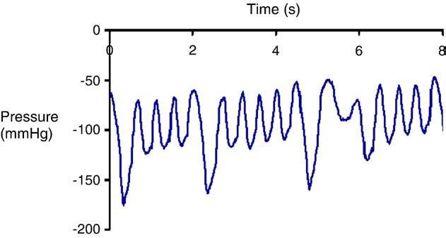
472                                                                                                          D.T. Geddes et al.

conserve energy reserves [2] and soothes the infants [3,4].       pressure transducer (Cobe Laboratories, Frenchs Forest, NSW
Furthermore, breastfeeding impacts upon the normal oral           2086, Australia). The transducer was connected via an
facial development of the infant [5]. However, one of the         interconnect cable (Cobe Laboratories) to an amp bridge
primary roles of suckling is to remove milk from the mammary      (ADInstruments, Castle Hill, NSW 2154, Australia) and the
gland and adequate milk removal is essential to sustain milk      output was recorded using computer hardware, Power Lab
synthesis for a successful lactation.                             (ADInstruments) and software package Chart v5.0.2 (ADInstru-
   Historically two theories have evolved, through observa-       ments) on a laptop computer (Mac OS X v10.3.8). This method
tion, to explain how the infant removes milk from the breast.     allowed for the direct measurement of the vacuums during
One theory suggests that compression of the breast by the         each suck cycle over the course of the breastfeed. No fluid was
infant's jaw and a peristaltic action of the tongue to express    removed from the SNS line during the breastfeed thus
milk into the mouth is the major factor in milk removal           measured vacuum was not affected by additional milk flow.
during breastfeeding [6,7,8]. and this theory presupposes
the existence of lactiferous sinuses. Since these are not         2.4. Ultrasound equipment and imaging
present in the human breast [9] this theory must be re-
examined. The second theory suggests that vacuum applied
                                                                  Submental scans of the midline of the infant's oral cavity were
by the infant results in efficient milk removal from the breast
                                                                  performed [11,13] by an experienced ultrasonographer. For
[10]. The few studies that have tested these theories [7,8]
                                                                  10 infants an Acuson, XP10 (Siemens, Mountain View,
have still not clarified the sucking mechanism in detail. Most
                                                                  California, USA) with an endocavity convex transducer
of the studies performed on breastfeeding infants have
                                                                  (EC7) was used to acquire ultrasound images. The remaining
involved imaging in order to determine the tongue motion in
                                                                  10 infants were scanned using Toshibia SSA-770A/80, Aplio 80
relation to the nipple. One early study used fluoroscopy (X-
                                                                  (Tokyo, Japan) using the PVT-661VT transducer. Parker
Radiation) to image the infant breastfeeding [7]. The mother
                                                                  Ultrasonic Gel (Fairfield, New Jersey, USA) was used. This
and infant were unable to feed in a ‘normal position’ and
                                                                  transducer has a long handle which is both easy to manipulate
milk flow was not identified. In subsequent studies ultra-
                                                                  and is less invasive than bulkier transducers. Further more
sound imaging was the modality of choice due to being non-
                                                                  this transducer provides a wide convex field of view (160°)
invasive and safe (compared to X-radiation). However, large
                                                                  facilitating a panoramic view of the hard and soft palate. The
transducers with limited image resolution compared to
                                                                  transducer was positioned along the midsagittal line of the
current technology were used. Improved image resolution
                                                                  infant's body and light pressure was used to maintain contact
of current machines allows real-time imaging of infant oral
                                                                  with the infant's chin. The transducer was rotated until the
structures such as the tongue, hard and soft palate [11]. The
                                                                  image of the nipple was both at its maximum length and
aim of the study was to examine the relationship between
                                                                  widest diameter and a clear view of the hard–soft palate
tongue movement and intra-oral vacuum generated during
                                                                  junction was achieved (Fig. 1A, B). Since the supply line (SNS)
breastfeeding by the infant.
                                                                  measuring intra-oral pressure was positioned alongside the
                                                                  nipple this was not visible in the midline. However the
2. Methods                                                        position of the supply line in the infant's mouth could be
                                                                  confirmed by moving the transducer laterally and visualizing
2.1. Participants                                                 the tube as hyperechoic (white) lines. The position of the
                                                                  transducer was altered accordingly with infant movement to
Twenty mothers and infants (3–24 weeks old) were recruited        maintain a midline sagittal view. Average setting values for
through either the Australian Breastfeeding Association or        the Acuson, XP10 were gained: 50 db, dynamic range: 57 db,
                                                                  frequency: 7 MHz and for the Toshiba, Aplio 80 they were gain
community health centres. All infants were successfully
                                                                  55 db, dynamic range: 60 db, frequency: 8.8 MHz. Two focal
breastfed and mothers supplied written, informed consent to
                                                                  zones were used to narrow the ultrasound beam and hence
participate in the study, which was approved by the Human
                                                                  improve image resolution. One was placed at the hard palate
Research Ethics Committee of The University of Western
                                                                  and the other at the nipple–tongue apposition. Further ad-
Australia.
                                                                  justments were made to the gain, dynamic range and time
                                                                  gain compensation to optimise the image during scanning. In
2.2. Determination of milk intake                                 some cases lowering of the frequency of the probe from
                                                                  8.8 MHz to 7.3 or 5.8 MHz was necessary to improve imaging at
The amount of milk consumed during a breastfeed was               depth in older and larger infants. All ultrasound scans began
determined by test weighing the baby using an electronic          when the infant attached to the breast and ended when the
balance (Medela Electronic Baby-Weigh Scales, Medela AG,          infant finished feeding. The scan was videotaped for later
Switzerland) prior to and after breastfeeding. No correction      analysis. The ultrasound images and the intra-oral pressure
for infant insensible water loss was made; therefore milk         were recorded simultaneously.
intake may be underestimated by 10 ± 12% (mean ± SD) [12].
                                                                  2.5. Statistical analysis
2.3. Intra-oral pressure
                                                                  The tongue motion was linked to pressure measurement and
A supply line (SNS) filled with the mother's expressed            frame by frame analysis of tongue position was performed for
breastmilk was placed alongside the nipple and connected via      at least three full suck cycles during which milk flow was
a silicon tube (650 ×4 mm) and three-way tap to a disposable      evident on the ultrasound image (Fig. 1C). Further ultrasound
Tongue movement and intra-oral vacuum in breastfeeding infants                                                                   473

measurements were made of the nipple diameter, nipple to
hard–soft palate junction (N-HSPJ) and depth of the milk flow
area.
   For all mothers descriptive statistics of the breastfeed
were calculated. The statistics: mean vacuum, peak vacuum

                                                                 Figure 2 A typical infant intra-oral pressure trace during a
                                                                 breastfeed. Peak vacuum (mean minimum pressure) ranges from
                                                                 −110 to − 170 mmHg. Baseline pressure (mean maximum
                                                                 pressure) ranges from −50 to − 60 mmHg.

                                                                 (mean minimum pressure; Fig. 2) and baseline vacuum (mean
                                                                 maximum pressure; Fig. 1) was generated by the Chart
                                                                 v5.0.2 analysis software.
                                                                    Paired t-tests were performed and two sided P-values are
                                                                 quoted. A P-value b 0.05 was regarded as statistically
                                                                 significant. Results are presented as mean ± SD.

                                                                 3. Results

                                                                 The mean breastfeed duration was 8 min 16 s ± 2 min 45 s with
                                                                 a mean milk intake of 63 ± 31 g. The mothers regarded the
                                                                 behaviour of the infant during the test breastfeed as normal.

                                                                 3.1. Intra-oral pressure

                                                                 The mean vacuum was −114 ± 50 mmHg. The peak vacuum
                                                                 was − 145 ± 58 mmHg and baseline vacuum was − 64 ±
                                                                 45 mmHg. Milk intake was not related to the peak vacuum,
                                                                 baseline vacuum or duration of the breastfeed.

                                                                 3.2. Ultrasound

                                                                 On ultrasound imaging the hard palate appears as an echogenic
                                                                 (white) line whereas the soft palate appears as a grey structure
                                                                 (3–4 mm) with an echogenic border. The nipple on the left side
                                                                 of the image appears as a grey soft tissue structure and milk
                                                                 ducts within the nipple appear as hypoechoic (black) thin
                                                                 tubular structures. Milk flow was visualised as hypoechoic fluid
                                                                 containing echogenic flecks. The echogenic flecks are the fat
                                                                 globules in the milk (Fig. 1) [14].
                                                                    Tongue motion during a suck cycle showed that at baseline
                                                                 vacuum the posterior tongue was in apposition with the

                                                                 Figure 1 Submental ultrasound images of the intra-oral cavity
                                                                 of an infant during breastfeeding. A, B. The infants tongue (T) is up
                                                                 in apposition with the hard palate (H). The end of the nipple is not
                                                                 at the hard soft palate junction (HSPJ). The soft palate (SP) is in
                                                                 line with the hard palate. C. The infants tongue (T) is down and
                                                                 milk ducts (MD) are visible within the nipple (N). Milk flow (MF) is
                                                                 visible within the oral cavity and the nipple has moved closer to the
                                                                 hard soft palate junction (HSPJ).
474 D.T. Geddes et al.
was greater when the tongue was up compared to when it was
down. The maximum distance of the posterior tongue from the
hard palate (Table 1) coincided with peak vacuum.
4. Discussion
Recent advances in the resolution of ultrasound imaging have
allowed more detailed examination of the sucking dynamics
of the infant, most notably the identification of milk flow. In
addition, the use of a long-handled convex transducer has
minimised interference during a breastfeed. With the
synchronisation of ultrasound imaging of the infant's oral
structures and the measurement of vacuum applied by the
infant to the breast this study found that when the infant
Figure 3 Schematic representation of tongue movement
lowered its tongue the intra-oral vacuum increased. Peak
through one suck cycle. Note that the border of the nipple is not
vacuum coincided with the tongue in its lowermost position
markedly indented by the tongue.
and subsequently as the infant raised its tongue the vacuum
decreased and milk flow ceased (Fig. 4). This study has shown
that milk only flows into the infant oral cavity when the
palate (up position) and the anterior tongue did not markedly downward movement of the posterior tongue creates an
indent the nipple border (Fig. 3). Vacuum was generated as intra-oral vacuum, highlighting the importance of vacuum in
the posterior (proximal) tongue and soft palate moved milk removal rather than the stripping action of the tongue in
downward (down position), coinciding with observed milk milk removal by the breastfeeding infant. The mechanism of
flow (Fig. 4). In addition milk ducts became visible within the milk removal used by the breastfeeding infant has remained
nipple as thin black lines (Fig. 1C). Peak vacuum occurred controversial for many years. Generally it is believed that
when the posterior tongue was maximally lowered (down the infant sucking dynamic consists of a stripping action by
position). As the vacuum was released the anterior tongue upward movement of the tongue considered to be essential
momentarily rose slightly and milk was seen to pass under for milk removal and the application of vacuum which is
the soft palate. Both the posterior tongue and soft palate believed to create a pressure gradient between the positive
then rose and milk continued to flow toward the pharyngeal pressure in the milk ducts (generated by milk ejection) to
region (swallow). When the posterior tongue reached the refill the milk ducts in the breast [8].
palate (up position) the vacuum returned to baseline levels The role of intra-oral negative and positive pressure in
(Fig. 4). infant feeding has been based on cineradiographic studies of
Nipple diameter was greater when the tongue was in the bottle-feeding infants. These studies showed that the infant
down position than when the tongue was up. The distance from occluded the teat with the front of the tongue and
the tip of the nipple to the hard/soft palate junction (N-HSPJD) compressed the teat expressing a radio-opaque mixture of
Figure 4 The changes in infant tongue position during one suck cycle.Tongue movement and intra-oral vacuum in breastfeeding infants 475
Table 1 Ultrasound measurements of the nipple, nipple ing. They examined six infants between 2 and 6 days of age
hard soft palate junction distance and height of the cavity and did not measure the milk intake of the breastfeed. The
into which milk flowed during breastfeeding (n = 20 infants) discrepancy between the ages of the infants we examined
may explain some differences in the tongue action. The
Tongue up Tongue down
peristaltic action is believed to strip milk from the breast [8].
N-HSPJD 6.9 ± 1.3 mm 5.2 ± 1.6 mm⁎⁎ Indeed some authors have speculated that positive pressure
Nipple diameter 7.5 ± 0.3 mm 9.8 ± 1.2 mm⁎ produced by the tongue and upper gum on either the base of
Tongue distance 4.2 ± 1.2 mm the nipple or areola pushes milk forward into the oral cavity.
Ultrasound measurements made during a suck cycle. Nipple hard This speculation is based on the ability to express milk by
soft palate junction distance = N-HSPJD, Tongue distance is the squeezing the breast between the thumb and fingers to elicit
distance from the hard palate to the surface of the posterior a jet of milk and presumes that the proximity of the jaw to
tongue ⁎⁎denotes significance P b 0.01 ⁎P b 0.05. the location of the milk ducts has the same effect. Ardran
and Kemp [22] noted that the tip of the tongue rises slightly
just before the distal part of the tongue lowers, however
they are cautious about the significance of this motion
formula and barium into the oral cavity. The teat then re- stating that evidence is lacking to support compression of the
filled as the mandible lowered and intra-oral negative milk ducts under the areola during breastfeeding. Therefore
pressure decreased [7,15]. Despite the inability to demon- the role of the tip of the tongue in milk removal to date is
strate milk flow on both cineradiographic and previous speculative. A stripping motion is more feasible during
ultrasound studies during breastfeeding it has been assumed bottle-feeding as the teat can be compressed and milk
that “The mechanism of breastfeeding is probably similar to squeezed from the teat. This is illustrated well in premature
bottle-feeding” [15]. Bottles and teats are remarkably infants who are able to express milk from a bottle without
different to the breast in both design and flow-rate, there- exerting vacuum [23]. However they become more efficient
fore it is not unreasonable to expect that removal of milk when they use both expression and vacuum to remove milk
would be different to breastfeeding. Indeed studies measur- [24]. Furthermore premature infants may be able to feed
ing orofacial muscle function have shown significantly effectively from a bottle but have difficulty removing
reduced masseter muscle activity in bottle-fed infants [16]. adequate amounts of milk from the breast suggesting that
Furthermore differences in perioral muscle function between there are different mechanisms of milk removal between
breastfed and bottle-fed infants appear to persist into the bottle and breastfeeding [25].
second and third year of life [17]. It is interesting to note that The position of the nipple in the infant's mouth has been
significant reduction in growth of the mandible has been of great interest and it is widely accepted that the nipple
observed in bottle-fed mice compared to those suckled by the should be in close proximity to the hard–soft palate junction
dam [18]. These differences in oral motor function and for breastfeeding to be comfortable and effective [8,26].
mandibular growth are highly likely to be a response to a non- Measurements performed on ultrasound scans showed that
physiological feeding method, thus differences in the the nipple rarely reaches the hard–soft palate junction
mechanics of milk removal during breastfeeding compared (Fig. 2; Table 1). These results agree with the findings of
to bottle-feeding would be expected. Jacobs et al. [11] although their measurements include both
The importance of vacuum in milk removal from the the tongue up and tongue down phase of the suck cycle which
breast is further supported by data gathered during the does not account for movement of the nipple. In addition we
expression of milk using an electric breast pump. This study found that increasing vacuum was associated with shorter N-
showed that using the mother's maximum comfortable HSPJ distances and wider nipple diameter. Therefore both
vacuum resulted in the expression of significantly more the peak and baseline vacuum applied by the infant may at
milk than using vacuums of lower magnitude [19]. In addition least in part determine the position of the nipple. Should the
the pump (using vacuum only) removed on average as much nipple be positioned at the hard–soft palate junction the
milk as the breastfeeding infant. Our results therefore lend application of vacuum would cause the soft palate to
weight to the theory of both Waller [10] and Smith et al. [20] descend to the tip of the nipple and effectively there
that vacuum applied to the breast facilitates milk removal. would be no space for the milk to flow into the oral cavity. In
During this study neither milk flow nor milk ducts within addition gag sensors are present on the surface of the soft
the nipple were observed when the posterior tongue was palate and if milk is ejected directly onto the soft palate the
squeezing the nipple or when it was in apposition with the gag reflex may be stimulated [26,27]. In this respect it may
palate. In periods of milk flow, during the application of be possible to position the nipple too close to the soft palate
increasing vacuum by the infant (as the tongue was lowered) during breastfeeding. The reason why the infant places the
the nipple expanded rendering milk ducts visible and the nipple close to the hard–soft palate junction is still not clear.
nipple moved closer to the hard–soft palate junction Some investigators have assumed that if vacuum were the
(Fig. 1C). The expansion of the nipple diameter as the only mechanism involved in the removal of milk the infant
tongue moved down is in contrast to the observations during would feed by placing the nipple at the lips [15]. This
studies of bottle-feeding where the teat is compressed and assumption does not take into account the interaction of
milk expressed [21]. The tongue movement demonstrated in swallowing. Furthermore clinically it has been well docu-
this ultrasound study was not consistent with a marked mented that nipple pain is associated with poor positioning
peristaltic action (Fig. 3), which is in agreement with results and attachment of the infant to the breast (not enough of
of Smith et al. [20]. Weber et al [13] using ultrasound the breast in the infant's mouth) [26,27]. The study of all
described a “rolling” peristaltic motion during breastfeed- three components of breastfeeding: sucking, swallowing and476 D.T. Geddes et al.
breathing is essential to gain a complete understanding of [3] Christensson K, Cabrera T, Christensson E, Uvnas-Moberg K,
the mechanisms of sucking during breastfeeding. Winberg J. Separation distress call in the human neonate in
The intra-oral pressure measurements in this study were the absence of maternal body contact. Acta Paediatr 1995;84:
468—73.
comparable to that of other studies of babies of similar age
[4] Michelsson K, Christensson K, Rothganger H, Winberg J. Crying
(peak vacuum − 197 ± 10 mmHg [28]; −150 mmHg [15]).
in separated and non-separated newborns: sound spectro-
Younger infants of 4–5 days old exert lower vacuums on graphic analysis. Acta Paediatr 1996;85:471—5.
average (peak vacuum −112 mmHg) [29] and may be due to [5] Labbok MH, Hendershot GE. Does breast-feeding protect against
the maturity of the infant. In this study we were unable to malocclusion? An analysis of the 1981 Child Health Supplement to
find a relationship between the volume of milk consumed by the National Health Interview Survey. Am J Prev Med
the infants and the intra-oral vacuum parameters measured. 1987;4:227—32.
This suggests that other factors such as either infant appetite [6] Cooper AP. The anatomy of the breast. Longman, Orme, Green,
or the amount of milk available in the breast may indeed Browne and Longmans; 1840.
influence milk intake and sucking parameters. As noted [7] Ardran GM, Kemp PH, Lind J. A cineradiographic study of
bottlefeeding. Br J Radiol 1958;31:156—62.
previously the duration of the feed is not solely indicative
[8] Woolridge MW. The anatomy of infant sucking. Midwifery
of milk intake [14]. Infants applied a baseline vacuum of
1986;4:164—71.
approximately –60 mmHg over the whole breastfeed (Fig. 2). [9] Ramsay DT, Kent JC, Hartmann RL, Hartmann PE. The anatomy
Previously baseline vacuums have not been reported in of the lactating breast redefined with ultrasound imaging.
breastfeeding infants; however, similar vacuums have been J Anat 2005;206:525—34.
described in the piglet [30]. It is likely this vacuum reflects [10] Waller H. The force exerted by the baby. Clinical studies in
the seal formed on the breast by the infant prior to active lactation. William Heinemann Ltd; 1936.
sucking and it is reflected in the small amount of movement of [11] Jacobs LA, Dickinson JE, Hart PD, Doherty DA, Faulkner SJ.
the nipple when the infant applies vacuum by the downward Normal nipple position in term infants measured on breastfeed-
movement of the tongue. Furthermore this vacuum may be ing ultrasound. J Hum Lact 2007;23:52.
[12] Arthur PG, Hartmann PE, Smith M. Measurement of milk intake
important in instances where infants have difficulty latching
of breastfed infants. J Pediatr Gastroenterol Nutr 1987;6:
to the breast resulting in poor milk transfer. For example it
758—63.
would be of interest to determine if infants with ankyloglossia [13] Weber F, Woolridge MW, Baum JD. An ultrasonographic study of
and pre-term infants who find it difficult to remain at the the organization of sucking and swallowing by newborn infants.
breast have low or absent baseline vacuums. Dev Med Chil Neurol 1986;28:19—24.
[14] Ramsay DT, Kent JC, Owens RA, Hartmann PE. Ultrasound
5. Conclusion imaging of milk ejection in the breast of lactating women.
Pediatrics 2004;113:361—7.
[15] Ardran GM, Kemp FH. A correlation between suckling pressures
This study demonstrated that during breastfeeding milk flow and the movements of the tongue. Acta Paediatr
occurred when the infant's mid-to posterior portion of the 1959;48:261—72.
tongue was lowered and increasing vacuum was applied [16] Inoue N, Sakashita R, Kamegai T. Reduction of masseter muscle
without accentuated peristaltic action. This suggests that activity in bottle-fed babies. Early Hum Dev 1995;42:185—93.
vacuum plays an integral role in the removal of milk from the [17] Jacinto-Goncalves SR, Gaviao MB, Berzin F, de Oliveriera AS,
breast by the infant. The role of the movement of the tip of Semeguini TA. Electromyographic activity of perioral muscle in
tongue in milk removal still remains speculative requiring breastfed and non-breastfed children. J Clin Pediatr Dent
2004;29:57—62.
further investigation. Furthermore a lack of baseline vacuum
[18] Oseko T, Kuroe K, Matsuo H, Utsu H, Ono H, Oshikawa S, et al.
(the seal applied to the breast by the infant) may be An experimental study on feeding style and development of
important in conditions where the infant is unable to remain jaws. J Orthod 1988;33:33—8.
attached to the breast. [19] Kent JC, Cregan MD, Doherty DA, Mitoulas LR, Ramsay DT,
Hartmann PE. The effect of vacuum on the removal of milk using
an electric breast pump. 12th International Conference of the
Acknowledgments
International Society for Research in Human Milk and Lactation
(ISRHML). Cambridge, UK: Queens' College; September 10 – 14,
Funding for this study was provided by a research grant from 2004.
Medela AG. Ultrasound support was provided by Acuson [20] Smith WL, Erenberg A, Nowak AJ. Imaging evaluation of the human
(Siemens, USA) and Toshiba (Japan). Research assistance nipple during breast-feeding. Am J Dis Child 1988;142:76—8.
from Tracey Williams was greatly appreciated. Also Philip [21] Hayashi Y, Hoashi E, Nara T. Ultrasonographic analysis of sucking
Hart for help with ultrasound measurements. behavior of newborn infants: the driving force of sucking
vacuum. Early Hum Dev 1997;49:33—8.
[22] Ardran GM, Kemp PH, Lind J. A cineradiographic study of
References breastfeeding. Br J Radiol 1958;31:156—62.
[23] Lau C, Sheena HR, Shulman RJ, Schanler RJ. Oral feeding in low
birth weight infants. J Pediatr 1997;130:561—9.
[1] Winberg J. Mother and newborn baby: mutual regulation of [24] Lau C, Kusnierczyk I. Quantitative evaluation of infant's
physiology and behavior – a selective review. Dev Psychiatry nonnutritive and nutritive sucking. Dyspahagia 2001;16:58—67.
2005;47:217—29. [25] Meier PP. Supporting lactation in mothers with very low birth
[2] Christensson K, Siles C, Moreno L, Belaustequi A, De La Fuente weight infants. Pediatr Ann 2003;32:317—25.
P, Lagercranz H, et al. Temperature, metabolic adaptation, and [26] Neifert MR. Breastmilk transfer: positioning, latch-on, and
crying healthy full-term newborns cared for skin-to-skin or in a screening for problems in milk transfer. Clin Obstet Gynecol
cot. Acta Paediatr 1992;81:488—93. 2004;47:656—75.Tongue movement and intra-oral vacuum in breastfeeding infants 477
[27] Miller AJ. Oral and pharyngeal reflexes in the mammalian nervous [29] Mathew OP, Bhatia J. Sucking and breathing patterns during breast-
system: their diverse range in complexity and the pivotal role of and bottle-feeding term neonates. AJDC 1989;143:588—92.
the tongue. Crit Rev Oral Biol Med 2002;13:409—25. [30] Thexton AJ, Crompton AW, Owerkowicz T, German RZ.
[28] Prieto CR, Cardenas H, Salvatierra AM, Boza C, Montes CG, Correlation between intra-oral vacuums and tongue move-
Croxatto HB. Sucking vacuum and its relationship to milk transfer ments in the suckling pig. Arch Oral Biol 2004;49:567—75.
during breastfeeding in humans. J Reprod Fertil 1996;108:69—74.You can also read