Extra-Medullary Hematopoiesis in Sickle Cell Disease Presenting as a Right Adrenal Mass - Cureus
←
→
Page content transcription
If your browser does not render page correctly, please read the page content below
Open Access Case
                               Report                                                  DOI: 10.7759/cureus.21334

                                                Extra-Medullary Hematopoiesis in Sickle Cell
                                                Disease Presenting as a Right Adrenal Mass
Review began 01/03/2022
                                                Folasade Ajayi 1 , Meenakshi S. Nali 1 , Ruhma Ali 1 , Aditya Patel 1 , Hamid Shaaban 2
Review ended 01/09/2022
Published 01/17/2022                            1. Internal Medicine, Saint Michael's Medical Center, Newark, USA 2. Hematology/Oncology, Saint Michael's Medical
© Copyright 2022                                Center, Newark, USA
Ajayi et al. This is an open access article
distributed under the terms of the Creative     Corresponding author: Ruhma Ali, raoruhma@gmail.com
Commons Attribution License CC-BY 4.0.,
which permits unrestricted use, distribution,
and reproduction in any medium, provided
the original author and source are credited.
                                                Abstract
                                                Extramedullary hematopoiesis can occur during normal fetal development or as a compensatory mechanism
                                                in chronic anemia when the primary sites of hematopoiesis fail. When it occurs, it is mostly seen in sites
                                                such as the liver, spleen, and lymph nodes. Extramedullary hematopoiesis is seen in patients with abnormal
                                                morphology of red blood cells like thalassemia and abnormal red blood cell shape like hereditary
                                                spherocytosis. The incidence of extramedullary hematopoiesis in sickle cell disease is very rare. We report a
                                                case of focal adrenal extramedullary hematopoiesis in a 21-year-old patient with sickle cell disease who
                                                presented with pain in the right thorax. A retroperitoneal mass was seen in the MRI of the abdomen of the
                                                patient suggestive of extramedullary hematopoiesis. Our case highlights the importance of physician
                                                awareness of this rare pathology for proper diagnosis and management.

                                                Categories: Internal Medicine, Anatomy, Hematology
                                                Keywords: mri images, adrenal mass, sickle cell anemia, extramedullary hematopoiesis, adrenal

                                                Introduction
                                                Extramedullary hematopoiesis is defined as the production of erythroid and myeloid progenitor cells in
                                                organs outside the bone marrow. It occurs in diverse conditions, including fetal development, normal
                                                immune responses, and pathological circumstances. During fetal development, before the formation of
                                                mature marrow, extramedullary hematopoiesis occurs in the yolk sac, fetal liver, and spleen. Most
                                                frequently, this response occurs in the spleen and liver for the production of antigen-presenting and
                                                phagocytes. Extramedullary hematopoiesis (EMH) can also occur in pathological conditions when bone
                                                marrow is inhabitable for stem and progenitor cells, including myelofibrosis, lymphoma, leukemia, or with
                                                marrow hyperactivity in conditions such as congenital hemolytic anemia like thalassemia and spherocytic
                                                anemia. Common known sites for extramedullary hematopoiesis are the liver, spleen, lymph nodes, and
                                                mediastinum. We report the first case of focal retroperitoneal EMH in sickle cell anemia depicted on MRI.

                                                Case Presentation
                                                A 21-year-old African American male with a past medical history of homozygous sickle cell disease
                                                presented to the emergency department with a complaint of pain over the right thorax for the last two days.
                                                Patient described his pain as a clawing sensation that is worse on inspiration and movement. Patient has
                                                had frequent hospitalizations for sickle cell crisis. He denied any headache, shortness of breath,
                                                palpitations, or abdominal pain. He takes folic acid regularly. On admission, the patient was afebrile,
                                                normotensive, and saturating 96% on room air. Physical examination including cardiac and respiratory
                                                examination was normal. Initial laboratory workup revealed an elevated reticulocyte count, total bilirubin,
                                                and lactate dehydrogenase (LDH). The initial laboratory values are shown in Table 1.

                                How to cite this article
                                Ajayi F, Nali M S, Ali R, et al. (January 17, 2022) Extra-Medullary Hematopoiesis in Sickle Cell Disease Presenting as a Right Adrenal Mass.
                                Cureus 14(1): e21334. DOI 10.7759/cureus.21334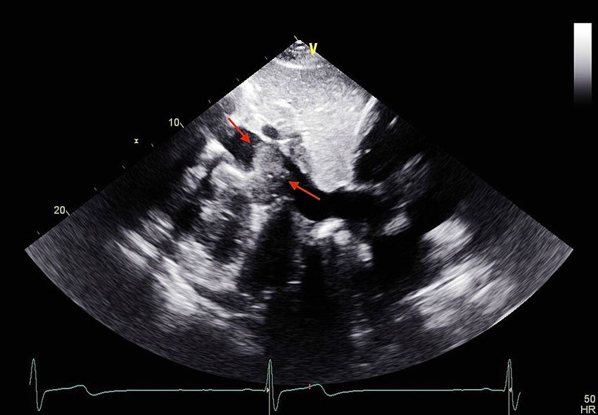
Laboratory Parameters Values Reference Range
Sodium 138 136-145 mmol/L
Potassium 4.3 3.5-5.3 mmol/L
Chloride 110 98-110 mmol/L
Blood urea nitrogen (BUN) 7 6-24 mg/dL
Creatinine 0.7 0.6-1.2 mg/dL
Aspartate transaminase (AST) 33 10-36 U/L
Alanine transaminase (ALT) 27 9-46 U/L
White blood cell (WBC) 9.4 4.4-11 x 103 /uL
Hemoglobin 7.8 13.5-17.5 g/dL
Platelets 311 150-450 x 103 /uL
Lactate dehydrogenase (LDH) 874 122-222 U/L
Reticulocyte count 7.8 0.5-1.5%
Total bilirubin 2 0.2-1.2 mg/dL
TABLE 1: Initial laboratory values.
Peripheral blood smear showed few giant cells, anisocytosis, and sickle cells. Chest x-ray revealed no
evidence of cardiopulmonary disease. Echocardiogram showed a 2.8 x 3.2 cm cystic mass compressing the
inferior vena cava with overlying fibrinous material (Figure 1). Ultrasound of the abdomen showed a mass
impressing on the posterior wall of the suprarenal inferior vena cava extending into the lumen. Magnetic
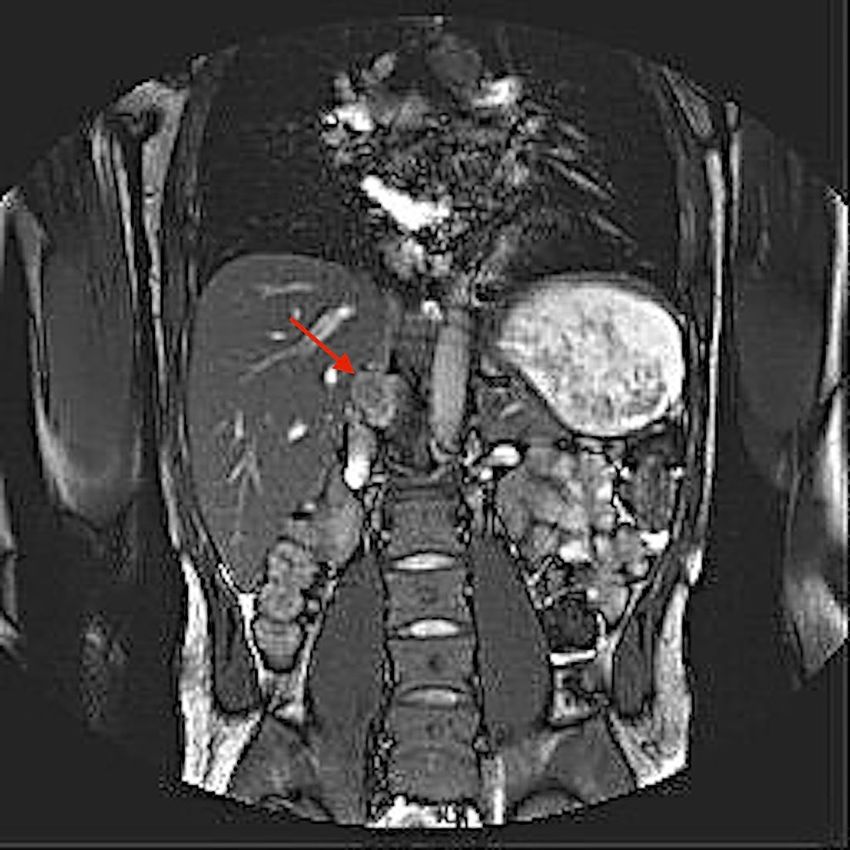
resonance imaging (MRI) of the abdomen with and without contrast revealed a 4.7 x 2.7 cm progressively
enhancing right retroperitoneal mass (Figure 2).
FIGURE 1: Cystic mass of 2.8 x 3.2 cm compressing the inferior vena
cava on ECHO.
ECHO: echocardiogram
2022 Ajayi et al. Cureus 14(1): e21334. DOI 10.7759/cureus.21334 2 of 4FIGURE 2: Progressively enhancing mass of 4.7 x 2.7 cm on MRI of the
abdomen.
The patient was started on symptomatic management and close imaging surveillance for the mass was
recommended. Follow-up after two months in the outpatient hematology clinic showed a stable mass with
no symptoms.
Discussion
Sickle cell disease is a common, congenital blood disorder that is characterized by abnormally shaped red
blood cells [1]. This deformation impairs the ability of the cell to pass through the small vascular channels
resulting in congestion of the vascular beds. These sickle-shaped erythrocytes then undergo intravascular
hemolysis and are destroyed at an increased rate leading to chronic anemia [2]. In these circumstances,
extramedullary production of erythrocytes can be initiated to supplement the increased destruction of red
blood cells [3].
Extramedullary hematopoiesis (EMH) is a reactive mechanism that can be seen in congenital hemolytic
anemias. EMH can be divided into two main groups. The first group is seen in hemolytic disorders where the
marrow shows tremendous activity and paraosseous foci which may result from herniation of the medullary
tissue from the underlying bone. The second group shows extraosseous foci and is seen when marrow
activity is ineffective [4]. Common sites of EMH include the reticuloendothelial system such as spleen,
lymph node, and mediastinum; however, the process can include virtually any tissue of the body [5]. Cases
have been reported of EMH in the bowel, breast, pleura, and brain [6]. In our literature review, we found
cases of retroperitoneal EMH after thalassemia and hereditary spherocytosis [7]. EMH presenting as
retroperitoneal mass in a patient with sickle cell disease has rarely been presented. We report a case of EMH
found as a retroperitoneal mass in a patient with sickle cell disease. Our patient presented with pain in the
right thorax with MRI abdomen findings suggestive of an adrenal retroperitoneal mass. Patient was also
found to have periportal and left retroperitoneal lymph nodes on diffusion-weighted imaging. Patient was
normotensive with a normal metanephrine level. This incidental adrenal mass along with lymph node
proliferation in the retroperitoneum is highly suggestive of EMH.
The prevalence of adrenal incidentaloma is about 0.5-2% on the abdominal computed tomography (CT) scan
2022 Ajayi et al. Cureus 14(1): e21334. DOI 10.7759/cureus.21334 3 of 4in the general population [8]. EMH is a rare cause of incidentaloma but may be seen in patients with
hematological disorders like thalassemia. The exact mechanism of EMH is unknown but several hypotheses
have been suggested. It is suggested that since adrenal glands have hematopoietic capacity during the fetal
period, anomalous activation of these embryonic stem cells can occur during the disease state. Another
hypothesis suggests that embolization of the hematopoietic stem cells and homing in the adrenal gland may
play a role in this phenomenon. Chronic hypoxia has also been reported as a presumptive cause of EMH [9].
EMH is normally asymptomatic and discovered as an incidental finding. Symptomatic cases can be seen due
to the mass effect created by compression of the adjacent organs [10].
Diagnosis of EMH is based on the clinical picture, laboratory data, and the use of imaging modalities [4]. MRI
is considered to be the diagnostic modality of choice and is superior to CT scan. MRI can effectively
delineate the extent of the EMH mass and has no radiation exposure [11]. Biopsy is not always indicated but
can be utilized for definitive diagnosis. Treatment modalities for patients with EMH depend on the location
and symptoms. Surgery, local radiation, blood transfusion, and hydroxyurea are some treatment approaches.
Surgical excision is indicated if the mass is more than 6 cm in diameter [9]. In our case, the size of the tumor
was not large enough for excision. Hydroxyurea is used as an adjuvant therapy due to its myelosuppressive
properties [12]. Hypertransfusion is another rising therapy that is postulated to reduce the risk of EMH by
bone marrow suppression. In our patient imaging surveillance with close follow-up was advised since the
patient was asymptomatic.
Conclusions
This case suggests that EMH should be taken into consideration when retroperitoneal masses are identified
in patients with blood dyscrasias. This case is unique due to the location of the EMH. The patient was
followed closely by imaging surveillance to monitor the size of the mass. This case also suggests that
increased awareness of this rare entity is needed to avoid aggressive therapeutic measures in asymptomatic
patients. In such patients, conservative management techniques should be employed.
Additional Information
Disclosures
Human subjects: Consent was obtained or waived by all participants in this study. Conflicts of interest: In
compliance with the ICMJE uniform disclosure form, all authors declare the following: Payment/services
info: All authors have declared that no financial support was received from any organization for the
submitted work. Financial relationships: All authors have declared that they have no financial
relationships at present or within the previous three years with any organizations that might have an
interest in the submitted work. Other relationships: All authors have declared that there are no other
relationships or activities that could appear to have influenced the submitted work.
References
1. Saito N, Nadgir RN, Flower EN, Sakai O: Clinical and radiologic manifestations of sickle cell disease in the
head and neck. RadioGraphics. 2010, 30:1021-34. 10.1148/rg.304095171
2. Delicou S: Extramedullary haemopoiesis in hemoglobinopathies. J Hematol Transfus. 2017, 5:1066.
3. Lonergan GJ, Cline DB, Abbondanzo SL: Sickle cell anemia. RadioGraphics. 2001, 21:971-94.
10.1148/radiographics.21.4.g01jl23971
4. Jelali MA, Luciani A, Kobeiter H, et al.: MRI features of intrahepatic extramedullary haematopoiesis in sickle
cell anaemia. Cancer Imaging. 2006, 6:182-5. 10.1102/1470-7330.2006.0030
5. Tayari N, Ahrar MH, Jafarpishe MS: Case report of the extramedullary hematopoiesis presented as a
hypervascular intracranial mass. Adv Biomed Res. 2013, 2:34. 10.4103/2277-9175.109719
6. Clark CA, Worden CP, Thorp BD, et al.: Extramedullary hematopoiesis in the sinonasal cavity: a case report
and review of the literature. Allergy Rhinol (Providence). 2020, 11: 10.1177/2152656720918874
7. Hassanzadeh M: Extramedullary hematopoiesis in thalassemia. N Engl J Med. 2013,
369:10.1056/NEJMicm1303084
8. Azarpira N, Esfahani MH, Paydar S: Extramedullary hematopoiesis in adrenal gland. An uncommon cause of
adrenal incidentaloma in sickle cell disease. Iran J Pediatr. 2014, 24:784-6.
9. Barzon L, Sonino N, Fallo F, Palu G, Boscaro M: Prevalence and natural history of adrenal incidentalomas .
Eur J Endocrinol. 2003, 149:273-85. 10.1530/eje.0.1490273
10. Kolev NH, Genov PP, Dunev VR, Stoykov BA: A rare case of extramedullary hematopoiesis in adrenal mass .
Urol Case Rep. 2020, 30:10.1016/j.eucr.2020.101120
11. Verani R, Olson J, Moake JL: Intrathoracic extramedullary hematopoiesis: report of a case in a patient with
sickle-cell disease-beta-thalassemia. Am J Clin Pathol. 1980, 73:133-7. 10.1093/ajcp/73.1.133
12. Wang A, Carberry N, Solli E, Gillick J, Islam H, Hillard V: Spinal cord compression secondary to
extramedullary hematopoiesis: case report and review of the literature. Case Rep Oncol. 2016, 9:290-7.
10.1159/000446473
2022 Ajayi et al. Cureus 14(1): e21334. DOI 10.7759/cureus.21334 4 of 4You can also read